Review
. 2020 Nov 29;21(23):9086.
doi: 10.3390/ijms21239086.

Role of Regular Physical Activity in Neuroprotection against Acute Ischemia

Affiliations
Review

Role of Regular Physical Activity in Neuroprotection against Acute Ischemia

Domenico Di Raimondo et al. Int J Mol Sci. .

Abstract

One of the major obstacles that prevents an effective therapeutic intervention against ischemic stroke is the lack of neuroprotective agents able to reduce neuronal damage; this results in frequent evolution towards a long-term disability with limited alternatives available to aid in recovery. Nevertheless, various treatment options have shown clinical efficacy. Neurotrophins such as brain-derived neurotrophic factor (BDNF), widely produced throughout the brain, but also in distant tissues such as the muscle, have demonstrated regenerative properties with the potential to restore damaged neural tissue. Neurotrophins play a significant role in both protection and recovery of function following neurological diseases such as ischemic stroke or traumatic brain injury. Unfortunately, the efficacy of exogenous administration of these neurotrophins is limited by rapid degradation with subsequent poor half-life and a lack of blood-brain-barrier permeability. Regular exercise seems to be a therapeutic approach able to induce the activation of several pathways related to the neurotrophins release. Exercise, furthermore, reduces the infarct volume in the ischemic brain and ameliorates motor function in animal models increasing astrocyte proliferation, inducing angiogenesis and reducing neuronal apoptosis and oxidative stress. One of the most critical issues is to identify the relationship between neurotrophins and myokines, newly discovered skeletal muscle-derived factors released during and after exercise able to exert several biological functions. Various myokines (e.g., Insulin-Like Growth Factor 1, Irisin) have recently shown their ability to protects against neuronal injury in cerebral ischemia models, suggesting that these substances may influence the degree of neuronal damage in part via inhibiting inflammatory signaling pathways. The aim of this narrative review is to examine the main experimental data available to date on the neuroprotective and anti-ischemic role of regular exercise, analyzing also the possible role played by neurotrophins and myokines.

Keywords: brain-derived neurotrophic factor (BDNF); inflammation; ischemic stroke; myokines; neuronal recovery; neurotrophins; physical activity.


Conflict of interest statement

The authors declare no conflict of interest.

Figures

Figure 1
Figure 1
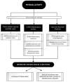
Flow chart of the literature search and selection.
Figure 2
Figure 2
The putative role of PA in the management of major neurological diseases. In this figure, we summarize the main mechanisms through which regular PA could lead to better management of the four major neurological diseases: stroke [127–131], Alzheimer’s disease, Parkinson’s disease and epilepsy [5,21–23]. PA’s role could be pivotal in the prevention as well as in the prognosis’ improvement.
Figure 3
Figure 3
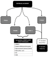
The attempt to improve neurological functions through regular PA: a three paths journey. PA achieves beneficial effects on brain function through its impact in these three main pathways whose modulation eventually leads to a neuroprotective effect. Regular aerobic training as opposed to acute bouts of exercise, seems to be more related to PA positive impact leading to antioxidant [47,48] and anti-inflammatory [51–53,56] effects and, through molecular and vascular changes, shapes neuronal and vascular plasticity [70,75,78,79]. See text for further information. ROS, Reactive Oxygen Species; CAMs, Cell Adhesion Molecules; DNA, DeoxyriboNucleic Acid.
Figure 4
Figure 4
The hormesis theory and the improved antioxidant activity of trained subjects [49,50]. The hormesis theory tries to explain the apparent paradox related to the relationship between oxidative stress and exercise (see text for more details); low-moderate regular exercise enhances endogenous antioxidant capacity resulting in an increased ability to handle acute oxidative stresses. ROS, reactive oxygen species.
Figure 5
Figure 5
Immuno-inflammatory activation during ischemic stroke. The figure briefly outlines the main determinants and characters involved in the “inflammatory face” of ischemic pathobiology. Chronic inflammatory diseases [98] and genetic predisposition [98,118] represent the key elements playing a leading role in determining the variability of immune-inflammatory activation [98,101,104,107–113] on which depends the patient’s outcome: the inflammatory background elements and the ischemic stroke subtypes (TOAST classification) [121]. CEI, CardioEmbolic Infarct; LAC, LACunar infarct; UDE, a stroke of UnDetermined Etiology; ODE, a stroke of Other Determined Etiology; LAAS, Large Artery AtheroSclerosis; IL, Interleukin; ICAM, Intercellular Cell Adhesion Molecule; TNF, Tumor Necrosis Factor.
Figure 6
Figure 6
PA: a neurovascular booster. During PA, the increased need for oxygen and energetic substrates leads to the activation of many mechanisms whose ultimate effect is to determine an increased cerebral blood flow and to maintain neurovascular integrity. This aim is exerted both by asserting a boosting of cardiac contractility (quantitative effect) [123–126] and by a local cerebral modulation of neurovascular dynamic (qualitative effect) through the recruitment of collateral vessels [127] and the stimulation of angiogenic processes [130]. VEGF, vascular endothelial growth factor; VEGFR-2, vascular endothelial growth factor receptor 2; ANG-2, Angiopoietin-2.
Figure 7
Figure 7
Preconditioning exercise: brain’s eustress. PA is a mild stressor for the organism. In particular, preconditioning exercise (see text for more information) can be defined as an “eustress” because it triggers cells to express multiple factors, thus helping the organism to acquire tolerance and self-defense against later possible damage, such as ischemic stroke. Preconditioning exercise triggers the brain to activate vascular and inflammatory mechanisms (seen in the figure) [134,141–146] whose production remodulates cerebral neurovascular structure so that later pathologic stimuli are less likely to act or less likely to exert important damage. Preconditioning exercise improves the function and structure of blood–brain barrier (BBB) [134] thus limiting the leakage of both fluids (edema) and proteins (cytokines) who play an important role in ischemic stroke pathobiology. VEGF, vascular endothelial growth factor; IGF-1, Insulin-like Growth factor 1; MK, midkine; BBB, blood–brain barrier.
Figure 8
Figure 8
Neurotrophins, Myokines and other muscle-derived factors involved in neuroprotection. Neuroprotective effects of PA result from multiple molecular changes operating directly in the cerebral tissue and indirectly via vascular modulation. These molecular targets interplay with each other achieving different outcomes. The BDNF-IGF1 network [89–194] and the VEGF-IGF1 network represent two main examples. BDNF-IGF1 network mediates the upregulation of genes mainly related to neuronal viability [78,79]. See text for further information. NAA, N-acetylaspartate; BDNF brain-Derived Neurotrophic Factor; IGF-1, Insulin-Like Growth Factor 1; VEGF, Vascular Endothelial Growth Factor; GABA, Gamma-AminoButyric Acid; NO, Nitric Oxyde; NMDA-R, N-methyl-d-aspartate receptor; CaM-KII, calcium/calmodulin protein kinase II; MAP-K/ERK I and II, mitogen-activated protein kinase; PK-C, protein kinase C; CREB, cAMP response element-binding protein.

References

    1. American College of Sports Medicine . In: ACM’s Guidelines for Exercise Testing and Prescription. 10th ed. Riebe D., Ehrman J.K., Liguori G., Magal M., editors. Wolters Kluwer; Philadelphia, PA, USA: 2018.
    1. Chodzko-Zajko W.J., Proctor D.N., Fiatarone Singh M.A., Minson C.T., Nigg C.R., Salem G.J., Skinner J.S. Exercise and physical activity for older adults. Med. Sci. Sports Exerc. 2009;41:1510–1530. doi: 10.1249/MSS.0b013e3181a0c95c. - DOI - PubMed
    1. Wahid A., Manek N., Nichols M., Kelly P., Foster C., Webster P., Kaur A., Friedemann Smith C., Wilkins E., Rayner M., et al. Quantifying the Association Between Physical Activity and Cardiovascular Disease and Diabetes: A Systematic Review and Meta-Analysis. J. Am. Heart Assoc. 2016;5:e002495. doi: 10.1161/JAHA.115.002495. - DOI - PMC - PubMed
    1. Sanchis-Gomar F., Fiuza-Luces C., Lucia A. Exercise as the master polypill of the 21st century for the prevention of cardiovascular disease. Int. J. Cardiol. 2015;181:360–361. doi: 10.1016/j.ijcard.2014.12.048. - DOI - PubMed
    1. Di Raimondo D., Musiari G., Miceli G., Arnao V., Pinto A. Preventive and Therapeutic Role of Muscle Contraction against Chronic Diseases. Curr. Pharm. Des. 2016;22:4686–4699. doi: 10.2174/1381612822666160510125011. - DOI - PubMed