. 2020 Nov 29;18(12):604.
doi: 10.3390/md18120604.

A New Bioassay Platform Design for the Discovery of Small Molecules with Anticancer Immunotherapeutic Activity

Affiliations

A New Bioassay Platform Design for the Discovery of Small Molecules with Anticancer Immunotherapeutic Activity

Carmela Gallo et al. Mar Drugs. .

Abstract

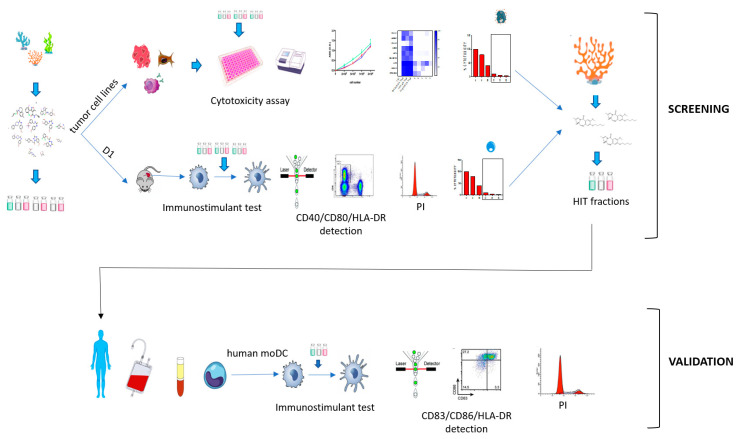
Immunotherapy takes advantage of the immune system to prevent, control, and eliminate neoplastic cells. The research in the field has already led to major breakthroughs to treat cancer. In this work, we describe a platform that integrates in vitro bioassays to test the immune response and direct antitumor effects for the preclinical discovery of anticancer candidates. The platform relies on the use of dendritic cells that are professional antigen-presenting cells (APC) able to activate T cells and trigger a primary adaptive immune response. The experimental procedure is based on two phenotypic assays for the selection of chemical leads by both a panel of nine tumor cell lines and growth factor-dependent immature mouse dendritic cells (D1). The positive hits are then validated by a secondary test on human monocyte-derived dendritic cells (MoDCs). The aim of this approach is the selection of potential immunotherapeutic small molecules from natural extracts or chemical libraries.

Keywords: anticancer; bioassay platform; chemical immunology; dendritic cell; drug discovery; high throughput; immunotherapy; screening guidelines.


Conflict of interest statement

The authors declare no competing interest.

Figures

Figure 1
Figure 1
(a) Mouse Dendritic cell line (D1) surface marker analysis of CD80, CD40, and MHC-II in each cell passage along 12 days (n = 6) from P0 to P5. The color bar on the right shows the MFI (mean fluorescence intensity) measured for each marker; (b) surface marker expression analysis of D1 untreated (Ctrl) and treated with LPS (10 µg/mL; 24 h) (n = 5); error bars indicate standard deviations; (c) MFI of CD80, CD40, and MHC-II in D1 cells treated with Sulfavant A compared with cells treated with vehicle (Ctrl = MeOH) (n = 7); error bars indicate standard deviations; asterisks indicate significant differences from Ctrl; * p < 0.5, ** p < 0.01, *** p < 0.001, **** p < 0.0001.
Figure 2
Figure 2
Overall scheme for hits selection. In the preliminary screening, natural products are tested on dendritic cell lines (D1) and for cytotoxicity on target lung carcinoma (LC), melanoma (Mel), and multiple myeloma (MM) cell lines. In the second step, the “candidate” samples are validated for immunomodulatory activity on human monocyte-derived dendritic cells (MoDCs).
Figure 3
Figure 3
(a) Surface expression analysis of CD80, CD40, and MHC-II and percentage of vitality on D1 treated with the extract (Ext) and four fractions (B, C, D, E) that were derived from T. weissflogii at 5 and 30 µg/mL; all data were compared to the cells treated only with the vehicle (Ctrl) and cells treated with positive controls (LPS, Sulfavant A); (b) heat map of cytotoxicity assays conducted on the nine different cell lines. Cells were treated with positive controls, the extract (Ext) and four fractions (B, C, D, E) were derived from the marine diatom. Values reported in the color bar legend on the right indicate the % of cytotoxicity. The color bar on the right shows the MFI (mean fluorescence intensity) measured for each marker and the % of cytotoxicity.
Figure 4
Figure 4
(a) Surface expression analysis of CD83, CD86, and HLA-DR in MoDCs treated with the fraction C derived from the extract of Thalassiosira weissflogii from 0.01 to 30 µg/mL; all data were compared to the cells treated only with the vehicle (Ctrl) and cells treated with Sulfavant A (30 µg/mL); (b) HLA-DR analysis of MoDCs treated with extract (Ext), SPE C fraction and purified α-SQDGs at concentration of 30 µg/mL; error bars indicate standard deviations; ** p < 0.01, **** p < 0.0001.
Figure 5
Figure 5
Screening platform workflow.

Similar articles

Cited by

References

    1. Dunn G.P., Bruce A.T., Ikeda H., Old L.J., Schreiber R.D. Cancer immunoediting: From immunosurveillance to tumor escape. Nat. Immunol. 2002;3:991–998. doi: 10.1038/ni1102-991. - DOI - PubMed
    1. De Visser K.E., Eichten A., Coussens L.M. Paradoxical roles of the immune system during cancer development. Nat. Rev. Cancer. 2006;6:24–37. doi: 10.1038/nrc1782. - DOI - PubMed
    1. Dougan M., Dranoff G., Dougan S.K. Cancer Immunotherapy: Beyond Checkpoint Blockade. Annu. Rev. Cancer Biol. 2019;3:55–75. doi: 10.1146/annurev-cancerbio-030518-055552. - DOI - PMC - PubMed
    1. Ribas A., Wolchok J.D. Cancer immunotherapy using checkpoint blockade. Science. 2018;359:1350–1355. doi: 10.1126/science.aar4060. - DOI - PMC - PubMed
    1. Wculek S.K., Cueto F.J., Mujal A.M., Melero I., Krummel M.F., Sancho D. Dendritic cells in cancer immunology and immunotherapy. Nat. Rev. Immunol. 2020;20:7–24. doi: 10.1038/s41577-019-0210-z. - DOI - PubMed

MeSH terms

Substances