Clinical Trial
. 2020 Nov 28;11(12):1434.
doi: 10.3390/genes11121434.

TYK2 Variants in B-Acute Lymphoblastic Leukaemia

Affiliations
Clinical Trial

TYK2 Variants in B-Acute Lymphoblastic Leukaemia

Edgar Turrubiartes-Martínez et al. Genes (Basel). .

Abstract

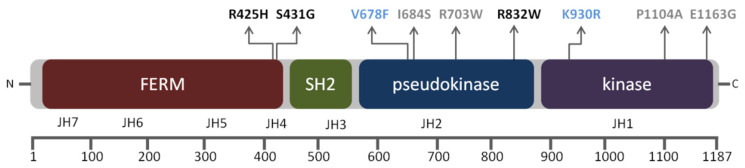
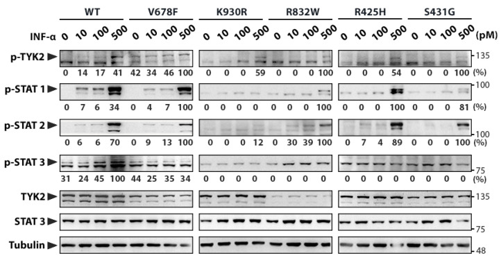
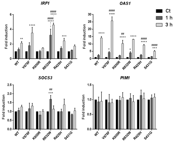
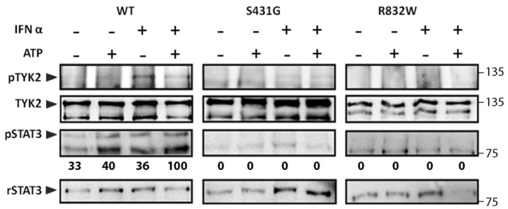
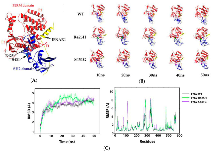
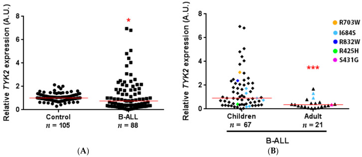
B-cell precursor acute lymphoblastic leukaemia (B-ALL) is a malignancy of lymphoid progenitor cells with altered genes including the Janus kinase (JAK) gene family. Among them, tyrosine kinase 2 (TYK2) is involved in signal transduction of cytokines such as interferon (IFN) α/β through IFN-α/β receptor alpha chain (IFNAR1). To search for disease-associated TYK2 variants, bone marrow samples from 62 B-ALL patients at diagnosis were analysed by next-generation sequencing. TYK2 variants were found in 16 patients (25.8%): one patient had a novel mutation at the four-point-one, ezrin, radixin, moesin (FERM) domain (S431G) and two patients had the rare variants rs150601734 or rs55882956 (R425H or R832W). To functionally characterise them, they were generated by direct mutagenesis, cloned in expression vectors, and transfected in TYK2-deficient cells. Under high-IFNα doses, the three variants were competent to phosphorylate STAT1/2. While R425H and R832W induced STAT1/2-target genes measured by qPCR, S431G behaved as the kinase-dead form of the protein. None of these variants phosphorylated STAT3 in in vitro kinase assays. Molecular dynamics simulation showed that TYK2/IFNAR1 interaction is not affected by these variants. Finally, qPCR analysis revealed diminished expression of TYK2 in B-ALL patients at diagnosis compared to that in healthy donors, further stressing the tumour immune surveillance role of TYK2.

Keywords: B-cell precursor acute lymphoblastic leukaemia; IFNα; IFNα/β receptor alpha chain (IFNAR1), next-generation sequencing; TYK2 expression; TYK2 variants; immune surveillance; molecular dynamics.


Conflict of interest statement

The authors declare no conflict of interest. The funders had no role in the design of the study; in the collection, analyses, or interpretation of data; in the writing of the manuscript; or in the decision to publish the results.

Figures

Figure 1
Figure 1
Schematic representation of structural and functional domains of tyrosine kinase 2 (TYK2) protein with the non-synonymous variants detected in this study (functional characterised variants in black; artificial mutant controls in blue: kinase-dead form of the protein (TYK2 K930R) and the catalytically hyperactive form (TYK2 V678F). Boxes illustrate functional domains from N- to C-terminus and the corresponding Janus kinase (JAK) structural homology domains (JH1–JH7) are marked below. Amino acid numbers are shown in the lower bar scale according to the human TYK2 protein sequence (RefSeq: NM_003331.4; FERM domain: 26–431, SH2 domain: 452–551, pseudokinase domain: 551–875, and kinase domain: 897–1176).
Figure 2
Figure 2
Functional analyses of TYK2 variants. Human TYK2-deficient U1A cell line was transfected with the different TYK2 variants and clones stably expressing these variants were selected. Clones were stimulated with the indicated doses of IFNα for 15 min, and the level of phosphorylation of TYK2 variants and STAT1–3 transcription factors was assessed by Western blot. Arrowheads point out the size of specific proteins and molecular weight markers (kDa) are noted on the right. Quantification of phosphorylated proteins is relative to TYK2 total protein (to account for TYK2 expression differences among cell lines) and then to Tubulin (shown under each lane/membrane). Values are expressed as percentage of the highest value of each membrane. WT: wild-type TYK2; V678F: catalytically hyperactive TYK2; K930R: kinase-dead TYK2 (ATP-binding site mutant); R832W, R425H, and S431G: disease-associated TYK2 variants. A representative result of three independent experiments is shown (Figure S2).
Figure 3
Figure 3
STAT-target gene expression induced by IFNα in U1A cells expressing TYK2 variants. Cells were treated with 500 pM IFNα. Total RNA was extracted, reverse-transcribed, and analysed by RT-qPCR for the indicated genes. Values were normalised to GAPDH and are expressed as n-fold versus untreated cells for each variant. Mean values of three independent experiments ± SD are shown. Two-way ANOVA was used for statistical analysis, performing post hoc Bonferroni tests for intergroup differences. * For each variant, comparison between the indicated time and untreated cells; # for each time condition, comparison between each variant and WT. For * and #: * p < 0.05; ** p < 0.01; *** p < 0.001; **** p < 0.0001.
Figure 4
Figure 4
In vitro kinase activity of TYK2 WT, TYK2 S431G, and TYK2 R832W proteins. TYK2 was immunoprecipitated with anti-TYK2 Ab from non-stimulated cells or cells stimulated with IFNα (500 pM) for 15 min and subjected to in vitro kinase reaction in the presence or absence of 30 μM ATP for 5 min at 30 °C. Recombinant STAT3 (rSTAT3) was added to the reaction as exogenous substrate. The phosphorylation level of the indicated proteins was analysed by immunoblotting. STAT3 phosphorylation was represented relative to rSTAT3 and then to TYK2, to account for differences in the amount of immunoprecipitated TYK2 in each sample. Values are expressed as percentage of the highest value of each membrane. A representative result of three independent experiments is shown.
Figure 5
Figure 5
Interaction of four-point-one, ezrin, radixin, moesin (FERM) and src-homology 2 (SH2) domains of TYK2 with IFNAR1−α/β receptor alpha chain (IFNAR1). (A) 3D representation of the interaction of TYK2 FERM and SH2 domains with IFNAR1 (ID PDB: 4PO6). FERM domain is represented by subdomains (F1, F2, and F3) in red, SH2 domain in blue, and IFNAR1 in yellow; the sites of TYK2 R425 and S431 are represented with spheres in the protein structure and are indicated with black arrows. (B) Structural models from 0 to 50 ns of molecular dynamics (MD) of the TYK2–IFNAR1 complex, using the WT, R425H and S431G mutants. (C) Root mean square deviation (RMSD) and root mean square fluctuation (RMSF) of the TYK2 WT–IFNAR1, the TYK2 R425H–IFNAR1, and TYK2 S431G–IFNAR1 complexes. RMSD as a function of time (left) and RMSF vs. residues (right).
Figure 6
Figure 6
TYK2 expression in B-cell precursor acute lymphoblastic leukaemia (B-ALL) patients. qPCR was performed on cDNA obtained from 1 μg of RNA isolated from the bone marrow of B-ALL patients and peripheral blood mononuclear cells (PBMCs) of healthy donors. For each sample, data were standardised against GAPDH expression. (A) Total B-ALL patients compared to healthy donors. Red line shows the median in each group (1.009 vs. 0.7594). (B) Age-related TYK2 expression in B-ALL patients. Children under 18 years compared to adults (median: 0.9147 vs. 0.3609). Patients with TYK2 variants are highlighted with different colours. * p < 0.05, *** p ≤ 0.001.

References

    1. Liu Y.F., Wang B.Y., Zhang W.N., Huang J.Y., Li B.S., Zhang M., Jiang L., Li J.F., Wang M.J., Dai Y.J., et al. Genomic profiling of adult and pediatric B-cell acute lymphoblastic leukemia. EBioMedicine. 2016;8:173–183. doi: 10.1016/j.ebiom.2016.04.038. - DOI - PMC - PubMed
    1. Iacobucci I., Mullighan C.G. Genetic basis of acutfe lymphoblastic leukemia. J. Clin. Oncol. 2017;35:975–983. doi: 10.1200/JCO.2016.70.7836. - DOI - PMC - PubMed
    1. Bhojwani D., Yang J.J., Pui C.H. Biology of childhood acute lymphoblastic leukemia. Pediatr. Clin. N. Am. 2015;62:47–60. doi: 10.1016/j.pcl.2014.09.004. - DOI - PMC - PubMed
    1. Mullighan C.G., Zhang J., Harvey R.C., Collins-Underwood J.R., Schulman B.A., Phillips L.A., Tasian S.K., Loh M.L., Su X., Liu W., et al. JAK mutations in high-risk childhood acute lymphoblastic leukemia. Proc. Natl. Acad. Sci. USA. 2009;106:9414–9418. doi: 10.1073/pnas.0811761106. - DOI - PMC - PubMed
    1. Forero-Castro M., Robledo C., Benito R., Bodega-Mayor I., Rapado I., Hernandez-Sanchez M., Abaigar M., Maria Hernandez-Sanchez J., Quijada-Alamo M., Maria Sanchez-Pina J., et al. Mutations in TP53 and JAK2 are independent prognostic biomarkers in B-cell precursor acute lymphoblastic leukaemia. Br. J. Cancer. 2017;117:256–265. doi: 10.1038/bjc.2017.152. - DOI - PMC - PubMed

Publication types

LinkOut - more resources