

Essential Oils of Alpinia nantoensis Retard Forskolin-Induced Melanogenesis via ERK1/2-Mediated Proteasomal Degradation of MITF
- PMID: 33260669
- PMCID: PMC7760488
- DOI: 10.3390/plants9121672
Essential Oils of Alpinia nantoensis Retard Forskolin-Induced Melanogenesis via ERK1/2-Mediated Proteasomal Degradation of MITF
Abstract
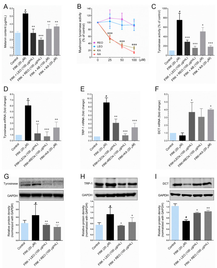
The anti-melanogenic activity of essential oils of Alpinia nantoensis and their bioactive ingredients were investigated in vitro. Treatment with leaf (LEO) and rhizome (REO) essential oils of A. nantoensis, significantly reduced forskolin-induced melanin production followed by down-regulation of tyrosinase (TYR) and tyrosinase related protein-1 (TRP-1) expression at both transcriptional and translational levels. Further studies revealed that down-regulation TYR and TRP-1 were caused by LEO/REO-mediated suppression of Microphthalmia-associated transcription factor (MITF), as evidenced by reduced nuclear translocation of MITF. Also, we found that LEO/REO induce the sustained activation of ERK1/2, which facilitate subsequent proteasomal degradation of MITF, as confirmed by that LEO/REO failed to inhibits MITF activity in ERK1/2 inhibitor treated cells. In addition, a significant increase of ubiquitinated MITF was observed after treatment with LEO and REO. Furthermore, the chemical composition of LEO and REO were characterized by gas chromatography-mass spectrometry (GC-MS) resulted that camphor, camphene, α-pinene, β-pinene, isoborneol and D-limonene were the major compounds in both LEO and REO. Further studies revealed that α-pinene and D-limonene were the active components responsible for the anti-melanogenic properties of LEO and REO. Based on the results, this study provided a strong evidence that LEO and REO could be promising natural sources for the development of novel skin-whitening agents for the cosmetic purposes.
Keywords: Alpinia nantoensis; MITF; Zingiberaceae; anti-melanogenesis; essential oil; forskolin.
Conflict of interest statement
The authors declare that there is no conflict of interest
Figures


Similar articles
-
Anti-Melanogenic Activity of Calocedrus formosana Wood Essential Oil and Its Chemical Composition Analysis.Plants (Basel). 2021 Dec 25;11(1):62. doi: 10.3390/plants11010062. Plants (Basel). 2021. PMID: 35009066 Free PMC article.
-
Anti-melanogenic activity of phytosphingosine via the modulation of the microphthalmia-associated transcription factor signaling pathway.J Dermatol Sci. 2017 Jul;87(1):19-28. doi: 10.1016/j.jdermsci.2017.03.011. Epub 2017 Mar 23. J Dermatol Sci. 2017. PMID: 28390782
-
Anti-Melanogenic Activities of Heracleum moellendorffii via ERK1/2-Mediated MITF Downregulation.Int J Mol Sci. 2016 Nov 4;17(11):1844. doi: 10.3390/ijms17111844. Int J Mol Sci. 2016. PMID: 27827938 Free PMC article.
-
Hesperidin, A Popular Antioxidant Inhibits Melanogenesis via Erk1/2 Mediated MITF Degradation.Int J Mol Sci. 2015 Aug 7;16(8):18384-95. doi: 10.3390/ijms160818384. Int J Mol Sci. 2015. PMID: 26262610 Free PMC article.
-
An overview of the chemical composition and biological activities of essential oils from Alpinia genus (Zingiberaceae).RSC Adv. 2021 Nov 23;11(60):37767-37783. doi: 10.1039/d1ra07370b. eCollection 2021 Nov 23. RSC Adv. 2021. PMID: 35498079 Free PMC article. Review.
Cited by
-
Oenothera laciniata Hill Extracts Exhibits Antioxidant Effects and Attenuates Melanogenesis in B16-F10 Cells via Downregulating CREB/MITF/Tyrosinase and Upregulating p-ERK and p-JNK.Plants (Basel). 2021 Apr 8;10(4):727. doi: 10.3390/plants10040727. Plants (Basel). 2021. PMID: 33917957 Free PMC article.
-
Anti-Melanogenic Activity of Calocedrus formosana Wood Essential Oil and Its Chemical Composition Analysis.Plants (Basel). 2021 Dec 25;11(1):62. doi: 10.3390/plants11010062. Plants (Basel). 2021. PMID: 35009066 Free PMC article.
-
Analysis of Chemical Composition and Biological Activities of Essential Oils from Different Parts of Alpinia uraiensis Hayata.Molecules. 2025 Mar 28;30(7):1515. doi: 10.3390/molecules30071515. Molecules. 2025. PMID: 40286082 Free PMC article.
-
Comparative Analysis of Chemical Profiles and Biological Activities of Essential Oils Derived from Torreya grandis Arils and Leaves: In Vitro and In Silico Studies.Plants (Basel). 2024 Sep 21;13(18):2640. doi: 10.3390/plants13182640. Plants (Basel). 2024. PMID: 39339615 Free PMC article.
-
Anti-Melanogenesis Effect of Polysaccharide from Saussurea involucrata on Forskolin-Induced Melanogenesis in B16F10 Melanoma Cells.Nutrients. 2022 Nov 27;14(23):5044. doi: 10.3390/nu14235044. Nutrients. 2022. PMID: 36501075 Free PMC article.
References
-
- Corre S., Primot A., Sviderskaya E., Bennett D.C., Vaulont S., Goding C.R., Galibert M.D. UV-induced expression of key component of the tanning process, the POMC and MC1R genes, is dependent on the p-38-activated upstream stimulating factor-1 (USF-1) J. Biol. Chem. 2004;279:51226–51233. doi: 10.1074/jbc.M409768200. - DOI - PubMed
Grants and funding
LinkOut - more resources
Full Text Sources
Research Materials
Miscellaneous
