. 2020 Nov 28;11(12):1050.
doi: 10.3390/mi11121050.

Synthesis of Nanoscale Liposomes via Low-Cost Microfluidic Systems

Affiliations

Synthesis of Nanoscale Liposomes via Low-Cost Microfluidic Systems

Andres Aranguren et al. Micromachines (Basel). .

Abstract

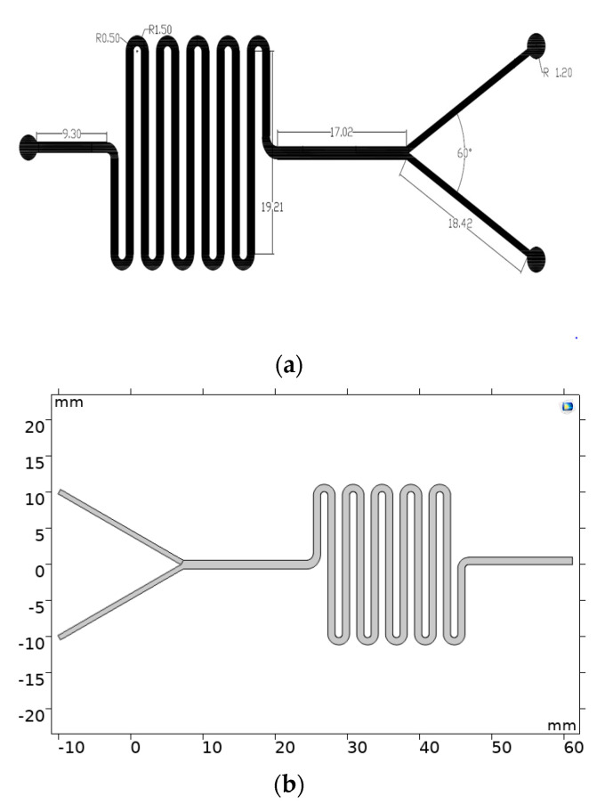
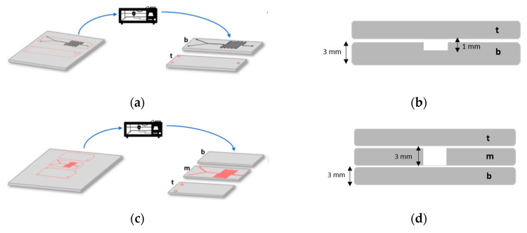
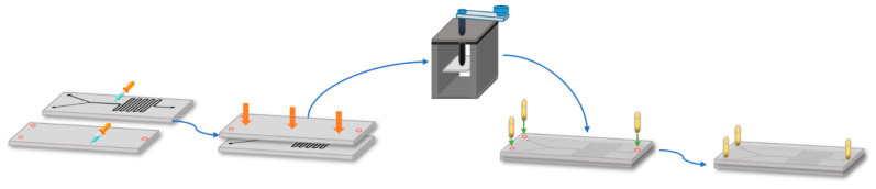
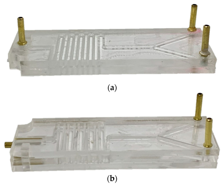
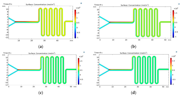
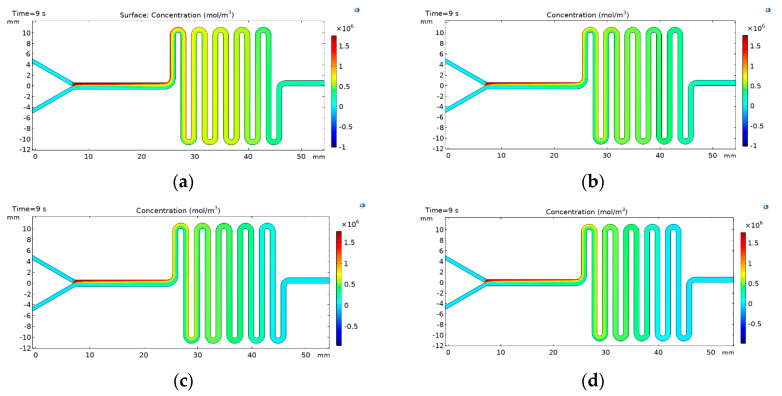
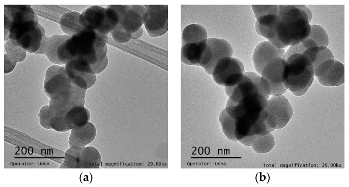
We describe the manufacture of low-cost microfluidic systems to produce nanoscale liposomes with highly uniform size distributions (i.e., low polydispersity indexes (PDI)) and acceptable colloidal stability. This was achieved by exploiting a Y-junction device followed by a serpentine micromixer geometry to facilitate the diffusion between the mixing phases (i.e., continuous and dispersed) via advective processes. Two different geometries were studied. In the first one, the microchannels were engraved with a laser cutting machine on a polymethyl methacrylate (PMMA) sheet and covered with another PMMA sheet to form a two-layer device. In the second one, microchannels were not engraved but through-hole cut on a PMMA sheet and encased by a top and a bottom PMMA sheet to form a three-layer device. The devices were tested out by putting in contact lipids dissolved in alcohol as the dispersed phase and water as the continuous phase to self-assemble the liposomes. By fixing the total flow rate (TFR) and varying the flow rate ratio (FRR), we obtained most liposomes with average hydrodynamic diameters ranging from 188 ± 61 to 1312 ± 373 nm and 0.30 ± 0.09 PDI values. Such liposomes were obtained by changing the FRR from 5:1 to 2:1. Our results approached those obtained by conventional bulk synthesis methods such as a thin hydration bilayer and freeze-thaw, which produced liposomes with diameters ranging from 200 ± 38 to 250 ± 38 nm and 0.30 ± 0.05 PDI values. The produced liposomes might find several potential applications in the biomedical field, particularly in encapsulation and drug delivery.

Keywords: liposomes; low-cost; microfluidic.


Conflict of interest statement

The authors declare no conflict of interest.

Figures

Figure 1
Figure 1
Design of geometries for manufacture and simulation of the microfluidic devices. (a) Microfluidic channel pattern designed for the two-layer device. The design was performed with the aid of AutoDesk® 2019 (AutoCAD) software. Measurements are presented in millimeters and degrees. (b) 2D design implemented in COMSOL Multiphysics ® for the finite elements simulations.
Figure 2
Figure 2
Microfluidic design pattern and laser cutting of device’s PMMA layer (t: top layer, m: middle layer, b: bottom layer). (a) Two-layers device. (b) Schematic of the cross-section view of the Two-layers device (channel width: 700 µm). (c) Three-layers device. (d) Schematic of the cross-section view of the Three-layers device (channel width: 700 µm).
Figure 3
Figure 3
Assembly method for the two-layer device. Gluing PMMA sheets with the aid of 70% (v/v) ethanol and a mechanical press, which was followed by placement on a hot plate (110 °C).
Figure 4
Figure 4
Actual pictures of the assembled devices. (a) Two-layers device. (b) Three-layers device.
Figure 5
Figure 5
Concentration profiles for the lipidic phase within the channel in the two-layers device (a) FRR 2:1, (b) FRR 3:1, (c) FRR 4:1, and (d) FRR 5:1.
Figure 6
Figure 6
Concentration profiles for the lipidic phase within the channel in the three-layers device (a) FRR 2:1, (b) FRR 3:1, (c) FRR 4:1, and (d) FRR 5:1. Selection of flow rate and solvent to an aqueous flow rate ratio (FRR).
Figure 7
Figure 7
(a). TEM microscopy analysis of the liposomes sample prepared using the two-layer device, PBS solution at a 4:1 FRR ratio. (b). TEM microscopy analysis of liposomes prepared using the three-layer device, solution (0.05 M) at a 2:1 FRR ratio.

References

    1. Van Swaay D., Demello A. Microfluidic methods for forming liposomes. Lab. Chip. 2013;13:752–767. doi: 10.1039/c2lc41121k. - DOI - PubMed
    1. Campaña A.L., Florez S.L., Noguera M.J., Fuentes O.P., Puentes P.R., Cruz J.C., Osma J.F. Enzyme-based electrochemical biosensors for microfluidic platforms to detect pharmaceutical residues in wastewater. Biosensors. 2019;9:41. doi: 10.3390/bios9010041. - DOI - PMC - PubMed
    1. Nunes J.K., Tsai S.S.H., Wan J., Stone H.A. Dripping and jetting in microfluidic multiphase flows applied to particle and fibre synthesis. J. Phys. D Appl. Phys. 2013;46:114002. doi: 10.1088/0022-3727/46/11/114002. - DOI - PMC - PubMed
    1. Peng Y., Zhao Y., Chen Y., Yang Z., Zhang L., Xiao W., Yang J., Guo L., Wu Y. Dual-targeting for brain-specific liposomes drug delivery system: Synthesis and preliminary evaluation. Bioorg. Med. Chem. 2018;26:4677–4686. doi: 10.1016/j.bmc.2018.08.006. - DOI - PubMed
    1. Carugo D., Bottaro E., Owen J., Stride E., Nastruzzi C. Liposome production by microfluidics: Potential and limiting factors. Sci. Rep. 2016;6:25876. doi: 10.1038/srep25876. - DOI - PMC - PubMed

LinkOut - more resources