. 2020 Nov 27;8(12):1878.
doi: 10.3390/microorganisms8121878.

Resolved Influenza A Virus Infection Has Extended Effects on Lung Homeostasis and Attenuates Allergic Airway Inflammation in a Mouse Model

Affiliations

Resolved Influenza A Virus Infection Has Extended Effects on Lung Homeostasis and Attenuates Allergic Airway Inflammation in a Mouse Model

Qingyu Wu et al. Microorganisms. .

Abstract

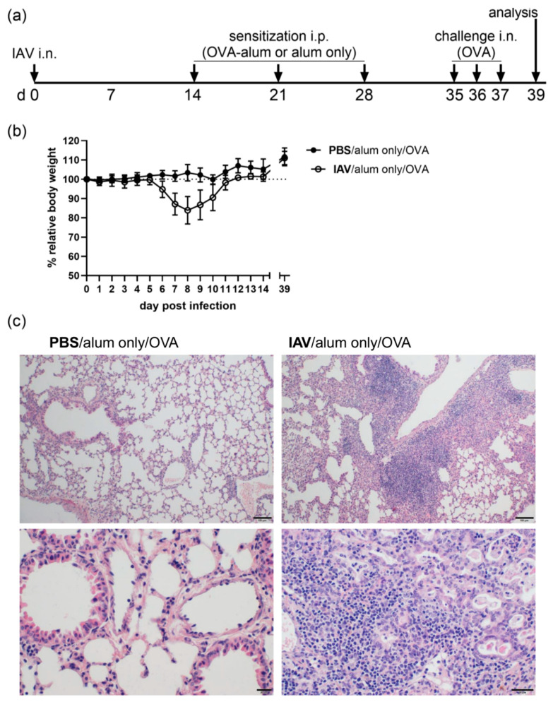
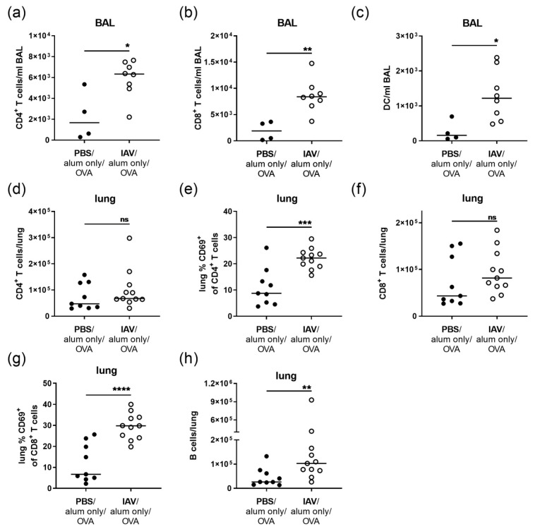
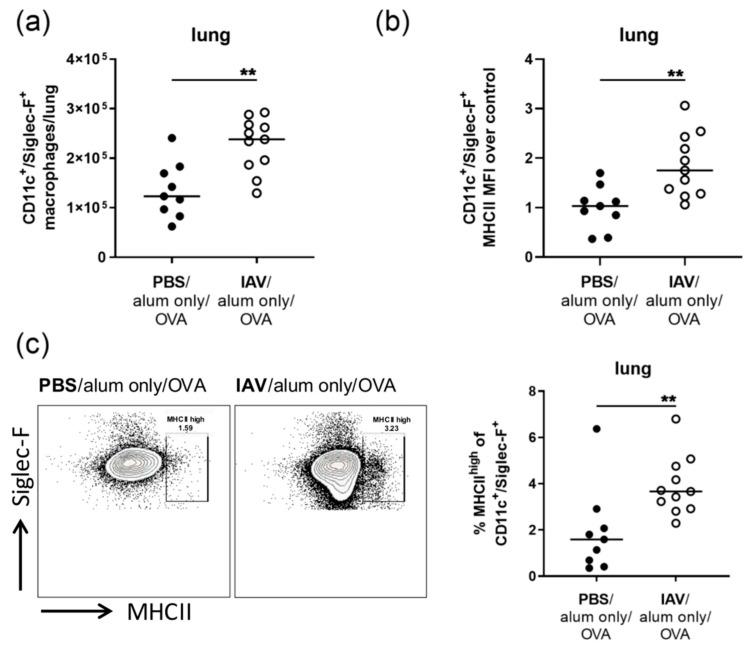
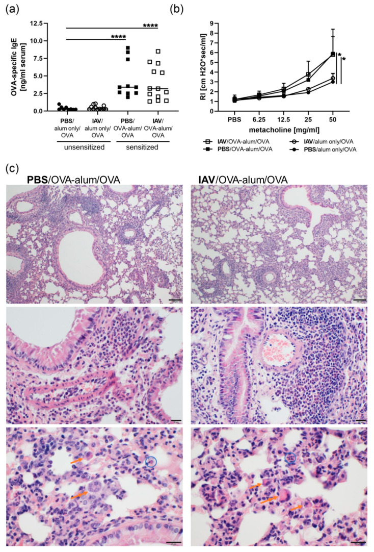
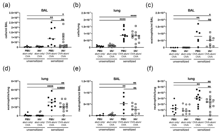
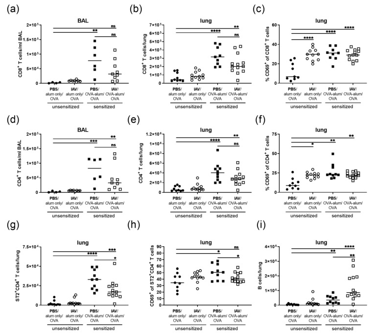
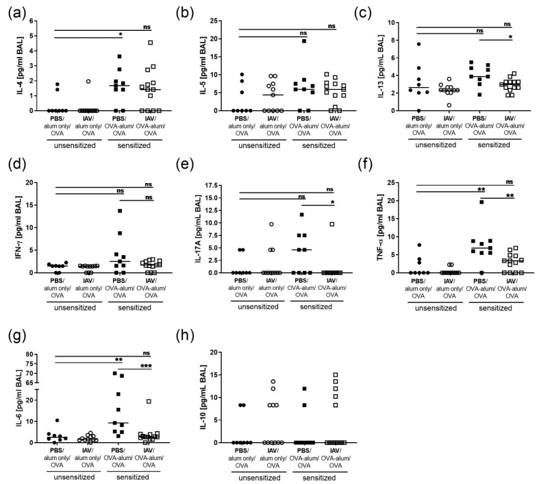
Allergic airway inflammation (AAI) involves T helper cell type 2 (Th2) and pro-inflammatory responses to aeroallergens and many predisposing factors remain elusive. Influenza A virus (IAV) is a major human pathogen that causes acute respiratory infections and induces specific immune responses essential for viral clearance and resolution of the infection. Beyond acute infection, IAV has been shown to persistently affect lung homeostasis and respiratory immunity. Here we asked how resolved IAV infection affects subsequently induced AAI. Mice infected with a sublethal dose of IAV were sensitized and challenged in an ovalbumin mediated mouse model for AAI after resolution of the acute viral infection. Histological changes, respiratory leukocytes, cytokines and airway hyperreactivity were analyzed in resolved IAV infection alone and in AAI with and without previous IAV infection. More than five weeks after infection, we detected persistent pneumonia with increased activated CD4+ and CD8+ lymphocytes as well as dendritic cells and MHCII expressing macrophages in the lung. Resolved IAV infection significantly affected subsequently induced AAI on different levels including morphological changes, respiratory leukocytes and lymphocytes as well as the pro-inflammatory cytokine responses, which was clearly diminished. We conclude that IAV has exceptional persisting effects on respiratory immunity with substantial consequences for subsequently induced AAI.

Keywords: allergic airway inflammation; allergic asthma; influenza A virus; macrophages; pro-inflammatory cytokines; respiratory immune regulation.


Conflict of interest statement

The authors declare no conflict of interest.

Figures

Figure 1
Figure 1
IAV (influenza A virus) infection leads to persistent histological changes in the lung. (a) Mice were infected i.n. with a sublethal dose of IAV or treated with PBS on day 0, were mock-sensitized (alum only i.p.; days 14, 21, 28) and treated i.n. with OVA (days 35, 36, 37). (b) Body weight was monitored daily following the infection. Relative body weights are shown as the mean ± SD of n = 19 controls (PBS/alum only/OVA) and n = 23 IAV infected mice (IAV/alum only/OVA) for days 0 to 14 and day 39 compiled from five experiments. (c) On day 39 post infection, mice were sacrificed and lung tissue was histologically analyzed. Representative images are shown for one out of 4 controls (PBS/alum only/OVA) and one out of 5 infected mice (IAV/alum only/OVA) analyzed. The upper images were prepared at 100× magnification (scale bar 100 µm), the lower images at 400× magnification (scale bar 20 µm).
Figure 2
Figure 2
Resolved IAV infection leads to elevated T cell numbers in the BAL and to persistent T cell activation as well as accumulation of B cells in the lung. Mice were infected i.n. with a sublethal dose of IAV or treated with PBS on day 0, were mock-sensitized (alum only i.p.; days 14, 21, 28) and treated i.n. with OVA (days 35, 36, 37). On day 39 post infection, CD4+ T cell (a), CD8+ T cell (b) and DC (c) numbers in the BAL (bronchoalveolar lavage) were flow cytometrically analyzed. In the lung, CD4+ T cell numbers (d), CD69-expression of CD4+ T cells (e), CD8+ T cell numbers (f), CD69-expression of CD8+ T cells (g) and B cell numbers (h) were analyzed. Data are shown for individual mice together with the group median. BAL data are compiled from at least two independent experiments and lung data are compiled from three independent experiments. * p < 0.05, ** p < 0.01, *** p < 0.005, **** p < 0.001, ns = not significant.
Figure 3
Figure 3
Resolved IAV infection leads to the accumulation of CD11c+/Siglec-F+ macrophages with increased MHCII expression the lung. Mice were infected i.n. with a sublethal dose of IAV or treated with PBS on day 0, were mock-sensitized (alum only i.p.; days 14, 21, 28) and treated i.n. with OVA (days 35, 36, 37). On day 39 post infection, CD11c+/Siglec-F+ macrophage numbers (a), CD11c+/Siglec-F+ macrophage MHCII expression (MFI, median fluorescence intensity) (b) and the frequency of MHCIIhigh CD11c+/Siglec-F+ macrophages in the CD11c+/Siglec-F+ macrophage population (c) were flow cytometrically analyzed. Data are shown for individual mice together with the group median. Representative flow cytometry plots show MHCII expression of the gated CD11c+/Siglec-F+ macrophage population of an uninfected and an infected mouse. Data are compiled from three independent experiments. ** p < 0.01.
Figure 4
Figure 4
Resolved IAV infection does not affect IgE production and airway hyperreactivity but affects histological changes in AAI. Mice were infected i.n. with a sublethal dose of IAV or treated with PBS on day 0, were sensitized against OVA i.p or mock-sensitized with alum only i.p. on days 14, 21 and 28 and all mice were challenged i.n. with OVA on days 35, 36 and 37. On day 39 post infection, OVA-specific IgE levels in the serum (a), airway hyperreactivity in terms of lung resistance (RI) in response to metacholine (b) and histological changes in hematoxylin and eosin stained lung tissue (c) were analyzed. In (a), data are shown for individual mice compiled from three independent experiments together with the group median. In (b) the mean + SD of n = 4 control mice (PBS/alum only/OVA) and n ≥ 5 IAV infected only (IAV/alum only/OVA), sensitized only (PBS/OVA-alum/OVA) or IAV infected and sensitized mice (IAV/OVA-alum/OVA) compiled from two independent experiments are shown. p-values in (b) refer to RI following administration of 50 mg/mL metacholine. In (c), representative images for one out of 5 analyzed mice/group are shown. The upper images were prepared at 100× magnification (scale bar 100 µm), the images of the middle panel at 400× magnification (scale bar 20 µm) and the lower panel at 600× magnification (scale bar 20 µm). Orange single arrows point at alveolar histiocytosis, double arrows at multinucleated macrophages and blue circles at eosinophils. * p < 0.05, **** p < 0.001.
Figure 5
Figure 5
Resolved IAV infection does not affect respiratory tract total cell and eosinophil numbers but reduces respiratory neutrophils in AAI. Mice were infected i.n. with a sublethal dose of IAV or treated with PBS on day 0, were sensitized against OVA i.p or mock-sensitized with alum only i.p. on days 14, 21 and 28 and all mice were challenged i.n. with OVA on days 35, 36 and 37. On day 39 post infection, total leukocyte numbers in the BAL (a) and lungs (b), eosinophil numbers in the BAL (c) and lungs (d) and neutrophil numbers in the BAL (e) and lungs (f) were flow cytometrically analyzed. Data are shown for individual mice together with the group median. BAL data are compiled from two to three independent experiments and lung data are compiled from three independent experiments. * p < 0.05, ** p < 0.01, **** p < 0.001, ns = not significant.
Figure 6
Figure 6
Resolved IAV infection significantly affects lung Th2 cell numbers, Th2 cell activation and B cell numbers in AAI. Mice were infected i.n. with a sublethal dose of IAV or treated with PBS on day 0, were sensitized against OVA i.p or mock-sensitized with alum only i.p. on days 14, 21 and 28 and all mice were challenged i.n. with OVA on days 35, 36 and 37. On day 39 post infection, CD8+ T cell numbers in the BAL (a) and lungs (b), the frequency of CD69-expressing CD8+ T cells in the lungs (c), CD4+ T cell numbers in the BAL (d) and lungs (e), the frequency of CD69-expressing CD4+ T cells in the lungs (f), Th2 cell (ST2+CD4+ T cells) numbers in the lungs (g), the frequency of CD69-expressing Th2 cells in the lung (h) and lung B cell numbers (i) were flow cytometrically analyzed. Data are shown for individual mice together with the group median. BAL data are compiled from two independent experiments and lung data are compiled from three independent experiments. * p < 0.05, ** p < 0.01, *** p < 0.005, **** p < 0.001, ns = not significant.
Figure 7
Figure 7
Resolved IAV infection significantly inhibits the pro-inflammatory cytokine response in AAI. Mice were infected i.n. with a sublethal dose of IAV or treated with PBS on day 0, were sensitized against OVA i.p or mock-sensitized with alum only i.p. on days 14, 21 and 28 and all mice were challenged i.n. with OVA on days 35, 36 and 37. On day 39 post infection, BAL levels of IL-4 (a), IL-5 (b), IL-13 (c), IFN-γ (d), IL-17A (e), TNF-α (f), IL-6 (g) and IL-10 (h) were analyzed. Data are shown for individual mice together with the group median. BAL samples were collected in three independent experiments. * p < 0.05, ** p < 0.01, *** p < 0.005, ns = not significant.

Similar articles

Cited by

References

    1. Holt P.G., Strickland D.H., Wikstrom M.E., Jahnsen F.L. Regulation of immunological homeostasis in the respiratory tract. Nat. Rev. Immunol. 2008;8:142–152. doi: 10.1038/nri2236. - DOI - PubMed
    1. Farne H.A., Johnston S.L. Immune mechanisms of respiratory viral infections in asthma. Curr. Opin. Immunol. 2017;48:31–37. doi: 10.1016/j.coi.2017.07.017. - DOI - PubMed
    1. Lloyd C.M., Marsland B.J. Lung homeostasis: Influence of age, microbes, and the immune system. Immunity. 2017;46:549–561. doi: 10.1016/j.immuni.2017.04.005. - DOI - PubMed
    1. Iwasaki A., Pillai P.S. Innate immunity to influenza virus infection. Nat. Rev. Immunol. 2014;14:315–328. doi: 10.1038/nri3665. - DOI - PMC - PubMed
    1. Braciale T.J., Sun J., Kim T.S. Regulating the adaptive immune response to respiratory virus infection. Nat. Rev. Immunol. 2012;12:295–305. doi: 10.1038/nri3166. - DOI - PMC - PubMed