Review
. 2020 Nov 27;10(12):1609.
doi: 10.3390/biom10121609.

Cannabinoid Receptor Interacting Protein 1a (CRIP1a) in Health and Disease

Affiliations
Review

Cannabinoid Receptor Interacting Protein 1a (CRIP1a) in Health and Disease

Emily E Oliver et al. Biomolecules. .

Abstract

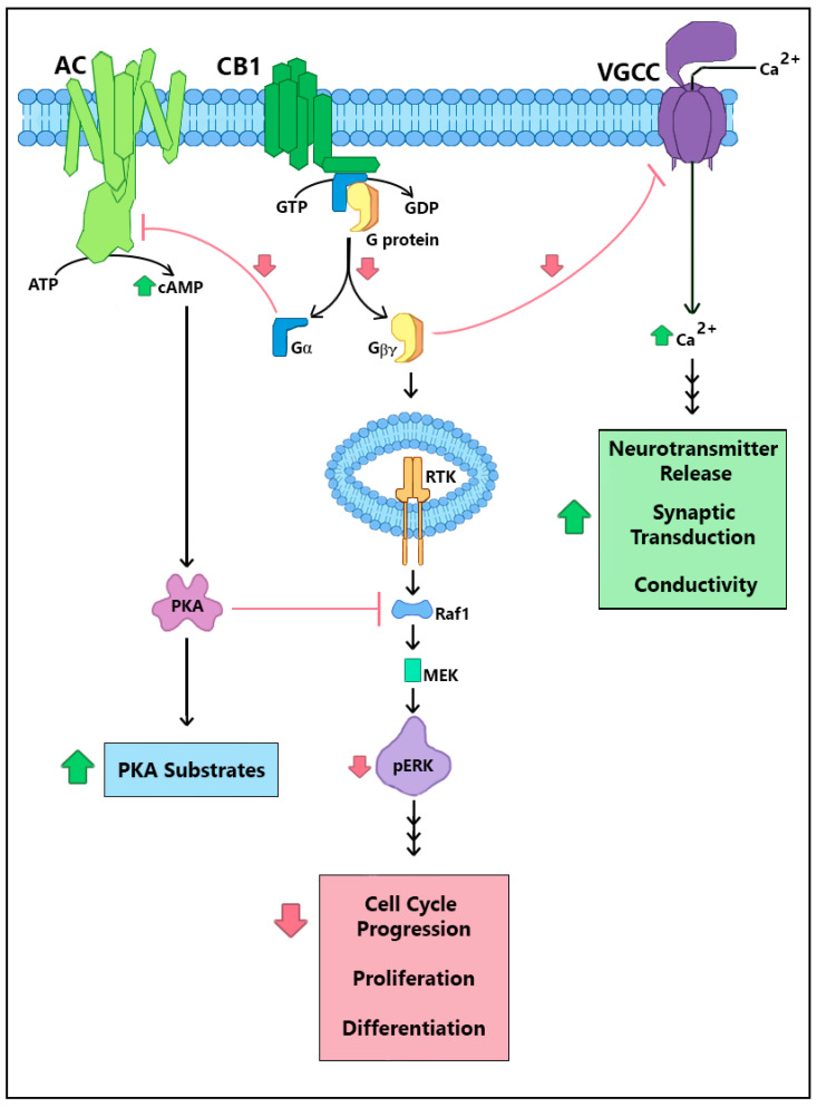
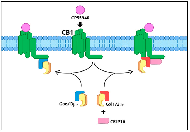
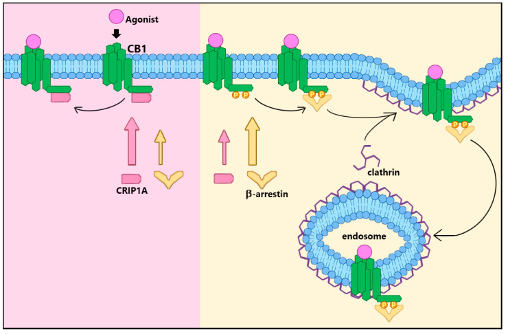
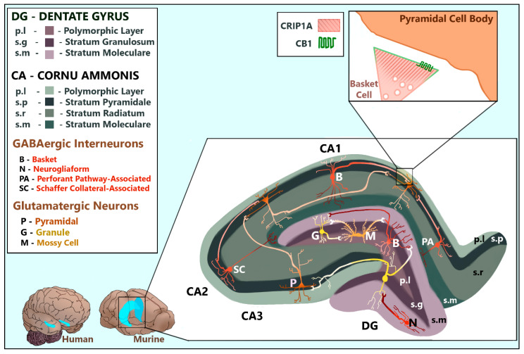
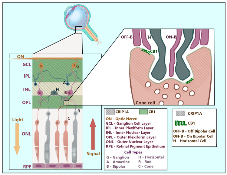
Endocannabinoid signaling depends upon the CB1 and CB2 cannabinoid receptors, their endogenous ligands anandamide and 2-arachidonoylglycerol, and intracellular proteins that mediate responses via the C-terminal and other intracellular receptor domains. The CB1 receptor regulates and is regulated by associated G proteins predominantly of the Gi/o subtypes, β-arrestins 1 and 2, and the cannabinoid receptor-interacting protein 1a (CRIP1a). Evidence for a physiological role for CRIP1a is emerging as data regarding the cellular localization and function of CRIP1a are generated. Here we summarize the neuronal distribution and role of CRIP1a in endocannabinoid signaling, as well as discuss investigations linking CRIP1a to development, vision and hearing sensory systems, hippocampus and seizure regulation, and psychiatric disorders including schizophrenia. We also examine the genetic and epigenetic association of CRIP1a within a variety of cancer subtypes. This review provides evidence upon which to base future investigations on the function of CRIP1a in health and disease.

Keywords: G protein-coupled receptors (GPCRs), hippocampus; cancer; embryonic development; endocannabinoids; epilepsy; retina; schizophrenia; seizures.


Conflict of interest statement

The authors declare no conflict of interest. The funders had no role in the design of the study; in the collection, analyses, or interpretation of data; in the writing of the manuscript, or in the decision to publish the results.

Figures

Figure 1
Figure 1
CB1R cellular signaling and points of CRIP1a-mediated suppression. Downstream effects of CB1R constitutive and agonist-mediated cascades include Gβγ-mediated inhibition of voltage-gated calcium channels (VGCC), Gαi-mediated inhibition of adenylyl cyclase (AC), and increased ERK1/2 phosphorylation through stimulation of receptor tyrosine kinase (RTK) and mitigation of PKA suppression. CRIP1a serves as a general negative-modulator, demonstrated to attenuate these basal and agonist-stimulated CB1R downstream actions: G protein activation, VGCC inhibition, cAMP diminution, and ERK1/2 phosphorylation. Green ‘up arrows’ reflect increased effects by CRIP1a expression; red ‘down arrows’ reflect decreased effects by CRIP1a expression.
Figure 2
Figure 2
Selective G protein subtype switching. Under the influence of CRIP1a, CP55940-mediated Go/i3 activation by CB1R C is attenuated while Gi1/2 activation is promoted. The binding of Go/i3 to CB1R requires a region in the C-terminus, while Gi1/2 binding utilizes a region of the third intracellular loop. This G protein selection by CRIP1a can hypothetically alter the downstream cascades in response to CP55940 activation of CB1R depending upon the availability of these Gα subtypes.
Figure 3
Figure 3
CRIP1a attenuation of β-arrestin/clathrin-dependent CB1R cell surface internalization. The mechanism of agonist-mediated, clathrin-dependent CB1R internalization from the cell membrane is dependent upon β-arrestin1/2 binding at the same C-terminal regions that bind CRIP1a. Preference for β-arrestin over CRIP1a is established by phosphorylation of specific Ser and Thr residues [20] within those binding domains. Preference for CRIP1a occurs when the same residues are not phosphorylated, attenuating agonist-stimulated CB1R internalization by precluding β-arrestin binding.
Figure 4
Figure 4
The mammalian hippocampal formation. CRIP1a is found dispersed in the presynaptic termini of GABAergic interneurons and glutamatergic neurons including DG granule cells, DG hilar mossy cells, and CA pyramidal cells. CB1R is localized to the presynaptic membranes of GABAergic interneurons and glutamatergic neurons including DG hilar mossy cells and CA pyramidal cells. This figure compares the relative size and location of the hippocampal formation in human and murine brains. The enlarged murine hippocampus depicts the shapes, positions, projections, and common synapses of relevant neurons within the layers of the dentate gyrus and hippocampus proper, viewed as a coronal slice. These cellular structures and positions were artistically rendered based upon data reviewed by [36,37,38,39,40]. The interneuron subtypes depicted were chosen based upon those with the strongest evidence of CB1R expression, as reviewed by Pelkey and colleagues [36]. The enlarged synapse depicts the subcellular localization of CB1R and CRIP1a in a CCK+ basket cell interneuron synapsing at a pyramidal soma, notably deficient of both proteins [36]. The synapse conformation was artistically rendered based upon data published by Dudok and colleagues [41].
Figure 5
Figure 5
Distribution of CB1R and CRIP1a in the retina. CRIP1a (shown here as diagonal lines) is found dispersed throughout presynaptic regions of glutamatergic and GABAergic retinal neurons spanning the ganglion cell layer through the outer plexiform layer. CB1R is localized to the presynaptic membranes of retinal synapses in the ganglion cell layer and both inner and outer plexiform layers. This figure artistically renders the positions and common synapses of relevant neurons within the layers of the retina while superimposing identified regions of CRIP1a and CB1R expression. The enlarged synapse depicts the known region of CRIP1a and CB1R co-expression at the presynaptic zone of a cone cell in the outer plexiform layer. This artistic rendering was designed based on the described interaction between cone cells and bipolar cells by Chapot and colleagues [63] and on the shape and location of retinal neuronal cell types described in Neuroscience, 2nd Edition [64].

Similar articles

Cited by

References

    1. Zou S., Kumar U. Cannabinoid Receptors and the Endocannabinoid System: Signaling and Function in the Central Nervous System. Int. J. Mol. Sci. 2018;19:833. doi: 10.3390/ijms19030833. - DOI - PMC - PubMed
    1. Kendall D.A., Yudowski G. Cannabinoid Receptors in the Central Nervous System: Their Signaling and Roles in Disease. Front. Cell. Neurosci. 2016;10:294. doi: 10.3389/fncel.2016.00294. - DOI - PMC - PubMed
    1. Lu Y., Anderson H.D. Cannabinoid signaling in health and disease. Can. J. Physiol. Pharmacol. 2017;95:311–327. doi: 10.1139/cjpp-2016-0346. - DOI - PubMed
    1. Ligresti A., De Petrocellis L., Di Marzo V. From Phytocannabinoids to Cannabinoid Receptors and Endocannabinoids: Pleiotropic Physiological and Pathological Roles through Complex Pharmacology. Physiol. Rev. 2016;96:1593–1659. doi: 10.1152/physrev.00002.2016. - DOI - PubMed
    1. Mechoulam R., Hanuš L.O., Pertwee R.G., Howlett A.C. Early phytocannabinoid chemistry to endocannabinoids and beyond. Nat. Rev. Neurosci. 2014;15:757–764. doi: 10.1038/nrn3811. - DOI - PubMed

Publication types

Substances

LinkOut - more resources