. 2020 Nov 27;12(12):3548.
doi: 10.3390/cancers12123548.

Serum-Derived Exosomal MicroRNA Profiles Can Predict Poor Survival Outcomes in Patients with Extranodal Natural Killer/T-Cell Lymphoma

Affiliations

Serum-Derived Exosomal MicroRNA Profiles Can Predict Poor Survival Outcomes in Patients with Extranodal Natural Killer/T-Cell Lymphoma

Kyung Ju Ryu et al. Cancers (Basel). .

Abstract

Exosomes containing microRNAs (miRNAs) might have utility as biomarkers to predict the risk of treatment failure in extranodal NK/T-cell lymphoma (ENKTL) because exosomal cargo miRNAs could reflect tumor aggressiveness. We analyzed the exosomal miRNAs of patients in favorable (n = 22) and poor outcome (n = 23) groups in a training cohort. Then, using the Nanostring nCounter® microRNA array, we compared them with miRNAs identified in human NK/T lymphoma (NKTL) cell line-derived exosomes to develop exosomal miRNA profiles. We validated the prognostic value of serum exosomal miRNA profiles with an independent cohort (n = 85) and analyzed their association with treatment resistance using etoposide-resistant cell lines. A comparison of the top 20 upregulated miRNAs in the training cohort with poor outcomes with 16 miRNAs that were upregulated in both NKTL cell lines, identified five candidate miRNAs (miR-320e, miR-4454, miR-222-3p, miR-21-5p, and miR-25-3p). Among these, increased levels of exosomal miR-4454, miR-21-5p, and miR-320e were associated with poor overall survival in the validation cohort. Increased levels were also found in relapsed patients post-treatment. These three miRNAs were overexpressed in NKTL cell lines that were resistant to etoposide. Furthermore, transfection of NKTL cell lines with miR-21-5p and miR-320e induced an increase in expression of the proinflammatory cytokines such as macrophage inflammatory protein 1 alpha. These studies show that serum levels of exosomal miR-21-5p, miR-320e, and miR-4454 are increased in ENKTL patients with poor prognosis. Upregulation of these exosomal miRNAs in treatment-resistant cell lines suggests they have a role as biomarkers for the identification of ENKTL patients at high risk of treatment failure.

Keywords: NK/T-cell lymphoma; biomarker; exosome; microRNA cancer.


Conflict of interest statement

The authors declare no conflict of interest.

Figures

Figure 1
Figure 1
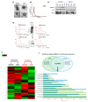
Characterization and miRNA analysis of exosomal miRNA in NK/T-cell lymphoma (NKTL) patients. (A) Exosomes were isolated by ExoQuick from equal volumes of serum of NKTL patients; their size and concentration were analyzed by transmission electron microscopy (TEM). Scale bar = 100 nm. (B) Nanoparticle tracking analysis. (C) Exosomes were analyzed by Western blot for the exosomal protein markers Alix and CD63. RNAs from NKTL serum-derived exosomes were submitted to exosomal miRNA analysis on a total RNA chip using an Agilent 2100 Bioanalyzer. (D) Study scheme for the training cohort. (E) Kaplan–Meier curves for overall survival of the training cohort. (F) Heatmap showing differential exosomal miRNA expression using the Nanostring nCounter® microRNA array between patients with favorable and poor clinical outcomes. (G) The top 20 upregulated and downregulated exosomal miRNAs in the group with poor clinical outcomes.
Figure 1
Figure 1
Characterization and miRNA analysis of exosomal miRNA in NK/T-cell lymphoma (NKTL) patients. (A) Exosomes were isolated by ExoQuick from equal volumes of serum of NKTL patients; their size and concentration were analyzed by transmission electron microscopy (TEM). Scale bar = 100 nm. (B) Nanoparticle tracking analysis. (C) Exosomes were analyzed by Western blot for the exosomal protein markers Alix and CD63. RNAs from NKTL serum-derived exosomes were submitted to exosomal miRNA analysis on a total RNA chip using an Agilent 2100 Bioanalyzer. (D) Study scheme for the training cohort. (E) Kaplan–Meier curves for overall survival of the training cohort. (F) Heatmap showing differential exosomal miRNA expression using the Nanostring nCounter® microRNA array between patients with favorable and poor clinical outcomes. (G) The top 20 upregulated and downregulated exosomal miRNAs in the group with poor clinical outcomes.
Figure 2
Figure 2
Characterization and miRNA analysis of exosomal and cellular miRNA in NKTL cell lines. (A) Exosomes were isolated from lymphoma cell lines by ExoQuick and the size and concentration of the exosomes were assessed by transmission electron microscopy. Scale bar = 100 nm. (B) Nanoparticle tracking analysis for NK92MI and SNK6. (C) Cell line-derived exosomes were analyzed by Western blot for the exosomal protein markers Alix and CD63. Calnexin, a marker for non-exosome components, was used as a negative control to ensure that there was no detectable cellular contamination. (D) NKTL cell-line-derived exosomal and cellular RNAs were measured using the small RNA chip. (E) Heatmap showing significantly upregulated and downregulated exosomal and cellular miRNAs. (F) The top 20 upregulated miRNAs in exosomes were compared with parent cells; 16 miRNAs were found to overlap between NK92MI and SNK6.
Figure 3
Figure 3
Exosomal miRNA markers correlated with clinical responses. (A) Venn diagrams show commonly upregulated miRNAs in both patient-serum-derived and NKTL cell-line-derived exosomes. (B) Overall survival of patients with extranodal NK/T-cell lymphoma (ENKTL) according to serum levels of exosomal miR-4454. (C) The pretreatment high miR-21-5p group showed poor overall survival compared with the low miR-21-5p group. (D) The pretreatment high exosomal miR-320e group showed a tendency of poor survival compared with the low exosomal miR-320e group. (E) Serial changes of miR-4454, miR-21-5p, and miR-302e during clinical course. Three relapsed patients showed increased exosomal miR-4454 read counts at the end of treatment (EOT). Patients who relapsed after treatment showed an increase in read counts of miR-21-5p at EOT compared with interim evaluation. Two relapsed patients showed a marked increase in exosomal miR-320e at EOT. (F) When the increase in each miRNA was counted as a risk factor for poor prognosis, patients were grouped according to the number of risk factors. Thus, ‘none’ represents the absence of risk factors whereas ‘three’ represents the presence of all risk factors (high miR-21-5p, high miR-320e, and high miR-4454). The overall survival of patients with all three risk factors was significantly worse than that of patients with ‘none’, as well as patients with one or two risk factors.
Figure 3
Figure 3
Exosomal miRNA markers correlated with clinical responses. (A) Venn diagrams show commonly upregulated miRNAs in both patient-serum-derived and NKTL cell-line-derived exosomes. (B) Overall survival of patients with extranodal NK/T-cell lymphoma (ENKTL) according to serum levels of exosomal miR-4454. (C) The pretreatment high miR-21-5p group showed poor overall survival compared with the low miR-21-5p group. (D) The pretreatment high exosomal miR-320e group showed a tendency of poor survival compared with the low exosomal miR-320e group. (E) Serial changes of miR-4454, miR-21-5p, and miR-302e during clinical course. Three relapsed patients showed increased exosomal miR-4454 read counts at the end of treatment (EOT). Patients who relapsed after treatment showed an increase in read counts of miR-21-5p at EOT compared with interim evaluation. Two relapsed patients showed a marked increase in exosomal miR-320e at EOT. (F) When the increase in each miRNA was counted as a risk factor for poor prognosis, patients were grouped according to the number of risk factors. Thus, ‘none’ represents the absence of risk factors whereas ‘three’ represents the presence of all risk factors (high miR-21-5p, high miR-320e, and high miR-4454). The overall survival of patients with all three risk factors was significantly worse than that of patients with ‘none’, as well as patients with one or two risk factors.
Figure 4
Figure 4
Expression of miR-21-5p, miR-320e, and miR-4454 in etoposide-resistant NKTL cell lines. (A) NK92MI (NK92MI_Etopo_R) cells showed significant resistance to etoposide compared with control cells. Cells were incubated with different concentrations of etoposide for 72 h and subjected to the CCK-8 assay. The expression levels of miR-21, miR-320e, and miR-4454 were increased in etoposide-resistant cells. (B) SNK6 (SNK6_Etopo_R) cells showed significant resistance to etoposide and increased expression levels of miR-21, miR-320e, and miR-4454 compared with control cells. (C) Nanoparticle tracking analysis showed no difference among control SNK6 cells and etoposide-resistant SNK6 cells. (D) Exosome amount and marker expression were not different between etoposide-resistant cell lines and control cells. (E) The expression levels of miR-21, miR-320e, and miR-4454 in exosomes isolated from SNK6 cells resistant to etoposide 200 nM were higher than in those of control cells.
Figure 5
Figure 5
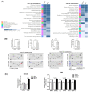
Effect of upregulated miR-21-5p and miR-320e on cytokine production. (A) KEGG (Kyoto Encyclopedia of Genes and Genomes) pathway analysis of dysregulated miRNAs was performed for NK92MI and SNK6 cells that were transfected with miR-21-5p mimic and miR-320e mimic. The list of target genes corresponding to up- and downregulated miR-21-5p and miR-320e. (B) After transfection of miR-21-5p and miR-320e in NK92MI and SNK6 cells, human cytokine array analysis using a culture supernatant of transfected cell lines was performed. Individual cytokines were spotted in duplicate. Cytokines induced by transfection of cells with miR21-5p are indicated by red squares, while those induced by transfection with miR320e are indicated by blue squares. Positive control spots are located at the corners of the human cytokine array. (C) The measurement of pixel densitometry confirmed the increased expression of several cytokines, mainly in miR-320e-transfected SNK6 cells. miR-N.C (negative control); miR-21-5p (miR-21-5p transfection); miR-320e (miR-320e transfection). (D) The co-culture of human monocyte THP-1 cells and SNK6 cells transfected with miR-21 showed the trend of overexpression of TGF-β, IL-10, CD206, and CCL2 in THP-1 cells, although this was not statistically significant. (E) The co-culture of miR-320e-transfected SNK6 cells with THP-1 cells showed a significant increase in CD206, a marker for M2-like macrophages. (F) The co-culture of etoposide-resistant SNK6 cells with THP-1 for 48 h showed a significant increase in TGF-β and CCL2 expression in THP-1 cells. (G) SNK6 cells showed differential expression of the target genes, especially CD40L and osteopontin, by overexpression and knockdown of miR-320e.
Figure 5
Figure 5
Effect of upregulated miR-21-5p and miR-320e on cytokine production. (A) KEGG (Kyoto Encyclopedia of Genes and Genomes) pathway analysis of dysregulated miRNAs was performed for NK92MI and SNK6 cells that were transfected with miR-21-5p mimic and miR-320e mimic. The list of target genes corresponding to up- and downregulated miR-21-5p and miR-320e. (B) After transfection of miR-21-5p and miR-320e in NK92MI and SNK6 cells, human cytokine array analysis using a culture supernatant of transfected cell lines was performed. Individual cytokines were spotted in duplicate. Cytokines induced by transfection of cells with miR21-5p are indicated by red squares, while those induced by transfection with miR320e are indicated by blue squares. Positive control spots are located at the corners of the human cytokine array. (C) The measurement of pixel densitometry confirmed the increased expression of several cytokines, mainly in miR-320e-transfected SNK6 cells. miR-N.C (negative control); miR-21-5p (miR-21-5p transfection); miR-320e (miR-320e transfection). (D) The co-culture of human monocyte THP-1 cells and SNK6 cells transfected with miR-21 showed the trend of overexpression of TGF-β, IL-10, CD206, and CCL2 in THP-1 cells, although this was not statistically significant. (E) The co-culture of miR-320e-transfected SNK6 cells with THP-1 cells showed a significant increase in CD206, a marker for M2-like macrophages. (F) The co-culture of etoposide-resistant SNK6 cells with THP-1 for 48 h showed a significant increase in TGF-β and CCL2 expression in THP-1 cells. (G) SNK6 cells showed differential expression of the target genes, especially CD40L and osteopontin, by overexpression and knockdown of miR-320e.

Similar articles

Cited by

References

    1. Raposo G., Stoorvogel W. Extracellular vesicles: Exosomes, microvesicles, and friends. J. Cell Biol. 2013;200:373–383. doi: 10.1083/jcb.201211138. - DOI - PMC - PubMed
    1. Théry C., Witwer K.W., Aikawa E., Alcaraz M.J., Anderson J.D., Andriantsitohaina R., Antoniou A., Arab T., Archer F., Atkin-Smith G.K., et al. Minimal information for studies of extracellular vesicles 2018 (MISEV2018): A position statement of the International Society for Extracellular Vesicles and update of the MISEV2014 guidelines. J. Extracell. Vesicles. 2018;7:1535750. doi: 10.1080/20013078.2018.1535750. - DOI - PMC - PubMed
    1. El Andaloussi S., Mäger I., Breakefield X.O., Wood M.J.A. Extracellular vesicles: Biology and emerging therapeutic opportunities. Nat. Rev. Drug Discov. 2013;12:347–357. doi: 10.1038/nrd3978. - DOI - PubMed
    1. Yeh Y.-Y., Ozer H.G., Lehman A.M., Maddocks K., Yu L., Johnson A.J., Byrd J.C. Characterization of CLL exosomes reveals a distinct microRNA signature and enhanced secretion by activation of BCR signaling. Blood. 2015;125:3297–3305. doi: 10.1182/blood-2014-12-618470. - DOI - PMC - PubMed
    1. Azmi A.S., Bao B., Sarkar F.H. Exosomes in cancer development, metastasis, and drug resistance: A comprehensive review. Cancer Metastasis Rev. 2013;32:623–642. doi: 10.1007/s10555-013-9441-9. - DOI - PMC - PubMed