Case Reports
. 2020 Dec 1;20(1):540.
doi: 10.1186/s12887-020-02436-8.

Ecthyma gangrenosum due to Pseudomonas aeruginosa sepsis as initial manifestation of X-linked agammaglobulinemia: a case report

Affiliations
Case Reports

Ecthyma gangrenosum due to Pseudomonas aeruginosa sepsis as initial manifestation of X-linked agammaglobulinemia: a case report

Haixia Huang et al. BMC Pediatr. .

Abstract

Background: X-linked agammaglobulinemia (XLA, OMIM#300,300), caused by mutations in the Bruton tyrosine kinase (BTK) gene, is a rare monogenic inheritable immunodeficiency disorder. Ecthyma gangrenosum is a cutaneous lesion caused by Pseudomonas aeruginosa that typically occurs in patients with XLA and other immunodeficiencies.

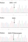
Case presentation: We report the case of a 20-month-old boy who presented with fever, vomiting, diarrhea, and ecthyma gangrenosum. Blood, stool, and skin lesion culture samples were positive for P. aeruginosa. A diagnosis of XLA was established, and the c.262G > T mutation in exon 4 of BTK was identified with Sanger sequencing. Symptoms improved following treatment with antibiotics and immunoglobulin infusion.

Conclusions: Primary immunodeficiency (i.e., XLA) should be suspected in male infants with P. aeruginosa sepsis, highlighting the importance of genetic and immune testing in these patients.

Keywords: Ecthyma gangrenosum; Pseudomonas aeruginosa; X-linked agammaglobulinemia; male infants.


Conflict of interest statement

The authors declare that they have no competing interests.

Figures
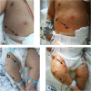
Fig. 1
Fig. 1
General appearance of the patient showing multiple purple necrotic lesions. (A) Day 1, ICU: Initial appearance of the abdominal lesions of ecthyma gangrenosum (a). (B) Day 2, ICU: The black central eschar in the lesion was deep seated and large (a`). (C) Day 1, ICU: Initial appearance of the lesions over the right lower leg (b&c).(D) Day 2, ICU: The erythematous lesions appeared as gangrenous ulcers (b`); The black central eschar was deep seated and large (c`)
Fig. 2
Fig. 2
Serial CT and MRI images showing changes in the brain. a Day 19 of admission: Lateralcranial CT showing diffuse brain edema, hypodensity of the brain parenchyma, and subdural effusion in the bilateral frontal area (red arrow). b Day 25 of admission (one day before Ommaya reservoir implantation): Lateral T2-weighted MRI showing enlarged lateral ventricles and hydrops with peripheral white matter edema. c Day 27 of admission (one day after Ommaya reservoir implantation): Lateral cranial CT showing the drainage tube in the anterior horn of the left lateral ventricle. d Day 70 of admission (one day before ventriculoperitoneal shunt): Lateral T2-weighted MRI showing hydrocephalus in the ventricles was not obviously aggravated. e Day 72 of admission (one day after ventriculoperitoneal shunt): Lateral cranial CT showing the drainage tube in the lateral ventricle. e 20 months after discharge: Lateral T2-weighted MRI showing less hydrocephalus and improved interstitial cerebral edema
Fig. 3
Fig. 3
Results of Sanger sequencing for blood samples from the patient (upper panel) and parents (bottom panel)
Fig. 4
Fig. 4
X-ray and MRI images showing changes around the left elbow. (A) Day 29 of admission: X-ray showing swelling of soft tissue around the left elbow (arrow). (B) Day 47 of admission: Coronal T2-weighted MRI showing patchy hyperintensity in the left humerus and proximal ulna (a) accompanied by soft tissue edema and abscess formation around the elbow joint (b).(C) Day 96 of admission: X-ray showing low-density shadows in the left humerus, suggesting bone destruction (c). Similar less severe changes were observed in the proximal left ulna (d). Soft tissue swelling was reduced

Similar articles

Cited by

References

    1. Soresina A, Moratto D, Chiarini M, Paolillo C, Baresi G, Focà E, Bezzi M, Baronio B, Giacomelli M, Badolato R. Two X-linked agammaglobulinemia patients develop pneumonia as COVID-19 manifestation but recover. Pediatr Allergy Immunol. 2020;31(5):565–9. - PMC - PubMed
    1. Qin X, Jiang LP, Tang XM, Wang M, Liu EM, Zhao XD. Clinical features and mutation analysis of X-linked agammaglobulinemia in 20 Chinese patients. World J Pediatrics. 2013;9(3):273–7. doi: 10.1007/s12519-013-0400-x. - DOI - PubMed
    1. Yang MA, Lee J, Choi EH, Lee HJ. Pseudomonas aeruginosa bacteremia in children over ten consecutive years: analysis of clinical characteristics, risk factors of multi-drug resistance and clinical outcomes. J Korean Med Sci. 2011;26(5):612–8. doi: 10.3346/jkms.2011.26.5.612. - DOI - PMC - PubMed
    1. Vaiman M, Lazarovitch T, Heller L, Lotan G. Ecthyma gangrenosum and ecthyma-like lesions: review article. Eur J Clin Microbiol Infect Dis. 2015;34(4):633–9. doi: 10.1007/s10096-014-2277-6. - DOI - PubMed
    1. Lackey AE, Ahmad F. In: X-linked Agammaglobulinemia. StatPearls, editor. Treasure Island: StatPearls Publishing, StatPearls Publishing LLC.; 2020. - PubMed

Publication types

Supplementary concepts