

NOD1/RIP2 signalling enhances the microglia-driven inflammatory response and undergoes crosstalk with inflammatory cytokines to exacerbate brain damage following intracerebral haemorrhage in mice
- PMID: 33261639
- PMCID: PMC7708246
- DOI: 10.1186/s12974-020-02015-9
NOD1/RIP2 signalling enhances the microglia-driven inflammatory response and undergoes crosstalk with inflammatory cytokines to exacerbate brain damage following intracerebral haemorrhage in mice
Abstract
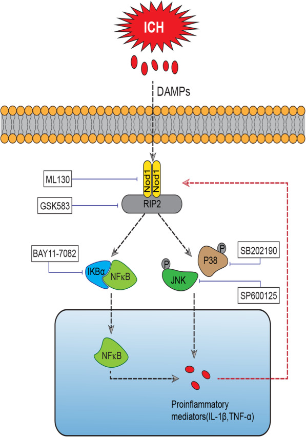
Background: Secondary brain damage caused by the innate immune response and subsequent proinflammatory factor production is a major factor contributing to the high mortality of intracerebral haemorrhage (ICH). Nucleotide-binding oligomerization domain 1 (NOD1)/receptor-interacting protein 2 (RIP2) signalling has been reported to participate in the innate immune response and inflammatory response. Therefore, we investigated the role of NOD1/RIP2 signalling in mice with collagenase-induced ICH and in cultured primary microglia challenged with hemin.
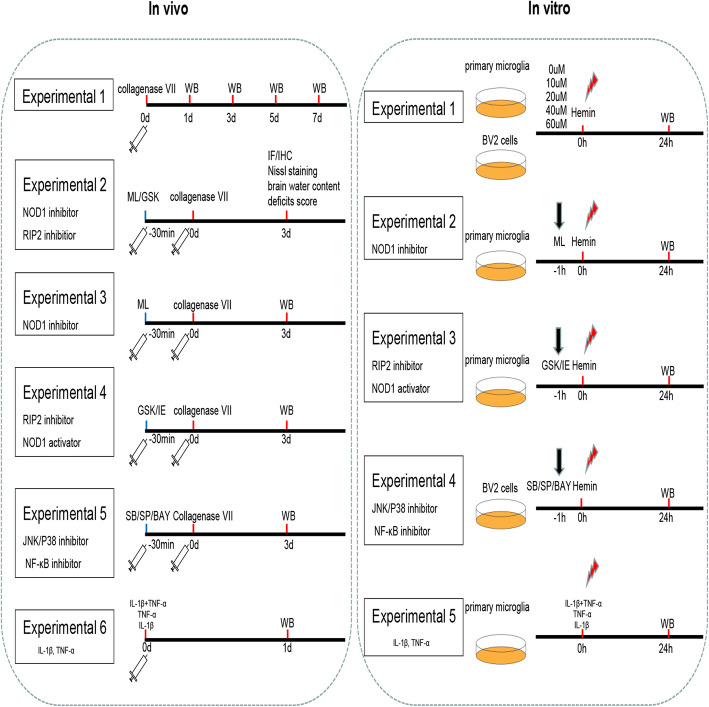
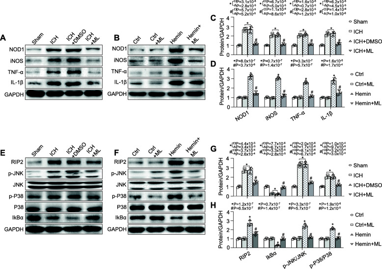
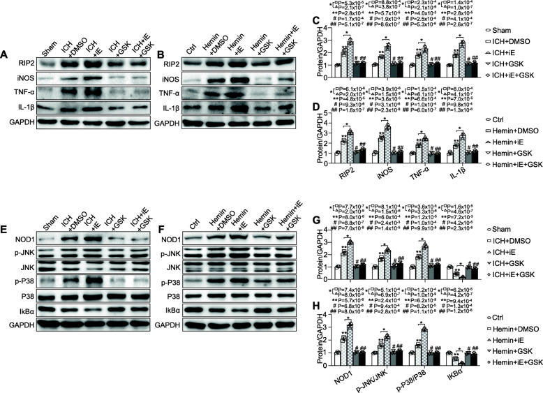
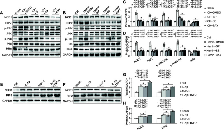
Methods: Adult male C57BL/6 mice were subjected to collagenase for induction of ICH model in vivo. Cultured primary microglia and BV2 microglial cells (microglial cell line) challenged with hemin aimed to simulate the ICH model in vitro. We first defined the expression of NOD1 and RIP2 in vivo and in vitro using an ICH model by western blotting. The effect of NOD1/RIP2 signalling on ICH-induced brain injury volume, neurological deficits, brain oedema, and microglial activation were assessed following intraventricular injection of either ML130 (a NOD1 inhibitor) or GSK583 (a RIP2 inhibitor). In addition, levels of JNK/P38 MAPK, IκBα, and inflammatory factors, including tumour necrosis factor-α (TNF-α), interleukin (IL)-1β, and inducible nitric oxide synthase (iNOS) expression, were analysed in ICH-challenged brain and hemin-exposed cultured primary microglia by western blotting. Finally, we investigated whether the inflammatory factors could undergo crosstalk with NOD1 and RIP2.
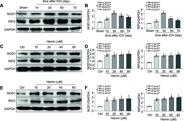
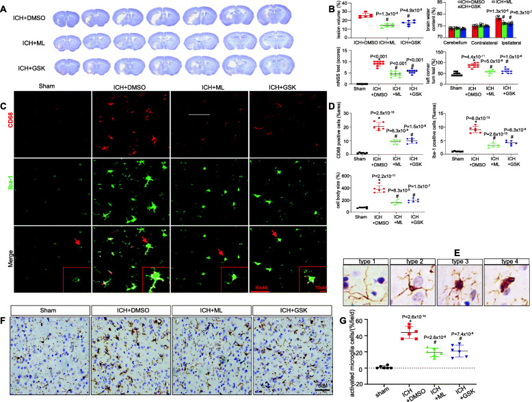
Results: The levels of NOD1 and its adaptor RIP2 were significantly elevated in the brains of mice in response to ICH and in cultured primary microglia, BV2 cells challenged with hemin. Administration of either a NOD1 or RIP2 inhibitor in mice with ICH prevented microglial activation and neuroinflammation, followed by alleviation of ICH-induced brain damage. Interestingly, the inflammatory factors interleukin (IL)-1β and tumour necrosis factor-α (TNF-α), which were enhanced by NOD1/RIP2 signalling, were found to contribute to the NOD1 and RIP2 upregulation in our study.
Conclusion: NOD1/RIP2 signalling played an important role in the regulation of the inflammatory response during ICH. In addition, a vicious feedback cycle was observed between NOD1/RIP2 and IL-1β/TNF-α, which could to some extent result in sustained brain damage during ICH. Hence, our study highlights NOD1/RIP2 signalling as a potential therapeutic target to protect the brain against secondary brain damage during ICH.
Keywords: Inflammatory response; Intracerebral haemorrhage; Microglial activation; NOD1; RIP2.
Conflict of interest statement
The authors declare that they have no competing interests.
Figures
Similar articles
-
Excretory/secretory proteins of adult Toxocara canis induce changes in the expression of proteins involved in the NOD1-RIP2-NF-κB pathway and modulate cytokine production in mouse macrophages.Exp Parasitol. 2021 Oct;229:108152. doi: 10.1016/j.exppara.2021.108152. Epub 2021 Aug 19. Exp Parasitol. 2021. PMID: 34419413
-
[Analgesic effect of "Zhibian" (BL54)-toward-"Shuidao" (ST28) needling technique of acupuncture on primary dysmenorrhea based on NOD1/RIP2/NF-κB signaling pathway in the rats].Zhongguo Zhen Jiu. 2025 Feb 12;45(2):209-16. doi: 10.13703/j.0255-2930.20240331-k0001. Zhongguo Zhen Jiu. 2025. PMID: 39943763 Chinese.
-
Genetic deletion or pharmacological inhibition of soluble epoxide hydrolase reduces brain damage and attenuates neuroinflammation after intracerebral hemorrhage.J Neuroinflammation. 2017 Nov 25;14(1):230. doi: 10.1186/s12974-017-1005-4. J Neuroinflammation. 2017. PMID: 29178914 Free PMC article.
-
Modulators of microglial activation and polarization after intracerebral haemorrhage.Nat Rev Neurol. 2017 Jul;13(7):420-433. doi: 10.1038/nrneurol.2017.69. Epub 2017 May 19. Nat Rev Neurol. 2017. PMID: 28524175 Free PMC article. Review.
-
RIPK2 NODs to XIAP and IBD.Semin Cell Dev Biol. 2021 Jan;109:144-150. doi: 10.1016/j.semcdb.2020.07.001. Epub 2020 Jul 3. Semin Cell Dev Biol. 2021. PMID: 32631784 Review.
Cited by
-
Perampanel, an AMPAR antagonist, alleviates experimental intracerebral hemorrhage‑induced brain injury via necroptosis and neuroinflammation.Mol Med Rep. 2021 Aug;24(2):544. doi: 10.3892/mmr.2021.12183. Epub 2021 Jun 3. Mol Med Rep. 2021. PMID: 34080030 Free PMC article.
-
MiR-200c-3p inhibits LPS-induced M1 polarization of BV2 cells by targeting RIP2.Genes Genomics. 2022 Apr;44(4):477-486. doi: 10.1007/s13258-021-01210-z. Epub 2022 Jan 10. Genes Genomics. 2022. PMID: 35013887 Free PMC article.
-
Intermittent fasting reduces neuroinflammation in intracerebral hemorrhage through the Sirt3/Nrf2/HO-1 pathway.J Neuroinflammation. 2022 May 27;19(1):122. doi: 10.1186/s12974-022-02474-2. J Neuroinflammation. 2022. PMID: 35624490 Free PMC article.
-
Effects of Global Ripk2 Genetic Deficiency in Aged Mice following Experimental Ischemic Stroke.bioRxiv [Preprint]. 2025 Feb 6:2025.02.05.636687. doi: 10.1101/2025.02.05.636687. bioRxiv. 2025. Update in: Aging Brain. 2025 Mar 29;7:100135. doi: 10.1016/j.nbas.2025.100135. PMID: 39974926 Free PMC article. Updated. Preprint.
-
Pharmacological inhibition of receptor-interacting protein kinase 2 (RIPK2) elicits neuroprotective effects following experimental ischemic stroke.Exp Neurol. 2024 Jul;377:114812. doi: 10.1016/j.expneurol.2024.114812. Epub 2024 May 9. Exp Neurol. 2024. PMID: 38729551 Free PMC article.
References
-
- van Asch CJ, Luitse MJ. al. e: Incidence, case fatality, and functional outcome of intracerebral haemorrhage over time, according to age, sex, and ethnic origin: a systematic review and meta-analysis. Lancet Neurol. 2010;9:167–176. - PubMed
-
- Wang X, Arima H, Yang J, Zhang S, Wu G, Woodward M, Munoz-Venturelli P, Lavados PM, Stapf C, Robinson T, et al. Mannitol and outcome in intracerebral hemorrhage: propensity score and multivariable intensive blood pressure reduction in acute cerebral hemorrhage trial 2 results. Stroke. 2015;46:2762–2767. - PubMed
-
- Xiong XY, Liu L, Yang QW. Functions and mechanisms of microglia/macrophages in neuroinflammation and neurogenesis after stroke. Prog Neurobiol. 2016;142:23–44. - PubMed
MeSH terms
Substances
Grants and funding
LinkOut - more resources
Full Text Sources
Research Materials
