Review
. 2021 Mar 1;81(5):1189-1200.
doi: 10.1158/0008-5472.CAN-20-0373. Epub 2020 Dec 1.

Preclinical Applications of Multi-Platform Imaging in Animal Models of Cancer

Affiliations
Review

Preclinical Applications of Multi-Platform Imaging in Animal Models of Cancer

Natalie J Serkova et al. Cancer Res. .

Abstract

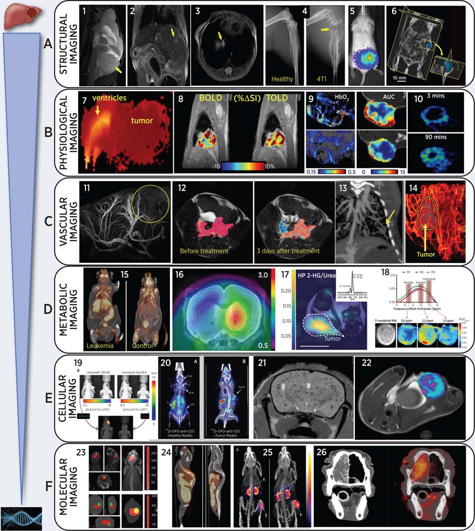
In animal models of cancer, oncologic imaging has evolved from a simple assessment of tumor location and size to sophisticated multimodality exploration of molecular, physiologic, genetic, immunologic, and biochemical events at microscopic to macroscopic levels, performed noninvasively and sometimes in real time. Here, we briefly review animal imaging technology and molecular imaging probes together with selected applications from recent literature. Fast and sensitive optical imaging is primarily used to track luciferase-expressing tumor cells, image molecular targets with fluorescence probes, and to report on metabolic and physiologic phenotypes using smart switchable luminescent probes. MicroPET/single-photon emission CT have proven to be two of the most translational modalities for molecular and metabolic imaging of cancers: immuno-PET is a promising and rapidly evolving area of imaging research. Sophisticated MRI techniques provide high-resolution images of small metastases, tumor inflammation, perfusion, oxygenation, and acidity. Disseminated tumors to the bone and lung are easily detected by microCT, while ultrasound provides real-time visualization of tumor vasculature and perfusion. Recently available photoacoustic imaging provides real-time evaluation of vascular patency, oxygenation, and nanoparticle distributions. New hybrid instruments, such as PET-MRI, promise more convenient combination of the capabilities of each modality, enabling enhanced research efficacy and throughput.


Conflict of interest statement

The authors declare no potential conflicts of interest

Figures

Figure 1:
Figure 1:
Representative multi-modality images of animal cancer models (from left to right): (A) Anatomical cancer detection in mouse models: (1) T2-weighted MRI of pediatric cerebellar brain tumor (medulloblastoma) pdx; (2) gadolinium-enhanced T1-MRI of an orthotopic liver HCC; (3) proton-density MRI of a lung metastasis from breast cancer; (4) microCT of bone metastasis from engrafted breast cancer cells, adapted from (62); (5) BLI of multi-organ breast cancer metastases, adapted from (62); (6) 3D-ultrasound of pancreatic cancer in a genetically modified LSL-Kras mouse, adapted from (80). (B) Physiology-based images in rodent cancer models: (7) high ADC (brain edema and ventricle hydrocephalus) and low ADC (highly proliferative medulloblastoma mouse pdx) from diffusion-weighted MRI; (8) blood and tissue oxygen level dependent (BOLD and TOLD) MRI in response to O2 gas breathing challenge in orthotopic human A549 lung tumor xenograft in nude rat, adapted from (23); (9) left: photoacoustic imaging of subcutaneous A549 human lung tumor growing in leg of nude rat showing endogenous HbO2 concentration before (upper) and 48 hrs after (lower) administration of vascular disrupting agent (VDA), based on multiple wavelengths (MSOT), while breathing O2 and right: corresponding DCE MRI showing area under the curve reflecting reduced perfusion after VDA (10) 18F-MISO (hypoxia tracer) PET in a syngeneic Dunning R3327-AT1 rat prostate tumor, adapted from (143) (C) Imaging tumor vasculature in-vivo: (11) high-resolution magnetic resonance angiography (MRA) after gadolinium injection in an orthotopic rat isograft C6 glioma model; (12) DCE-MRI during gadolinium injection in mouse TRAMP model for prostate cancer, adapted from (33); (13) contrast-enhanced microCT of lung vasculature and small lung tumor using liposomal-iodinated contrast agent, adapted from (72); (14) US enhanced with microbubbles reveals high perfusion in the rim of a flank pancreatic cancer xenograft in a mouse. (D) Imaging tumor metabolism non-invasively: (15) abnormal 18F-FDG uptake in spleen, liver, and lymph nodes in transgenic leukemic (left) vs. control mouse, adapted from (129); (16) increased GBM uptake of 18F-ethyltyrosine (18F-FET) without (left) and with bevacizumab treatment in an orthotopic U87 glioma mouse model, adapted from (138); (17) Representative heatmap of spectral data from a mouse with a mutant IDH1 tumor xenograft following injection of hyperpolarized [1-13C]-glutamine showing accumulation of 2-HG in the tumor region only, which was referenced and normalized to a 5 mM [1-13C] urea phantom. Dotted lines highlight the tumor, and the white line at the bottom represents 10 mm for scaling, adapted from (46); (18) In-vivo CEST-MRI of MDA-MB-231 breast tumor xenografts showing representative CEST MRI maps (top row, A), T1-weighted RARE MRI (bottom left, B), and MTRasym for three individual mice with orthotopic human MDA-MB-231 breast tumor xenografts, which were labeled M1 for mouse 1, M2 for mouse 2, and M3 for mouse 3. CEST shifts of amide, amine, and hydroxyl resonances are highlighted in C, adapted from (52); (E) Cellular tracking using non-invasive imaging in mouse cancer models: (19) Dual reporter bioluminescence imaging using spectral unmixing algorithm. CBG99 cells were transplanted into the right striatum, PpyRE9 cells into the left striatum of the nude mouse on the right. A spectral unmixing algorithm was applied in order to select green light from CBG99 and red light from PpyRE9, adapted from (191); (20) immune-PET to image T-lymphocytes using 89Zr-anti-CD3 in normal and BBN975 bladder cancer tumor-bearing mice, adapted from (147); (21) T2-weighted brain MRI of ferumoxytol-labeled breast cancer cells after intra-cardiac injection, adapted from (35); (22) T2-weighted maps for macrophage imaging after ferumoxytol injection in inflamed mammary gland tumor mouse model, adapted from (37); (F) Molecular imaging of tumor-specific molecules: (23) tracking fluorescent micelles (red signal) to bioluminescent brain tumors (green) in anatomical context (124); (24) whole-body 18F-estradiol (FES) PET/CT of estrogen receptor in ER positive and negative bone metastases in mouse models of breast cancer; (25) whole body SPECT/CT with 111In-MSH peptide (melanocyte stimulating hormone) to image melanocortin-1 receptor in mouse B16/F1 melanoma model, adapted from (154); (26) CT (left) and 18FDG-PET of nasal adenocarcinoma in a canine cancer patient (a 10-year old standard poodle), adapted from (187).

References

    1. de Jong M, Essers J, van Weerden WM. Imaging preclinical tumour models: improving translational power. Nature reviews Cancer. 2014;14(7):481–93. - PubMed
    1. Jardim-Perassi BV, Huang S, Dominguez-Viqueira W, Poleszczuk J, Budzevich MM, Abdalah MA, et al. Multiparametric MRI and Coregistered Histology Identify Tumor Habitats in Breast Cancer Mouse Models. Cancer research. 2019;79(15):3952–64. - PMC - PubMed
    1. Weissleder R, Schwaiger MC, Gambhir SS, Hricak H. Imaging approaches to optimize molecular therapies. Science translational medicine. 2016;8(355):355ps16. - PubMed
    1. Condeelis J, Weissleder R. In vivo imaging in cancer. Cold Spring Harb Perspect Biol. 2010;2(12):a003848. - PMC - PubMed
    1. Hausner SH, Bold RJ, Cheuy LY, Chew HK, Daly ME, Davis RA, et al. Preclinical Development and First-in-Human Imaging of the Integrin alphavbeta6 with [(18)F]alphavbeta6-Binding Peptide in Metastatic Carcinoma. Clinical cancer research : an official journal of the American Association for Cancer Research. 2019;25(4):1206–15. - PMC - PubMed

Publication types

MeSH terms

Substances