. 2021 Jan 1;14(1):dmm045963.
doi: 10.1242/dmm.045963. Epub 2021 Jan 7.

Regulation of murine copper homeostasis by members of the COMMD protein family

Affiliations

Regulation of murine copper homeostasis by members of the COMMD protein family

Amika Singla et al. Dis Model Mech. .

Abstract

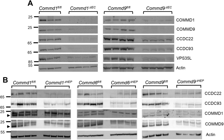
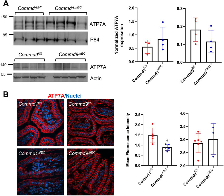
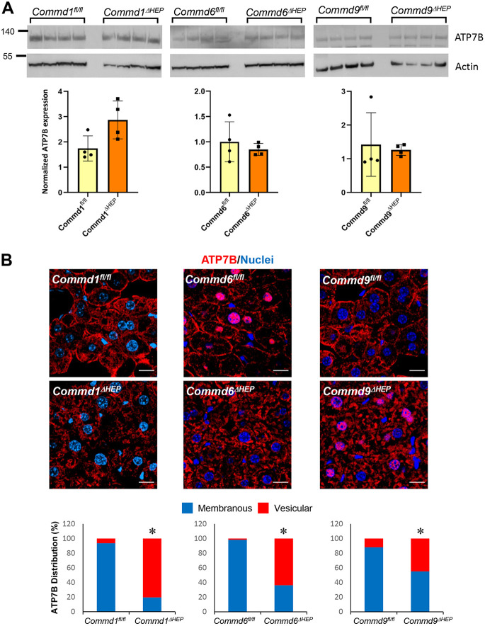
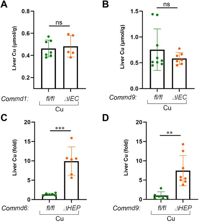
Copper is an essential transition metal for all eukaryotes. In mammals, intestinal copper absorption is mediated by the ATP7A copper transporter, whereas copper excretion occurs predominantly through the biliary route and is mediated by the paralog ATP7B. Both transporters have been shown to be recycled actively between the endosomal network and the plasma membrane by a molecular machinery known as the COMMD/CCDC22/CCDC93 or CCC complex. In fact, mutations in COMMD1 can lead to impaired biliary copper excretion and liver pathology in dogs and in mice with liver-specific Commd1 deficiency, recapitulating aspects of this phenotype. Nonetheless, the role of the CCC complex in intestinal copper absorption in vivo has not been studied, and the potential redundancy of various COMMD family members has not been tested. In this study, we examined copper homeostasis in enterocyte-specific and hepatocyte-specific COMMD gene-deficient mice. We found that, in contrast to effects in cell lines in culture, COMMD protein deficiency induced minimal changes in ATP7A in enterocytes and did not lead to altered copper levels under low- or high-copper diets, suggesting that regulation of ATP7A in enterocytes is not of physiological consequence. By contrast, deficiency of any of three COMMD genes (Commd1, Commd6 or Commd9) resulted in hepatic copper accumulation under high-copper diets. We found that each of these deficiencies caused destabilization of the entire CCC complex and suggest that this might explain their shared phenotype. Overall, we conclude that the CCC complex plays an important role in ATP7B endosomal recycling and function.

Keywords: ATP7A; ATP7B; COMMD proteins; Copper homeostasis; Copper transporters; Endosomal trafficking.


Conflict of interest statement

Competing interests The authors declare no competing or financial interests.

Figures

Fig. 1.
Fig. 1.
COMMD protein deficiency causes the destabilization of the CCC complex in enterocytes and hepatocytes. (A) Expression of CCC complex subunits was examined by immunoblotting in enterocytes isolated from intestinal-specific Commd1 and Commd9 knockout mice (Commd1ΔIEC and Commd9ΔIEC, respectively). Animals carrying the floxed alleles served as controls (Commd1fl/fl and Commd9fl/fl, respectively). β-Actin served as a loading control. (B) Same as in A, but using liver lysates from hepatocyte-specific COMMD-deficient mice (Commd1ΔHEP, Commd6ΔHEP and Commd9ΔHEP) and corresponding controls. Arrowheads indicate the band specific to the antibody.
Fig. 2.
Fig. 2.
ATP7A expression is diminished in Commd1 but not Commd9 intestinal knockout mice. (A) Immunoblot analysis for ATP7A expression in Commd1 and Commd9 intestinal knockout mice (top). ATP7A quantification after normalization by the loading control (P84 or actin) (bottom). (B) Representative images of immunofluorescence staining for ATP7A (red) and nuclei (Hoechst, blue) in intestinal tissues of Commd1ΔIEC (n=5) or Commd9ΔIEC (n=3) mice (bottom row) or corresponding control floxed animals, Commd1fl/fl (n=4) and Commd9fl/fl (n=6) (top row). Scale bar: 25 µm. Bar graphs represent quantification of ATP7A signal intensity in immunofluorescence staining of intestinal tissues, expressed as the fold change over the nuclear staining. Results for individual mice are plotted along with the mean and s.e.m. for each group; *P<0.05 (unpaired two-tailed Student's t-test).
Fig. 3.
Fig. 3.
ATP7B localization is altered in hepatic Commd1, Commd6 or Commd9 knockout mice. (A) Immunoblot analysis for ATP7B expression in hepatocyte-specific Commd1, Commd6 or Commd9 knockout mice or corresponding floxed control animals (top). Quantification after normalization by the loading control (actin) (bottom). None of the knockout groups exhibited a statistically significant difference from the corresponding control (unpaired two-tailed Student's t-test). (B) Representative images of immunofluorescence staining for ATP7B (red) and nuclei (Hoechst, blue) in liver tissues of Commd1ΔHEP, Commd6ΔHEP or Commd9ΔHEP and their respective control (floxed) mice (top); n=4 for each genotype. Scale bars: 15 µm. Quantification of ATP7B distribution pattern in immunofluorescence staining of liver tissues (bottom). Four images were acquired from each mouse, and eight to ten cells were analyzed from each image (n=32-40 cells per mouse). *P<0.05 (Chi square test).
Fig. 4.
Fig. 4.
Hepatic but not enteric knockout mice develop altered copper homeostasis. (A) Hepatic copper contents (μmol/g) were measured in dried liver tissue of Commd1ΔIEC mice (n=7) and corresponding floxed control animals (n=5) on a high-copper diet. Results for individual mice are plotted along with the mean and s.e.m. for each group; ns, not significant (unpaired two-tailed Student's t-test). (B) Same analysis as in A, but for Commd9ΔIEC (n=9) and the corresponding littermate controls (n=7). (C) Hepatic copper concentrations were measured in dried liver tissue of Commd6ΔHEP (n=6) and corresponding floxed control animals (n=6) on a high-copper diet. (D) Same analysis as in C, but for Commd9ΔHEP (n=6) and the corresponding littermate controls (n=8). Data are presented as the fold change relative to the average value in the control floxed mouse group. **P<0.01; ***P<0.001 (unpaired two-tailed Student's t-test).

References

    1. Alekhina O., Burstein E. and Billadeau D. D. (2017). Cellular functions of WASP family proteins at a glance. J. Cell Sci. 130, 2235-2241. 10.1242/jcs.199570 - DOI - PMC - PubMed
    1. Bartuzi P., Billadeau D. D., Favier R., Rong S., Dekker D., Fedoseienko A., Fieten H., Wijers M., Levels J. H., Huijkman N. et al. (2016). CCC- and WASH-mediated endosomal sorting of LDLR is required for normal clearance of circulating LDL. Nat. Commun. 7, 10961 10.1038/ncomms10961 - DOI - PMC - PubMed
    1. Biasio W., Chang T., McIntosh C. J. and McDonald F. J. (2004). Identification of Murr1 as a regulator of the human δ epithelial sodium channel. J. Biol. Chem. 279, 5429-5434. 10.1074/jbc.M311155200 - DOI - PubMed
    1. Burstein E., Hoberg J. E., Wilkinson A. S., Rumble J. M., Csomos R. A., Komarck C. M., Maine G. N., Wilkinson J. C., Mayo M. W. and Duckett C. S. (2005). COMMD proteins: a novel family of structural and functional homologs of MURR1. J. Biol. Chem. 280, 22222-22232. 10.1074/jbc.M501928200 - DOI - PubMed
    1. de Bie P., van de Sluis B., Burstein E., Duran K. J., Berger R., Duckett C. S., Wijmenga C. and Klomp L. W. J. (2006). Characterization of COMMD protein-protein interactions in NF-κB signalling. Biochem. J. 398, 63-71. 10.1042/BJ20051664 - DOI - PMC - PubMed

Publication types