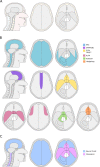
The spatial phenotype of genotypically distinct meningiomas demonstrate potential implications of the embryology of the meninges
- PMID: 33262459
- PMCID: PMC8440207
- DOI: 10.1038/s41388-020-01568-6
The spatial phenotype of genotypically distinct meningiomas demonstrate potential implications of the embryology of the meninges
Erratum in
-
Correction: The spatial phenotype of genotypically distinct meningiomas demonstrate potential implications of the embryology of the meninges.Oncogene. 2021 Oct;40(42):6139. doi: 10.1038/s41388-021-01700-0. Oncogene. 2021. PMID: 34522010 Free PMC article. No abstract available.
Abstract
Meningiomas are the most common primary brain tumor and their incidence and prevalence is increasing. This review summarizes current evidence regarding the embryogenesis of the human meninges in the context of meningioma pathogenesis and anatomical distribution. Though not mutually exclusive, chromosomal instability and pathogenic variants affecting the long arm of chromosome 22 (22q) result in meningiomas in neural-crest cell-derived meninges, while variants affecting Hedgehog signaling, PI3K signaling, TRAF7, KLF4, and POLR2A result in meningiomas in the mesodermal-derived meninges of the midline and paramedian anterior, central, and ventral posterior skull base. Current evidence regarding the common pathways for genetic pathogenesis and the anatomical distribution of meningiomas is presented alongside existing understanding of the embryological origins for the meninges prior to proposing next steps for this work.
Conflict of interest statement
The authors declare that they have no conflict of interest.
Figures

References
-
- Brodbelt AR, Barclay ME, Greenberg D, Williams M, Jenkinson MD, Karabatsou K. The outcome of patients with surgically treated meningioma in England: 1999–2013. A cancer registry data analysis The outcome of patients with surgically treated meningioma in England: 1999–2013. A cancer registry data analysis. 2019. 10.1080/02688697.2019.1661965. - PubMed
-
- Magill ST, Young JS, Chae R, Aghi MK, Theodosopoulos PV, McDermott MW. Relationship between tumor location, size, and WHO grade in meningioma. Neurosurg Focus. 2018;44. 10.3171/2018.1.FOCUS17752. - PubMed
Publication types
MeSH terms
Substances
LinkOut - more resources
Full Text Sources
Molecular Biology Databases

