Review
. 2020 Nov 11:11:541040.
doi: 10.3389/fphys.2020.541040. eCollection 2020.

The Functional Impact of Mitochondrial Structure Across Subcellular Scales

Affiliations
Review

The Functional Impact of Mitochondrial Structure Across Subcellular Scales

Brian Glancy et al. Front Physiol. .

Abstract

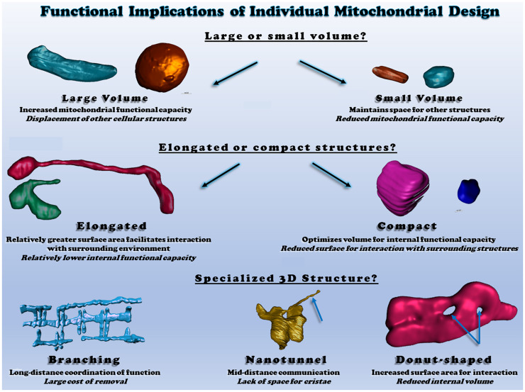
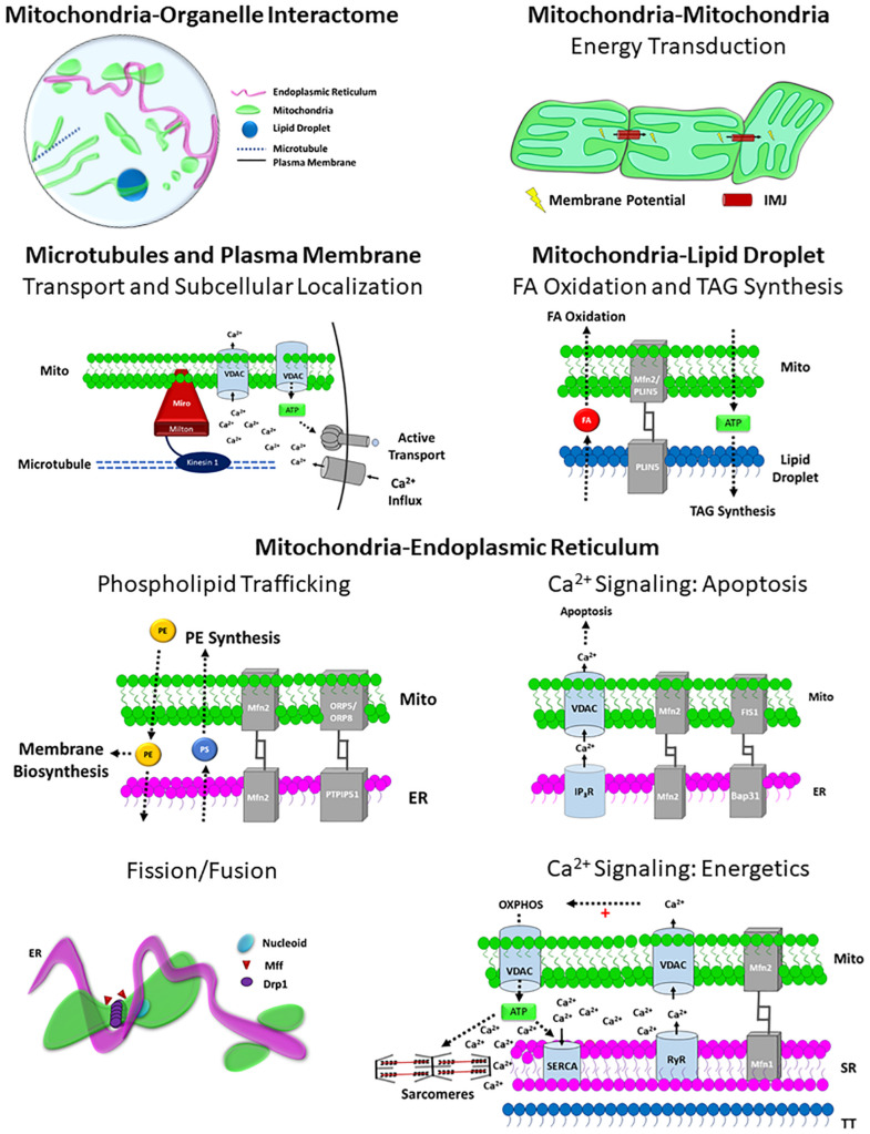
Mitochondria are key determinants of cellular health. However, the functional role of mitochondria varies from cell to cell depending on the relative demands for energy distribution, metabolite biosynthesis, and/or signaling. In order to support the specific needs of different cell types, mitochondrial functional capacity can be optimized in part by modulating mitochondrial structure across several different spatial scales. Here we discuss the functional implications of altering mitochondrial structure with an emphasis on the physiological trade-offs associated with different mitochondrial configurations. Within a mitochondrion, increasing the amount of cristae in the inner membrane improves capacity for energy conversion and free radical-mediated signaling but may come at the expense of matrix space where enzymes critical for metabolite biosynthesis and signaling reside. Electrically isolating individual cristae could provide a protective mechanism to limit the spread of dysfunction within a mitochondrion but may also slow the response time to an increase in cellular energy demand. For individual mitochondria, those with relatively greater surface areas can facilitate interactions with the cytosol or other organelles but may be more costly to remove through mitophagy due to the need for larger phagophore membranes. At the network scale, a large, stable mitochondrial reticulum can provide a structural pathway for energy distribution and communication across long distances yet also enable rapid spreading of localized dysfunction. Highly dynamic mitochondrial networks allow for frequent content mixing and communication but require constant cellular remodeling to accommodate the movement of mitochondria. The formation of contact sites between mitochondria and several other organelles provides a mechanism for specialized communication and direct content transfer between organelles. However, increasing the number of contact sites between mitochondria and any given organelle reduces the mitochondrial surface area available for contact sites with other organelles as well as for metabolite exchange with cytosol. Though the precise mechanisms guiding the coordinated multi-scale mitochondrial configurations observed in different cell types have yet to be elucidated, it is clear that mitochondrial structure is tailored at every level to optimize mitochondrial function to meet specific cellular demands.

Keywords: cristae; energetics; mitochondria; mitochondrial dynamics; mitochondrial networks; organelle interaction.


Figures

FIGURE 1
FIGURE 1
Functional consequences of different mitochondrial ultrastructure configurations. Top: the relative proportion of mitochondrial volume occupied by cristae or the matrix dictates space available to perform different functions. Scanning electron micrograph (SEM) of a liver mitochondrion from (Bochimoto et al., 2017). Middle right: the relative proportion of flat and curved cristae regions determines the space available for different oxidative phosphorylation enzymes. SEM of brown adipose tissue (Cinti, 2018) and heart mitochondria (Kanzaki et al., 2010) on the left and right, respectively. Middle left: mitochondrial nucleoids share the matrix space with enzymes and metabolites. Cryo-electron tomogram of a heart mitochondrion showing cristae (cyan) and nucleoids (green) from (Kanzaki et al., 2010; Kukat et al., 2015). Bottom left: the rate of cristae dynamics determines the frequency of remodeling and content sharing. Cristae remodeling in HeLa cells shown from (Kondadi et al., 2020). Bottom right: the relative electrical connectivity of cristae regulates distribution rate. Depolarization of some but not all HeLa cell cristae from (Wolf et al., 2019). All figures reproduced with permission.
FIGURE 2
FIGURE 2
Functional consequences of different individual mitochondrial structures. Top: individual mitochondrial volume determines the relative capacity for function but displaces other structures. Middle: Elongated or compact mitochondrial shapes determine the surface area available for interactions relative to mitochondrial volume. Bottom: irregular mitochondrial shapes allow for communication and interaction across various cellular distances. All heart and skeletal muscle mitochondrial 3D renderings shown were created from raw data available within (Glancy et al., 2017; Bleck et al., 2018).
FIGURE 3
FIGURE 3
Functional consequences of different mitochondrial network structures. Top left: the size of the mitochondrial network determines the relative mitochondrial functional capacity within the cell. Shown are individual mitochondria (various colors) within oxidative (left) and glycolytic (right) muscle mitochondrial networks from (Bleck et al., 2018). Top right: Connectivity of mitochondrial networks enables rapid distribution. Mitochondrial networks from healthy (left) and diabetic (right) pancreatic β-cells are shown from (Ježek and Dlasková, 2019). Bottom left: mitochondrial networks can be dynamic or relatively stable. Mitochondria in axons (left from Misgeld et al., 2007) and H9c2 cells (middle from Eisner et al., 2014) are highly dynamic, while skeletal muscle mitochondria (right from Eisner et al., 2014) are relatively stable. Bottom right: regional variations in mitochondrial distribution allows for subcellular specification of mitochondrial function. Neurons (left from Gao et al., 2019) and salivary acinar cells (right from Porat-Shliom et al., 2019) both display regional heterogeneity of mitochondrial networks. All images reproduced with permission.
FIGURE 4
FIGURE 4
Structure-Function relationships of the Mitochondrial Interactome. Mitochondria-organelle interactions support cellular communication and facilitate many different processes related to energy metabolism, biosynthesis, and mitochondrial division through specialized structure-function relationships, and the topology of the overall mitochondrial-organelle can be specialized according to cell type and adaptable to changes in cellular energy homeostasis. The specific functions of mitochondria-organelle interactions are regulated by the transmembrane channels and tethering proteins that comprise the physical structure of the interorganelle contact site. Intermitochondrial junction (IMJ); Voltage-dependent anion channel (VDAC); Adenosine Triphosphate (ATP); Mitofusin 1 (MFN1); Mitofusin 2 (MFN2); Perilipin 5 (PLIN5); Phosphatidylserine (PS); Phosphatidylethanolamine (PE); Fatty acid (FA); Oxysterol-binding protein–related protein 5/8 (ORP5/8); Protein tyrosine phosphatase-interacting protein 51 (PTPIP51); Inositol-1,4,5-trisphosphate receptors (IP3R); β-cell receptor-associated protein 31 (Bap31); Mitochondrial fission 1 protein (Fis1); Dynamin related protein 1 (DRP1); Mitochondria fission factor (Mff); Ryanodine receptor (RyR); Sarco/endoplasmic reticulum Ca2+-ATPase (SERCA).

References

    1. Ahmad T., Aggarwal K., Pattnaik B., Mukherjee S., Sethi T., Tiwari B. K., et al. (2013). Computational classification of mitochondrial shapes reflects stress and redox state. Cell Death Dis. 4:e461. 10.1038/cddis.2012.213 - DOI - PMC - PubMed
    1. Al-Mehdi A. B., Pastukh V. M., Swiger B. M., Reed D. J., Patel M. R., Bardwell G. C., et al. (2012). Perinuclear mitochondrial clustering creates an oxidant-rich nuclear domain required for hypoxia-induced transcription. Sci. Signal 5:ra47. 10.1126/scisignal.2002712 - DOI - PMC - PubMed
    1. Amati-Bonneau P., Guichet A., Olichon A., Chevrollier A., Viala F., Miot S., et al. (2005). OPA1 R445H mutation in optic atrophy associated with sensorineural deafness. Ann. Neurol. 58 958–963. 10.1002/ana.20681 - DOI - PubMed
    1. Amchenkova A. A., Bakeeva L. E., Chentsov Y. S., Skulachev V. P., Zorov D. B. (1988). Coupling membranes as energy-transmitting cables. I. Filamentous mitochondria in fibroblasts and mitochondrial clusters in cardiomyocytes. J. Cell Biol. 107 481–495. 10.1083/jcb.107.2.481 - DOI - PMC - PubMed
    1. Anderson G. R., Wardell S. E., Cakir M., Yip C., Ahn Y. R., Ali M., et al. (2018). Dysregulation of mitochondrial dynamics proteins are a targetable feature of human tumors. Nat. Commun. 9:1677. - PMC - PubMed

LinkOut - more resources