. 2020 Nov 12:11:548220.
doi: 10.3389/fneur.2020.548220. eCollection 2020.

Concussion Disrupts Normal Brain White Matter Microstructural Symmetry

Affiliations

Concussion Disrupts Normal Brain White Matter Microstructural Symmetry

Jun Maruta et al. Front Neurol. .

Abstract

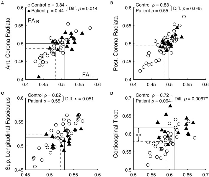
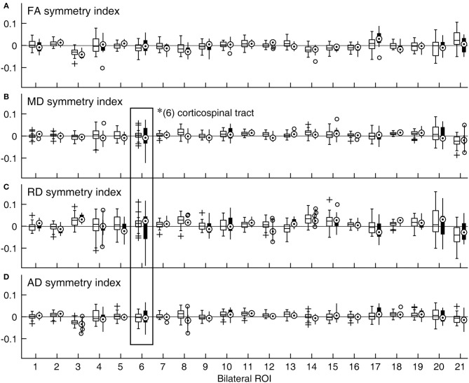
Injuries and illnesses can alter the normal bilateral symmetry of the brain, and determining the extent of this disruption may be useful in characterizing the pathology. One way of quantifying brain symmetry is in terms of bilateral correlation of diffusion tensor metrics between homologous white matter tracts. With this approach, we hypothesized that the brains of patients with a concussion are more asymmetrical than those of healthy individuals without a history of a concussion. We scanned the brains of 35 normal individuals and 15 emergency department patients with a recent concussion. Fractional anisotropy (FA), mean diffusivity (MD), axial diffusivity (AD), and radial diffusivity (RD) were determined for regions of interest (ROI) defined by a standard white-matter atlas that included 21 bilateral ROIs. For each ROI pair, bilateral correlation coefficients were calculated and compared between the two subject groups. A symmetry index, defined as the ratio between the difference and the sum of bilateral measures, was also calculated for each ROI pair and compared between the groups. We found that in normal subjects, the extent of symmetry varied among regions and individuals, and at least subtle forms of structural lateralization were common across regions. In patients, higher asymmetry was found overall as well as in the corticospinal tract specifically. Results indicate that a concussion can manifest in brain asymmetry that deviates from a normal state. The clinical utility of characterizing post-concussion pathology as abnormal brain asymmetry merits further exploration.

Keywords: acute concussion; bilateral homolog; diffusion tensor imaging (DTI); magnetic resonance imaging (MRI); mild traumatic brain injury (mTBI).


Figures

Figure 1
Figure 1
Bilateral correlation of FA values. (A) anterior corona radiata, (B) posterior corona radiata, (C) superior longitudinal fasciculus, and (D) corticospinal tract. Open circles and filled triangles indicate control and patient subjects, respectively, with the left and right FA values represented by the abscissa and ordinate, respectively. Dashed lines indicate means of the control group and solid lines those of the patient group. *Significant difference (with correction for multiple comparison) between the ρ-values. **Significant difference (with correction for multiple comparison) between the means (see Table 1).
Figure 2
Figure 2
Distributions of symmetry indices across ROIs and DTI metrics expressed as boxplots. (A) FA, (B) MD, (C) RD, and (D) AD. A symmetry index value of zero indicates bilaterally equal DTI metric values (horizontal lines). For a given ROI, the data for the control group are shown with an open box on the right side, while those for the patient group are shown with a filled box on the left side. *Significant group difference (with correction for multiple comparison). (1) anterior corona radiata; (2) anterior limb of internal capsule; (3) cingulum-cingulate gyrus; (4) cingulum-hippocampus; (5) cerebral peduncle; (6) corticospinal tract; (7) external capsule; (8) fornix/stria terminalis; (9) inferior cerebellar peduncle; (10) medial lemniscus; (11) posterior corona radiata; (12) posterior limb of internal capsule; (13) posterior thalamic radiation; (14) retrolenticular part of internal capsule; (15) superior cerebellar peduncle; (16) superior corona radiata; (17) superior fronto-occipital fasciculus; (18) superior longitudinal fasciculus; (19) sagittal stratum; (20) tapetum; (21) uncinate fasciculus.

References

    1. Wahl M, Li YO, Ng J, Lahue SC, Cooper SR, Sherr EH, et al. . Microstructural correlations of white matter tracts in the human brain. Neuroimage. (2010) 51:531–41. 10.1016/j.neuroimage.2010.02.072 - DOI - PMC - PubMed
    1. Presson N, Krishnaswamy D, Wagener L, Bird W, Jarbo K, Pathak S, et al. . Quantifying white matter structural integrity with high-definition fiber tracking in traumatic brain injury. Mil Med. (2015) 180:109–21. 10.7205/MILMED-D-14-00413 - DOI - PubMed
    1. Lerma-Usabiaga G, Mukherjee P, Ren Z, Perry ML, Wandell BA. Replication and generalization in applied neuroimaging. Neuroimage. (2019) 202:116048. 10.1016/j.neuroimage.2019.116048 - DOI - PMC - PubMed
    1. Mayer AR, Ling J, Mannell MV, Gasparovic C, Phillips JP, Doezema D, et al. . A prospective diffusion tensor imaging study in mild traumatic brain injury. Neurology. (2010) 74:643–50. 10.1212/WNL.0b013e3181d0ccdd - DOI - PMC - PubMed
    1. Vakhtin AA, Zhang Y, Wintermark M, Massaband P, Robinson MT, Ashford JW, et al. . White matter asymmetry: a reflection of pathology in traumatic brain injury. J Neurotrauma. (2020) 37:373–81. 10.1089/neu.2019.6487 - DOI - PubMed