. 2020 Nov 11:11:587931.
doi: 10.3389/fimmu.2020.587931. eCollection 2020.

Characterization of miRNAs in Extracellular Vesicles Released From Atlantic Salmon Monocyte-Like and Macrophage-Like Cells

Affiliations

Characterization of miRNAs in Extracellular Vesicles Released From Atlantic Salmon Monocyte-Like and Macrophage-Like Cells

Nicole C Smith et al. Front Immunol. .

Abstract

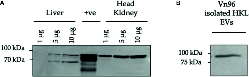
Cell-derived extracellular vesicles (EVs) participate in cell-cell communication via transfer of molecular cargo including genetic material like miRNAs. In mammals, it has previously been established that EV-mediated transfer of miRNAs can alter the development or function of immune cells, such as macrophages. Our previous research revealed that Atlantic salmon head kidney leukocytes (HKLs) change their morphology, phagocytic ability and miRNA profile from primarily "monocyte-like" at Day 1 to primarily "macrophage-like" at Day 5 of culture. Therefore, we aimed to characterize the miRNA cargo packaged in EVs released from these two cell populations. We successfully isolated EVs from Atlantic salmon HKL culture supernatants using the established Vn96 peptide-based pull-down. Isolation was validated using transmission electron microscopy, nanoparticle tracking analysis, and Western blotting. RNA-sequencing identified 19 differentially enriched (DE) miRNAs packaged in Day 1 versus Day 5 EVs. Several of the highly abundant miRNAs, including those that were DE (e.g. ssa-miR-146a, ssa-miR-155 and ssa-miR-731), were previously identified as DE in HKLs and are associated with macrophage differentiation and immune response in other species. Interestingly, the abundance relative of the miRNAs in EVs, including the most abundant miRNA (ssa-miR-125b), was different than the miRNA abundance in HKLs, indicating selective packaging of miRNAs in EVs. Further study of the miRNA cargo in EVs derived from fish immune cells will be an important next step in identifying EV biomarkers useful for evaluating immune cell function, fish health, or response to disease.

Keywords: Atlantic salmon; RNA-seq; RNA-sequencing; extracellular vesicles; head kidney culture; macrophage; microRNA.


Figures

Figure 1
Figure 1
Confirmation of extracellular vesicle (EV) release from Atlantic salmon HKLs. (A) Diagram of experimental workflow. (B) Transmission electron microscopy (TEM) images of EVs released into cell culture media by Day 1 HKLs (magnification 2700x and 6500x respectively; size of scale bar indicated on image). Area within the square of the left image is magnified in the right image. Arrows are pointing to double membranes. TEM images representative of n=3. NTA, Nanoparticle tracking analysis; WB, Western blot; RNA-seq, RNA-sequencing; RT-qPCR, reverse transcription quantitative polymerase chain reaction.
Figure 2
Figure 2
Characterization of Atlantic salmon extracellular vesicle (EV) size and quantity. Cell culture media containing EVs released from adherent Atlantic salmon HKLs was analyzed using nanoparticle tracking analysis (NTA). Five videos were captured per sample and results were reported as an average of the five videos. (A) Representative histogram of EV size profile (B) EV size distribution D10 (diameter where 10% of the population lies below the D10), D50 (diameter where 50% of the population lies below D50), and D90 (diameter where 90% of the population lies below D90) for EVs released at Day 1 and Day 5. Data reported as average mean +/- SE. (C) Mean size and mode size (+/- SE) of EVs released from Day 1 and Day 5 HKLs. (D) Concentration of EVs released from Day 1 and Day 5 HKLs. Scatterplots show data from individual fish (average of five videos); n=4; no statistical differences were observed as determined by a paired Student’s T-test.
Figure 3
Figure 3
HSP90 protein expression in Atlantic salmon HK, liver, and HKL derived extracellular vesicles (EVs). (A) Protein lysates from Atlantic salmon liver and head kidney tissue at 1, 5, and 10 μg were tested for cross-reactivity with anti-mouse HSP90. Wehi-231 murine B cells were used as a positive control (+ve). (B) HSP90 expression in Vn96 isolated EVs derived from Day 1 HKL culture media.
Figure 4
Figure 4
miRNA abundance (average normalized read counts) in extracellular vesicles (EVs) released from Day 1 and Day 5 Atlantic salmon HKLs. The top 20 most abundant miRNAs in Day 1 and Day 5 EVs are shown.
Figure 5
Figure 5
Heatmap illustration and hierarchical clustering analyses of differentially enriched miRNAs packaged in extracellular vesicles (EVs) released from Day 1 and Day 5 HKLs. The heatmap represents the normalized counts of DE miRNAs in EVs released from Day 1 HKLs and EVs released from Day 5 HKLs in each individual fish. miRNA normalized counts were median centred and clustered using Pearson correlation and complete linkage hierarchical clustering. Red indicates higher counts and green indicates lower counts. Integer adjusted to a maximum of 50 and a minimum of -50. F indicates fish number; D indicates Day 1 or Day 5 (i.e. F1D1 is Fish 1 Day 1).
Figure 6
Figure 6
RT-qPCR results. Scatterplots of the relative quantity (RQ) values of miRNAs determined by RNA sequencing to be DE between EVs released from Day 1 and Day 5 HKLs. Scatterplots show individual data with lines connecting data point from each individual fish, n=5. *p < 0.05; **p < 0.01. (A) miR-146a-5p (B) let-7a-5p (C) miR-16a-5p (D) miR-27d-5p (E) miR-210-3p (F) miR-221-5p (G) miR-21a-5p (H) miR-2188-3p (I) miR-150-5p.

References

    1. Robbins PD, Morelli AE. Regulation of immune responses by extracellular vesicles. Nat Rev Immunol (2014) 14:195–208. 10.1038/nri3622 - DOI - PMC - PubMed
    1. Colombo M, Raposo G, Thery C. Biogenesis, secretion, and intercellular interactions of exosomes and other extracellular vesicles. Annu Rev Cell Dev Biol (2014) 30:255–89. 10.1146/annurev-cellbio-101512-122326 - DOI - PubMed
    1. Longjohn MN, Hudson J-ABJ, Smith NC, Rise ML, Moorehead PC, Christian SL. Deciphering the messages carried by extracellular vesicles in hematological malignancies. Blood Rev (2020) 100734:1–11. 10.1016/j.blre.2020.100734 - DOI - PubMed
    1. Zhang Y, Liu Y, Liu H, Tang WH. Exosomes: biogenesis, biologic function and clinical potential. Cell Biosci (2019) 9:19. 10.1186/s13578-019-0282-2 - DOI - PMC - PubMed
    1. Gurunathan S, Kang M-H, Jeyaraj M, Qasim M, Kim J-H. Review of the Isolation, Characterization, Biological Function, and Multifarious Therapeutic Approaches of Exosomes. Cells (2019) 8:307. 10.3390/cells8040307 - DOI - PMC - PubMed

Publication types