. 2021 Jan;120(1):209-221.
doi: 10.1007/s00436-020-06968-x. Epub 2020 Dec 1.

Human serum activates the tegument of female schistosomes and supports recovery from Praziquantel

Affiliations

Human serum activates the tegument of female schistosomes and supports recovery from Praziquantel

Franziska Winkelmann et al. Parasitol Res. 2021 Jan.

Abstract

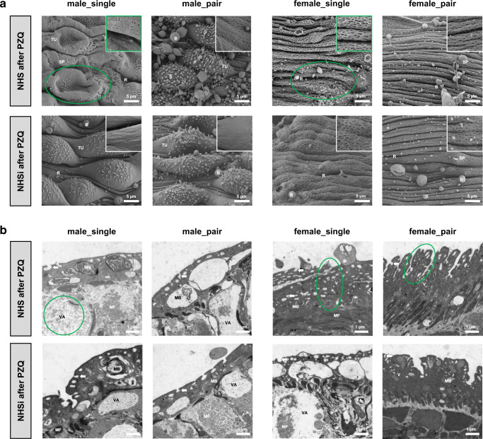
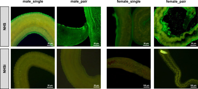
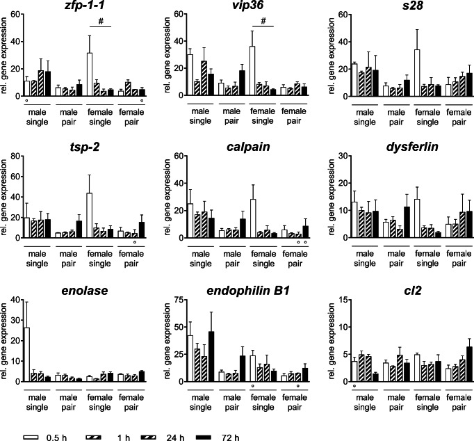
Schistosomiasis is one of the most devastating parasitic disease in the world. Schistosoma spp. survive for decades within the vasculature of their human hosts. They have evolved a vast array of mechanisms to avoid the immune reaction of the host. Due to their sexual dimorphism, with the female worm lying within the gynecophoric canal of the male worm, it is the male that is exposed to the immediate environment and the soluble parts of the host's immune response. To understand how the worms are so successful in fending off the immune attacks of the host, comparative analyses of both worm sexes in human serum (with or without Praziquantel) were performed using scanning electron microscopy, transmission electron microscopy, and immunohistochemistry. Further, gene expression analyses of tegument-specific genes were performed. Following the incubation in human serum, males and females out of pairs show morphological changes such as an altered structure of the pits below the surface and an increased number of pits per area. In addition, female schistosomes presented a marked tuft-like repulsion of their opsonized surface. The observed resistance of females to Praziquantel seemed to depend on active proteins in the human serum. Moreover, different expression profiles of tegument-specific genes indicate different functions of female_single and male_single teguments in response to human serum. Our results indicate that female schistosomes developed different evasion strategies toward the host's immune system in comparison to males that might lead to more robustness and has to be taken into account for the development of new anti-schistosomal drugs.

Keywords: Human serum; Schistosoma mansoni; Tegument integrity; Ultrastructural analysis.


Figures

Fig. 1
Fig. 1
Ultrastructural analysis of surface modifications in adult male and female S. mansoni after incubation in human serum. a Scanning electron microscopy (SEM; 2500x) with details of tegument structure (10,000×) and b transmission electron microscopy (TEM; 7100×) of the medial posterior portion of adult male (male_single, male_pair) and female (female_single, female_pair) S. mansoni after 0.5-h incubation in NHS and NHSi. Presence of tegumental ridges (R), tubercles (TU), spines (S), sensory papillae (SP), blebs (B), circular musculature (CM), longitudinal musculature (LM), cytoplasmic bridge (CB), membranous body (MB), vesicles (V), and pits of the outer surface (arrows) are indicated. Representative pictures out of five adult worms per group. Conspicuous areas as a result of NHS treatment encircled in green
Fig. 2
Fig. 2
Ultrastructural analysis of surface modifications in adult male and female S. mansoni after Praziquantel treatment. Scanning electron microscopy (SEM; 2500×) with details of tegument structure (10,000×) and transmission electron microscopy (TEM; 7100×) of the medial posterior portion of adult male (male_single, male_pair) and female (female_single, female_pair) S. mansoni after 0.5-h incubation in a NHS after PZQ and b NHSi after PZQ. Presence of tegumental ridges (R), tubercles (TU), spines (S), sensory papillae (SP), blebs (B), muscle fibers (MF), membranous body (MB), vacuoles (VA), vesicles (V), and pits of the outer surface (arrows) are indicated. Representative pictures out of five adult worms per group. Conspicuous areas as a result of NHS treatment encircled in green
Fig. 3
Fig. 3
Female schistosomes show a marked tuft-like repulsion of their opsonized surface after incubation in human serum in vitro. Fluorescence microscopy (100×: females in NHSi, 200×:) of adult male (male_single, male_pair) and female (female_single, female_pair) S. mansoni after 0.5-h incubation in NHS or NHSi with following immunohistochemical staining of human complement factor C3b
Fig. 4
Fig. 4
Human serum differentially impacts gene expression profiles of male and female S. mansoni. Relative gene expression of Calpain, SmCL2-like peptidase (CL2), Dysferlin, EndophilinB1, Enolase, Family S28 unassigned peptidase (S28), Tetraspanin-2 (TSP-2), Vesicular integral-membrane protein 36 (mannose-binding lectin 2)-related (Vip36), and Zinc finger protein-1-1 (ZFP-1-1) treated with normal human serum (NHS) at different time points was determined by real-time PCR. Data are presented as mean + SEMEAN; n = 3 (○ 2) each group; # pKruskal-Wallis < 0.05

Similar articles

Cited by

References

    1. Angeles JMM, van Mercado JP, Rivera PT. Behind enemy lines: immunomodulatory armamentarium of the Schistosome parasite. Front Immunol. 2020;11:1018. doi: 10.3389/fimmu.2020.01018. - DOI - PMC - PubMed
    1. Boissier J, Durand P, Moné H. PCR effectiveness for sexing Schistosoma mansoni cercariae: application for sexing clonal cercarial populations. Molecular and biochemical parasitology. 2001;112(1):139–141. doi: 10.1016/s0166-6851(00)00344-3. - DOI - PubMed
    1. Braschi S, Wilson RA. Proteins exposed at the adult schistosome surface revealed by biotinylation. Molecular & Cellular Proteomics : MCP. 2006;5(2):347–356. doi: 10.1074/mcp.M500287-MCP200. - DOI - PubMed
    1. Brindley PJ, Kalinna BH, Dalton JP, Day SR, Wong JYM, Smythe ML, McManus DP. Proteolytic degradation of host hemoglobin by schistosomes1Note: nucleotide sequences data reported in the paper have been submitted to the GenBank™ data base with the accession numbers L41346 and U77932.1. Mol Biochem Parasitol. 1997;89(1):1–9. doi: 10.1016/S0166-6851(97)00098-4. - DOI - PubMed
    1. Camacho M, Agnew A. Glucose uptake rates by Schistosoma mansoni, S. haematobium, and S. bovis adults using a flow in vitro culture system. J Parasitol. 1995;81(4):637. doi: 10.2307/3283866. - DOI - PubMed

LinkOut - more resources