. 2021 Jan:202:108325.
doi: 10.1016/j.exer.2020.108325. Epub 2020 Oct 22.

TGFβ1 and TGFβ2 proteins in corneas with and without stromal fibrosis: Delayed regeneration of apical epithelial growth factor barrier and the epithelial basement membrane in corneas with stromal fibrosis

Affiliations

TGFβ1 and TGFβ2 proteins in corneas with and without stromal fibrosis: Delayed regeneration of apical epithelial growth factor barrier and the epithelial basement membrane in corneas with stromal fibrosis

Rodrigo Carlos de Oliveira et al. Exp Eye Res. 2021 Jan.

Abstract

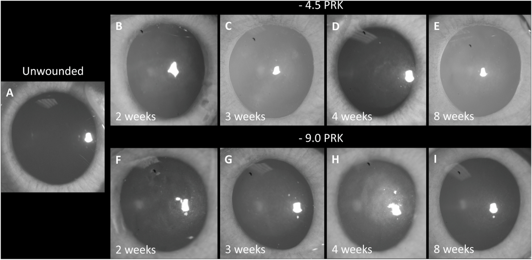
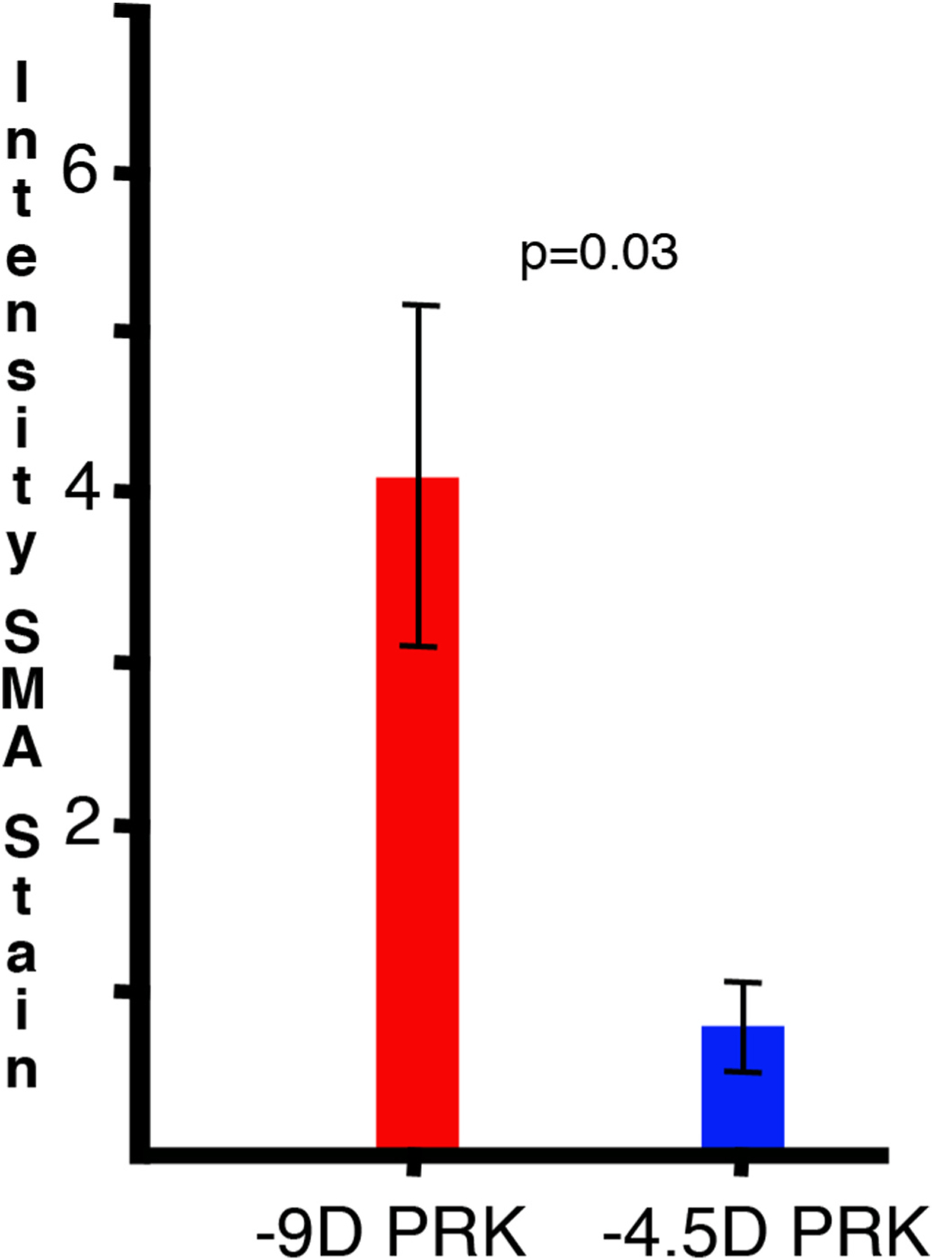
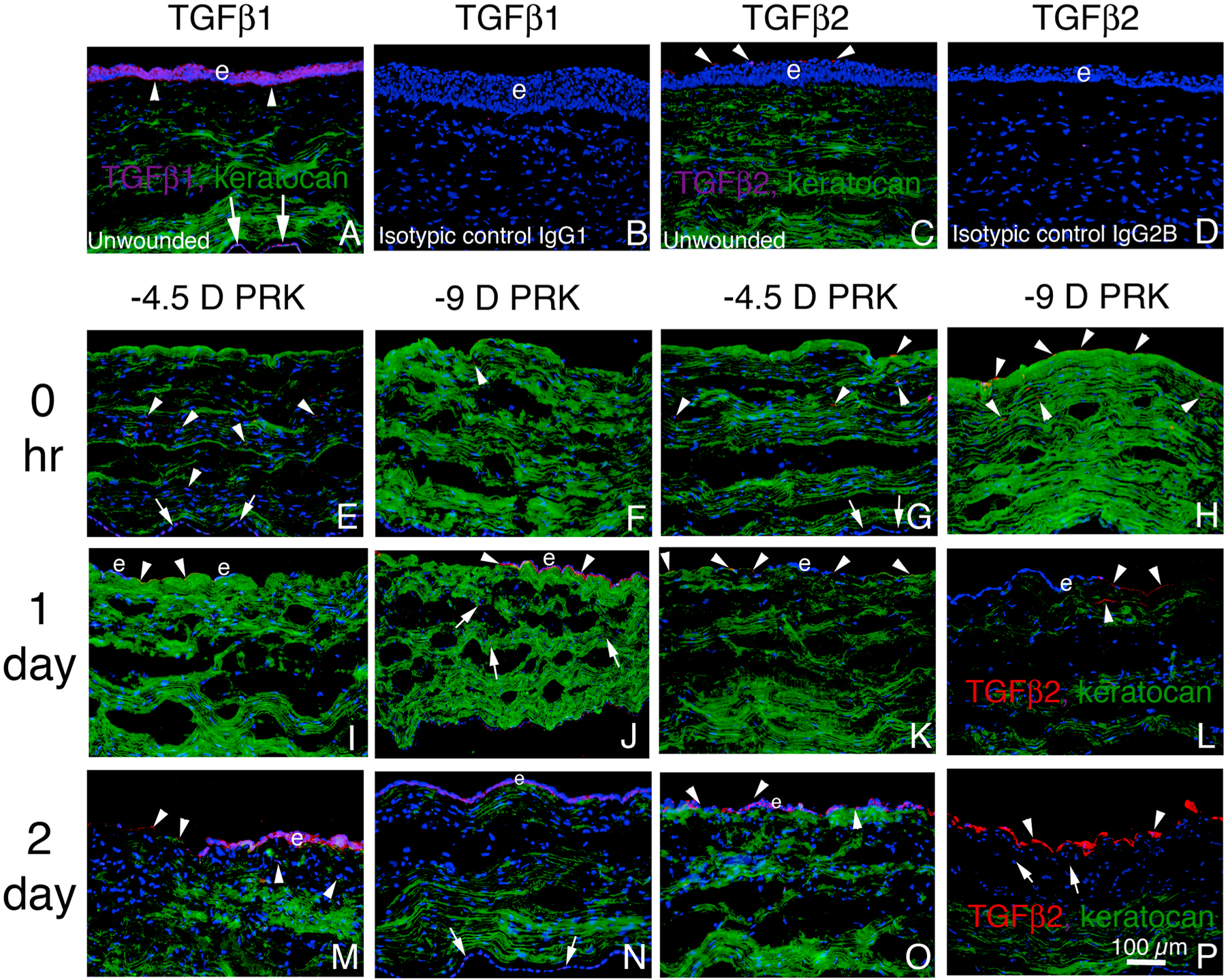
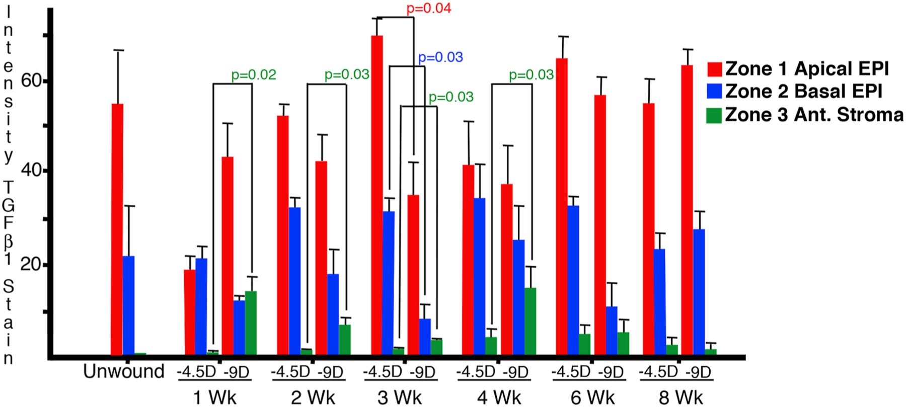
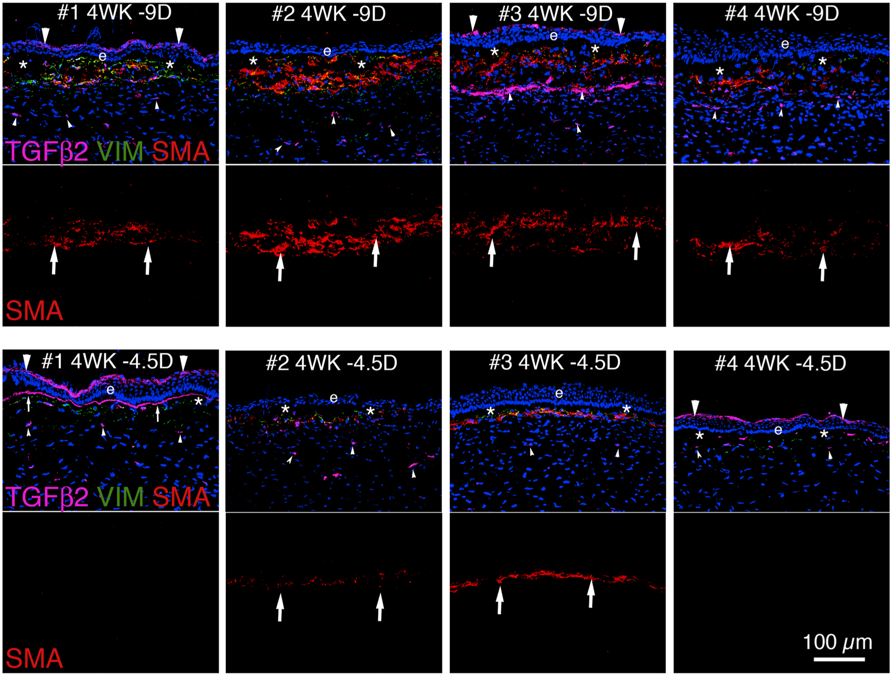
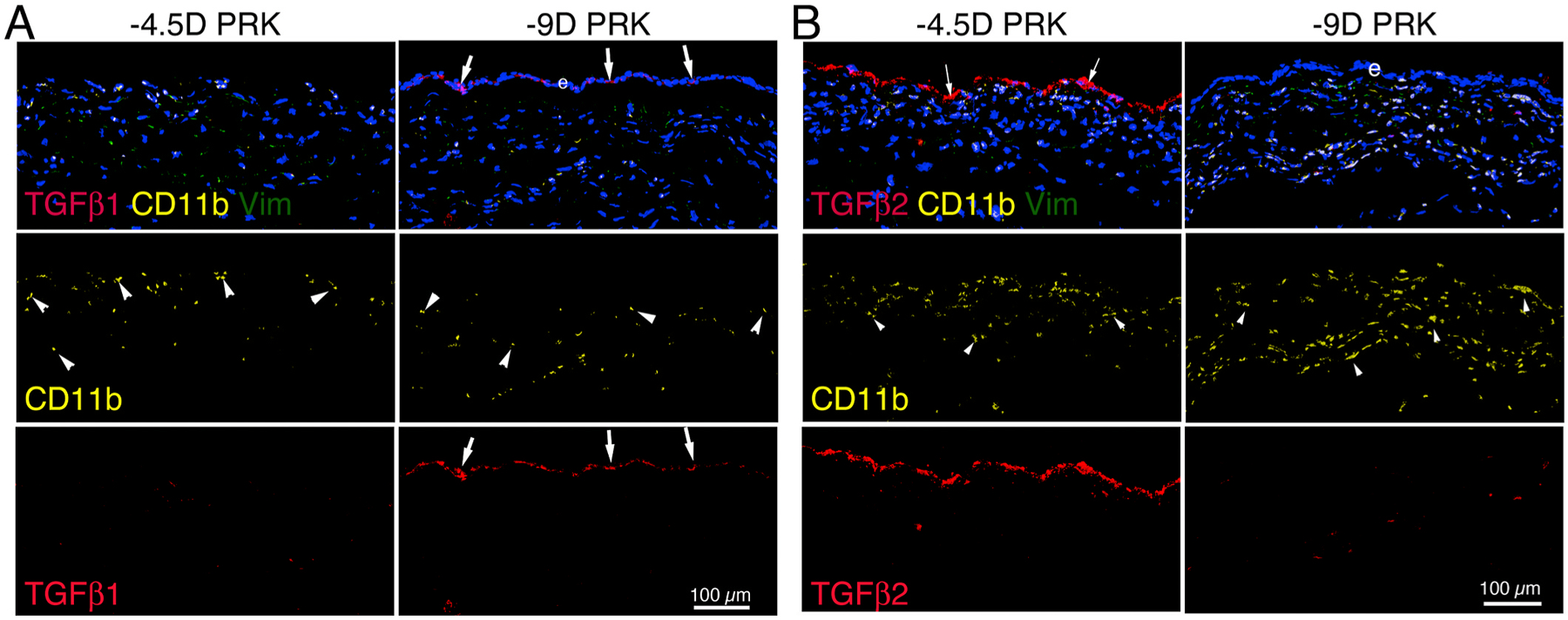
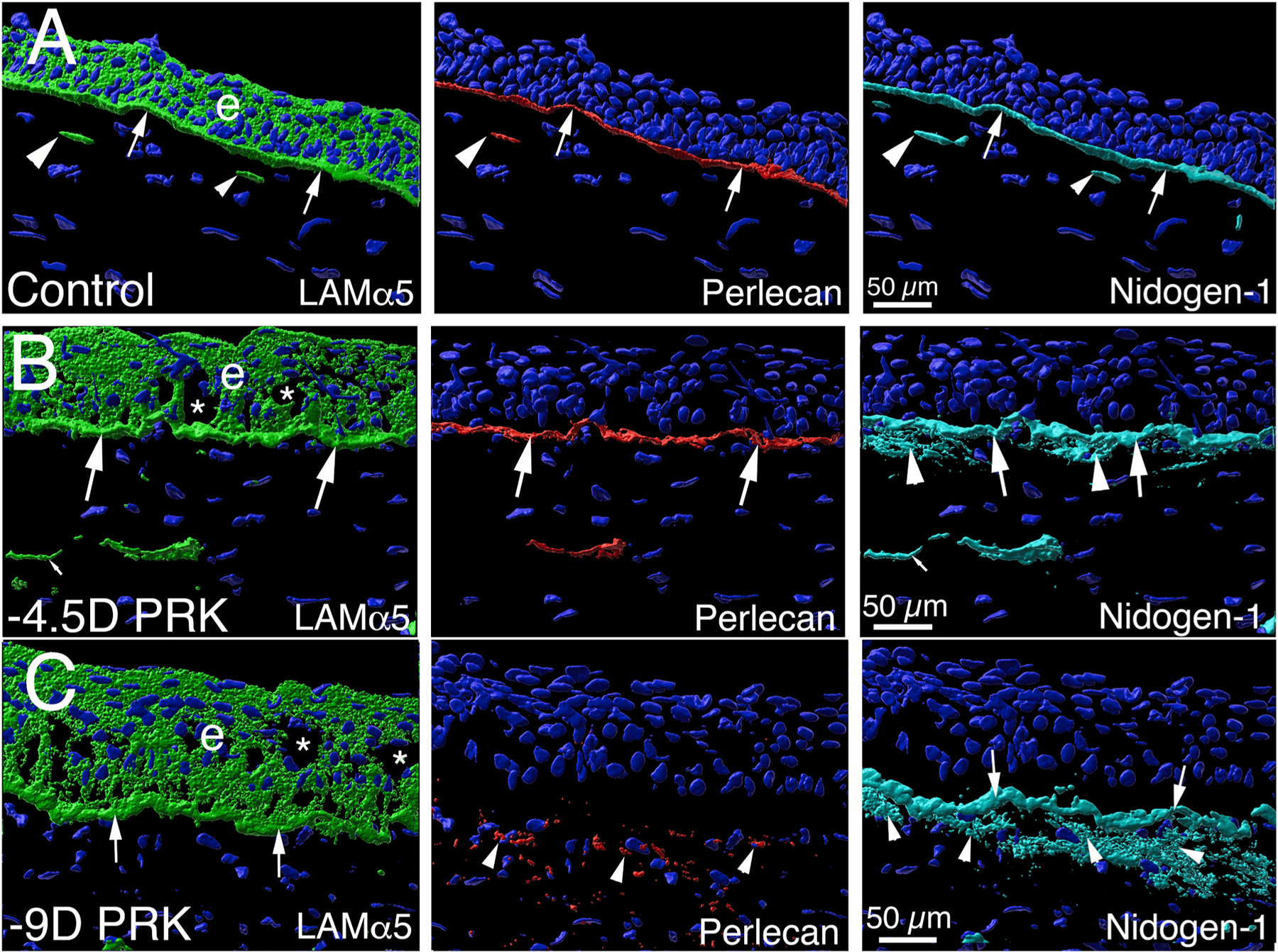
The purpose of this study was to investigate the expression and localization of transforming growth factor (TGF) β1 and TGFβ2 in rabbit corneas that healed with and without stromal fibrosis, and to further study defective perlecan incorporation in the epithelial basement membrane (EBM) in corneas with scarring fibrosis. A total of 120 female rabbits had no surgery, -4.5D PRK, or -9D PRK. Immunohistochemistry (IHC) was performed at time points from unwounded to eight weeks after surgery, with four corneas at each time point in each group. Multiplex IHC was performed for TGFβ1 or TGFβ2, with Image-J quantitation, and keratocan, vimentin, alpha-smooth muscle actin (SMA), perlecan, laminin-alpha 5, nidogen-1 or CD11b. Corneas at the four-week peak for myofibroblast and fibrosis development were evaluated using Imaris 3D analysis. Delayed regeneration of both an apical epithelial growth factor barrier and EBM barrier function, including defective EBM perlecan incorporation, was greater in high injury -9D PRK corneas compared to -4.5D PRK corneas without fibrosis. Defective apical epithelial growth factor barrier and EBM allowed epithelial and tear TGFβ1 and tear TGFβ2 to enter the corneal stroma to drive myofibroblast generation in the anterior stroma from vimentin-positive corneal fibroblasts, and likely fibrocytes. Vimentin-positive cells and unidentified vimentin-negative, CD11b-negative cells also produce TGFβ1 and/or TGFβ2 in the stroma in some corneas. TGFβ1 and TGFβ2 were at higher levels in the anterior stroma in the weeks preceding myofibroblast development in the -9D group. All -9D corneas (beginning two to three weeks after surgery), and four -4.5D PRK corneas developed significant SMA + myofibroblasts and stromal fibrosis. Both the apical epithelial growth factor barrier and/or EBM barrier functions tended to regenerate weeks earlier in -4.5D PRK corneas without fibrosis, compared to -4.5D or -9D PRK corneas with fibrosis. SMA-positive myofibroblasts were markedly reduced in most corneas by eight weeks after surgery. The apical epithelial growth factor barrier and EBM barrier limit TGFβ1 and TGFβ2 entry into the corneal stroma to modulate corneal fibroblast and myofibroblast development associated with scarring stromal fibrosis. Delayed regeneration of these barriers in corneas with more severe injuries promotes myofibroblast development, prolongs myofibroblast viability and triggers stromal scarring fibrosis.

Keywords: Corneal fibroblasts; Corneal scarring; Epithelial barrier function; Epithelial basement membrane; Fibrocytes; Fibrosis; Macrophages; Monocytes; Myofibroblasts; Perlecan; Secretory vesicles; Transforming growth factor beta-1; Transforming growth factor beta-2.


Conflict of interest statement

Proprietary interest statement

None of the authors have any commercial or proprietary interest in this study.

Figures

Fig. 1.
Fig. 1.
A representative IHC for TGFβ1 (purple), vimentin (green) and SMA (red) showing the ImageJ zones used to measure total pixels in the three areas of interest—1) apical epithelium, 2) posterior cornea including EBM (or location where EBM would be), and 3) anterior stroma just posterior to area 2. Area 3 was positioned to not include areas of artifactual dislocation of the epithelium from the anterior stromal surface that commonly occur during cryo-sectioning in the first few months after PRK, as shown in this example. Blue is DAPI staining of all nuclei. Original Mag. 400X.
Fig. 2.
Fig. 2.
Broad illumination slit lamp photographs at time points after −4.5D or −9D PRK. A. An unwounded control cornea shows normal transparency. At 2 weeks (B), 3 weeks (C), and 4 weeks (D) after −4.5 PRK. E. At 8 weeks after −4.5D PRK, a rabbit cornea has diffuse mild opacity despite having few vimentin-positive cells and no SMA-positive myofibroblasts. At two weeks (F) and 3 weeks (G) after −9D PRK there was increasing stromal opacity. At 4 weeks after −9D PRK (H) there was high opacity in the same cornea (Fig. 3. 4 week −9D far right column). At 8 weeks after −9D PRK (I) there is faint opacity in a cornea that had similar opacity to that shown in H at 4 weeks after the surgery. Original Mag. 40X.
Fig. 3.
Fig. 3.
Graph of mean ± SE intensity of stromal SMA in −4.5D PRK and −9D PRK corneas at 4 weeks after surgery determined by histogram analysis with ImageJ analysis software. n = 4 for each group. A 600 W × 200 H box in the stroma was used that encompassed all myofibroblasts in each cornea. The difference was significant at p = 0.03.
Fig. 4.
Fig. 4.
TGFβ1 or TGFβ2 and keratocan IHC from unwounded to two days after −4.5D PRK or −9D PRK. Purple or red indicates TGFβ1 or TGFβ2, depending on the secondary antibody used in IHC. Green is keratocan. Blue is DAPI staining of nuclei. e indicates epithelium in each panel where it is present. A. An unwounded cornea. Arrowheads indicate the EBM. Arrows indicate the corneal endothelium. Little TGFβ1 was detected in the stroma. TGFβ1 was also localized to the corneal endothelium (arrows). B. No non-specific staining was noted when isotypic control IgG1was included as the primary antibody with a 4 week −9D PRK cornea that had heavy TGFβ1 in the epithelium (see Fig. 5). C. An unwounded cornea with IHC for TGFβ2 (arrowheads) and keratocan. No TGFβ2 localized to the corneal endothelium (not shown). D. No non-specific staining was noted when isotypic control IgG2B was included as the primary antibody with a 4 week −4.5D PRK cornea that had heavy TGFβ2 in the epithelium (see Fig. 7). E. A cornea immediately after −4.5D PRK (0 h). TGFβ1 was detected associated with stromal cells (arrowheads). TGFβ1 was associated with corneal endothelial cells (arrows). F. Cornea immediately after −9D PRK (O hr). G. Cornea immediately after −4.5D PRK (0 h). TGFβ2 was associated with stromal cells and on the stromal surface (arrowheads). No TGFβ2 was associated with corneal endothelial cells (arrows). H. Cornea immediately after −9D PRK (0 h). TGFβ2 (arrowheads) was associated with stromal cells and on the stromal surface. I. Cornea one day after −4.5D PRK. TGFβ1 (arrowheads) was on the bare stromal surface and in islands of monolayered epithelium (e). J. Cornea at one day after −9D PRK. TGFβ1 (arrowheads) was associated with a monolayer of healing epithelial cells and some stromal cells (arrows). K. Cornea one day after −4.5D PRK. TGFβ2 (arrowheads) was detected on the stromal surface, but not in epithelial cells. L. Cornea at one day after −9D PRK. TGFβ2 was detected on the stromal surface (upper two arrowheads) and immediately beneath it (lower arrowhead), but was not associated with a monolayer of healing epithelium on the stromal surface. M. Cornea at two days after −4.5D PRK. TGFβ1 was associated with the stromal surface (upper two arrowheads) and a few stromal cells (lower two arrowheads). TGFβ1 was associated with the healing monolayer of epithelium. N. Cornea at two days after −9D PRK. TGFβ1 was associated with a monolayer of epithelium on the stromal surface. TGFβ1 was also associated with corneal endothelium (arrows). O. Cornea at two days after −4.5D PRK. TGFβ2 (arrowheads) was detected on the bare stromal surface and the surface of monolayered healing epithelium. P. Cornea at two days after −9D PRK. TGFβ2 (arrowheads) was present on the corneal surface and was also detected in the stroma (arrows). Original Mag. 200X.
Fig. 5.
Fig. 5.
TGFβ1 composite of all −4.5D and −9D PRK corneas at 4 weeks after surgery with the upper row for each group showing a composite triplex IHC for TGFβ1, vimentin and SMA and the middle row for each group showing the corresponding SMA (arrows) alone from the composite. Vimentin + cells are green. Perlecan IHC in the bottom rows for each group was from a subsequent IHC run from adjacent sections in the same blocks. TGFβ1 in the apical epithelium is indicated by small arrowheads in both groups. EBM TGFβ1 localization is indicated by arrows in both groups. Perlecan associated with EBM is indicated by arrows. Perlecan associated with anterior stromal cells is indicated by arrowheads. Little, if any, perlecan was associated with the EBM in −9D PRK corneas at 4 weeks after surgery. Blue is DAPI stained nuclei and e is epithelium in all panels. Original mag 200X.
Fig. 6.
Fig. 6.
A graph of Image J analysis of TGFβ1 intensity for the three anterior corneal zones (apical epithelium, basal epithelium with EBM, and anterior stroma shown in Fig. 1 for the −4.5D and −9D groups. n = 4 for each group. Each bar represents the mean ± S.E for the four corneas in that group at that time point. TGFβ1 was significantly greater in the anterior stroma, where myofibroblast precursors develop into myofibroblasts, during the first four weeks after corneal injury.
Fig. 7.
Fig. 7.
TGFβ2 composite for all −4.5D and −9D PRK corneas at 4 weeks after surgery. The upper row for each group showing a composite triplex IHC for TGFβ2 (purple), vimentin (green) and SMA (red). The middle row for each group showing the corresponding IHC of SMA (arrows) alone from the composite. The corresponding perlecan IHC for each of these corneas was shown in Fig. 5. Large arrowheads indicate apical epithelial TGFβ2 when present. Arrows indicate EBM associated TGFβ2 in −4.5D PRK cornea #1. Small arrowheads indicate stromal TGFβ2. Blue is DAPI stained nuclei and e is epithelium in all panels. * indicates artifactual displacement of the epithelium from the underlying stroma during sectioning. Original mag 200X.
Fig. 8.
Fig. 8.
A graph of Image J analysis of TGFβ2 intensity for the three anterior corneal zones shown in Fig. 1 for the −4.5D and −9D groups. Each bar represents the mean ± S.E for the four corneas in that group at that time point. TGFβ2 was significantly greater in the anterior stroma of −9D PRK corneas compared to −4.5D PRK corneas at 2 weeks and 3 weeks after corneal injury.
Fig. 9.
Fig. 9.
CD11b, vimentin and TGFβ1 or TGFβ2 IHC at 2 days after PRK in rabbits. A. The epithelium has not yet healed over the central cornea of this eye after −4.5D PRK. CD11b-positive cells (arrowheads) infiltrated the cornea, but none of these cells had associated TGFβ1. A monolayer of epithelium (e) had healed over the central cornea of the eye that had –9D PRK, and TGFβ1 was present in these epithelial cells. Large numbers of CD11b-positive cells (arrowheads) infiltrated the cornea, but none were producing TGFβ1. B. The epithelium is not yet healed over the central cornea of this eye at 2 days after −4.5D PRK and the bare stroma had high levels of associated TGFβ2 (arrows). A monolayer of epithelium (e) had healed over the central cornea of the eye that had –9D PRK, and small amounts of TGFβ2 were detected on the surface of the epithelium but not produced by epithelial cells. Large numbers of CD11b-positive cells were infiltrating both corneas, but none had associated TGFβ2. Blue is DAPI staining of nuclei and e is epithelium in all panels. Original Mag. 200X.
Fig. 10.
Fig. 10.
Imaris 3D constructions of confocal microscopy images of laminin alpha-5, perlecan and nidogen-1 in the epithelium, EBM and anterior stroma. A. In an unwounded control cornea, laminin alpha-5 (green) is detected throughout the epithelium (e), and in the EBM (arrows). Two fusiform DAPI-negative bodies with laminin alpha-5 (arrowheads) are present in the subepithelial stroma adjacent to the EBM. Perlecan (red) is detected in the EBM (arrows), and in bodies in the stroma (arrowhead). Nidogen-1 (blue gray) is also present in the EBM (arrows) and in bodies in the subepithelial stroma (arrowheads). B. In a −4.5D PRK cornea at 1 mo after surgery that did not develop stromal opacity or myofibroblasts (#1 in Fig. 5), the results were similar to the unwounded control cornea with laminin alpha-5, perlecan and nidogen-1 in the EBM (large arrows). Additional nidogen-1 (arrowheads) was present in the subepithelial stroma surrounding stromal cells detected via their nuclei. DAPI-negative bodies (small arrows) containing one or more of the EBM components were present in the anterior stroma. C. In a −9D PRK cornea at 1 mo after surgery that had severe stromal opacity and myofibroblasts (#3 in Fig. 5) there was laminin alpha-5 and nidogen-1 in the EBM (arrows) similar to the unwounded control. However, perlecan was not detected in the EBM, despite its presence in myofibroblasts in the anterior stroma (arrowheads, see Fig. 5). Stromal nidogen-1 (arrowheads) was also detected in the subepithelial cells that are myofibroblasts. Blue is DAPI-stained nuclei in all panels. e is epithelium. * indicates artifactual defects in the epithelium which are often seen in PRK corneas that are cryo-sectioned in the first one to two months after surgery. Original mag 630X.

References

    1. Annes JP, Rifkin DB, Munger JS, 2002. The integrin αVβ6 binds and activates latent TGFβ3. FEBS Lett 511, 65–68. - PubMed
    1. Azar DT, Hahn TW, Jain S, Yeh YC, Stetler-Stevensen WG, 1996. Matrix metalloproteinases are expressed during wound healing after excimer laser keratectomy. Cornea 15, 18–24. - PubMed
    1. Ban Y, Dota A, Cooper LJ, Fullwood NJ, Nakamura T, Tsuzuki M, Mochida C, Kinoshita S, 2003. Tight junction-related protein expression and distribution in human corneal epithelium. Exp. Eye Res 76, 663–669. - PubMed
    1. Blanco-Mezquita JT, Hutcheon AE, Stepp MA, Zieske JD, 2011. αVβ6 integrin promotes corneal wound healing. Invest. Ophthalmol. Vis. Sci 52, 8505–8513. - PMC - PubMed
    1. Brown JC, Sasaki T, Göhring W, Yamada Y, Timpl R, 1997. The C-terminal domain V of perlecan promotes beta1 integrin-mediated cell adhesion, binds heparin, nidogen and fibulin-2 and can be modified by glycosaminoglycans. Eur. J. Biochem 250, 39–46. - PubMed

Publication types

Substances