. 2020 Dec 1;76(Pt 12):1192-1200.
doi: 10.1107/S2059798320013170. Epub 2020 Nov 19.

Shift-field refinement of macromolecular atomic models

Affiliations

Shift-field refinement of macromolecular atomic models

K Cowtan et al. Acta Crystallogr D Struct Biol. .

Abstract

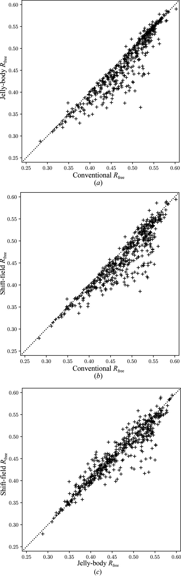
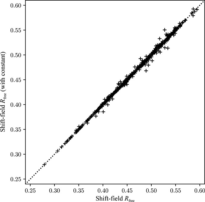
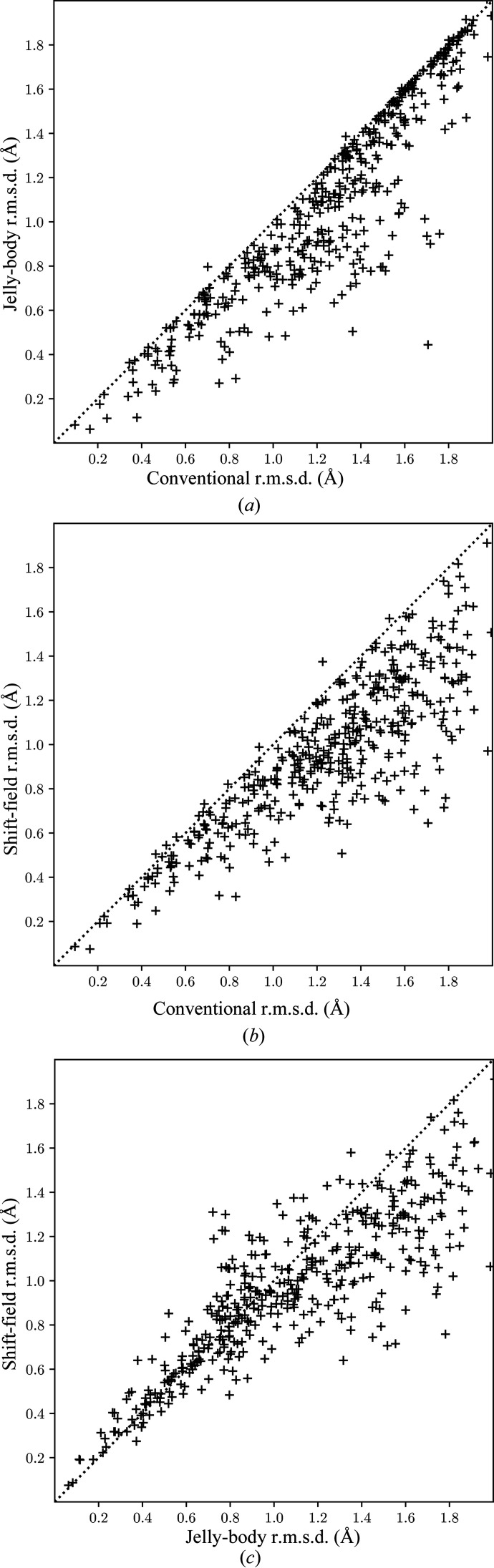
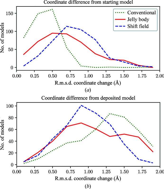
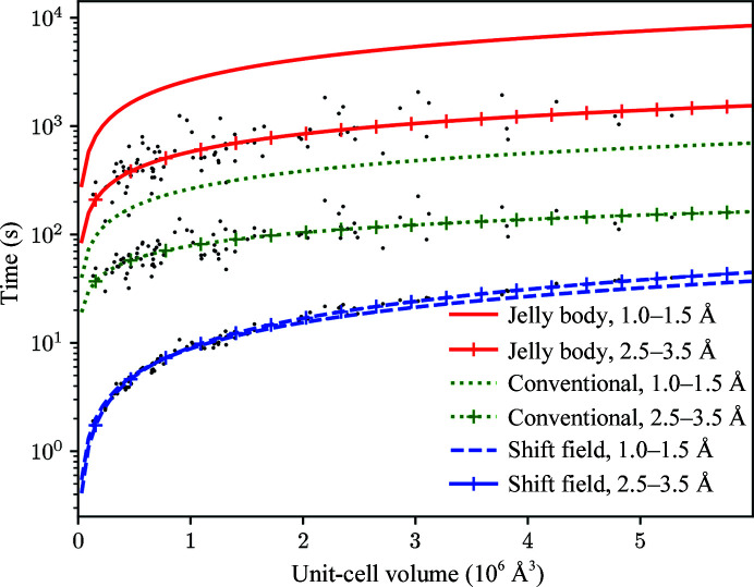
The aim of crystallographic structure solution is typically to determine an atomic model which accurately accounts for an observed diffraction pattern. A key step in this process is the refinement of the parameters of an initial model, which is most often determined by molecular replacement using another structure which is broadly similar to the structure of interest. In macromolecular crystallography, the resolution of the data is typically insufficient to determine the positional and uncertainty parameters for each individual atom, and so stereochemical information is used to supplement the observational data. Here, a new approach to refinement is evaluated in which a `shift field' is determined which describes changes to model parameters affecting whole regions of the model rather than individual atoms only, with the size of the affected region being a key parameter of the calculation which can be changed in accordance with the resolution of the data. It is demonstrated that this approach can improve the radius of convergence of the refinement calculation while also dramatically reducing the calculation time.

Keywords: computational methods; low resolution; refinement.


Figures

Figure 1
Figure 1
Comparison of the free R factor after the various refinement procedures. (a) Jelly-body refinement compared with conventional refinement alone. (b) Shift-field refinement compared with conventional refinement alone. (c) Shift-field refinement compared with jelly-body refinement. Points below the diagonal indicate a better result for the method on the y axis.
Figure 2
Figure 2
Comparison of the free R factor after shift-field plus conventional refinement with the shift-field regression calculation either including (y axis) or omitting (x axis) the constant term.
Figure 3
Figure 3
Comparison of the root-mean-square difference in Cα coordinate positions between matched residues of the refined molecular-replacement model and the refined deposited structure under different refinement protocols. (a) Jelly-body refinement compared with conventional refinement alone. (b) Shift-field refinement compared with conventional refinement alone. (c) Shift-field refinement compared with jelly-body refinement. Points below the diagonal indicate a better result for the method on the y axis.
Figure 4
Figure 4
Comparison of the distribution of r.m.s.d. values over the 452 test structures between the initial model and models refined using the conventional, jelly-body and shift-field refinement protocols (a) and between the refined models and the deposited structure (b). The distributions are binned in steps of 0.2 Å but are plotted with lines for ease of comparison.
Figure 5
Figure 5
Comparative CPU times (in seconds) for the three refinement protocols averaged over 160 structures between 1.0 and 1.5 Å resolution and 90 structures between 2.5 and 3.5 Å resolution. Results are given for shift-field refinement alone (without the conventional refinement step), for 200 cycles of jelly-body refinement in REFMAC5 and for 20 cycles of conventional refinement in REFMAC5. The least-squares linear fit of time against unit-cell volume is shown for each method and resolution range, with scatter points shown for the low-resolution subsets.
Figure 6
Figure 6
Comparison of a section through models for PDB entry 4l9m, showing the Cα trace of the jelly-body refined model (thick dark bonds), the shift-field refinement model (thick light bonds) and the deposited model (thin bonds).
Figure 7
Figure 7
Comparison of models for PDB entry 2d66 showing the Cα trace of the jelly-body refined model (thick dark bonds), the shift-field refinement model (thick light bonds) and the deposited model (thin bonds). Some loops have been removed for clarity. Symmetry contacts of the deposited model are also shown (thin light bonds).

References

    1. Afonine, P. V., Grosse-Kunstleve, R. W., Echols, N., Headd, J. J., Moriarty, N. W., Mustyakimov, M., Terwilliger, T. C., Urzhumtsev, A., Zwart, P. H. & Adams, P. D. (2012). Acta Cryst. D68, 352–367. - PMC - PubMed
    1. Agarwal, R., Lifchitz, A. & Dodson, E. (1981). In Proceedings of the CCP4 Study Weekend. Refinement of Protein Structures, edited by P. A. Machin, J. W. Campbell & M. Elder. Warrington: Daresbury Laboratory.
    1. Berman, H., Henrick, K., Nakamura, H. & Markley, J. (2007). Nucleic Acids Res. 35, D301–D303. - PMC - PubMed
    1. Blanc, E., Roversi, P., Vonrhein, C., Flensburg, C., Lea, S. M. & Bricogne, G. (2004). Acta Cryst. D60, 2210–2221. - PubMed
    1. Bond, P. S., Wilson, K. S. & Cowtan, K. D. (2020). Acta Cryst. D76, 713–723. - PMC - PubMed

MeSH terms

Substances