. 2020 Dec 11;6(12):3174-3189.
doi: 10.1021/acsinfecdis.0c00500. Epub 2020 Dec 2.

Comparative Multiplexed Interactomics of SARS-CoV-2 and Homologous Coronavirus Nonstructural Proteins Identifies Unique and Shared Host-Cell Dependencies

Affiliations

Comparative Multiplexed Interactomics of SARS-CoV-2 and Homologous Coronavirus Nonstructural Proteins Identifies Unique and Shared Host-Cell Dependencies

Jonathan P Davies et al. ACS Infect Dis. .

Abstract

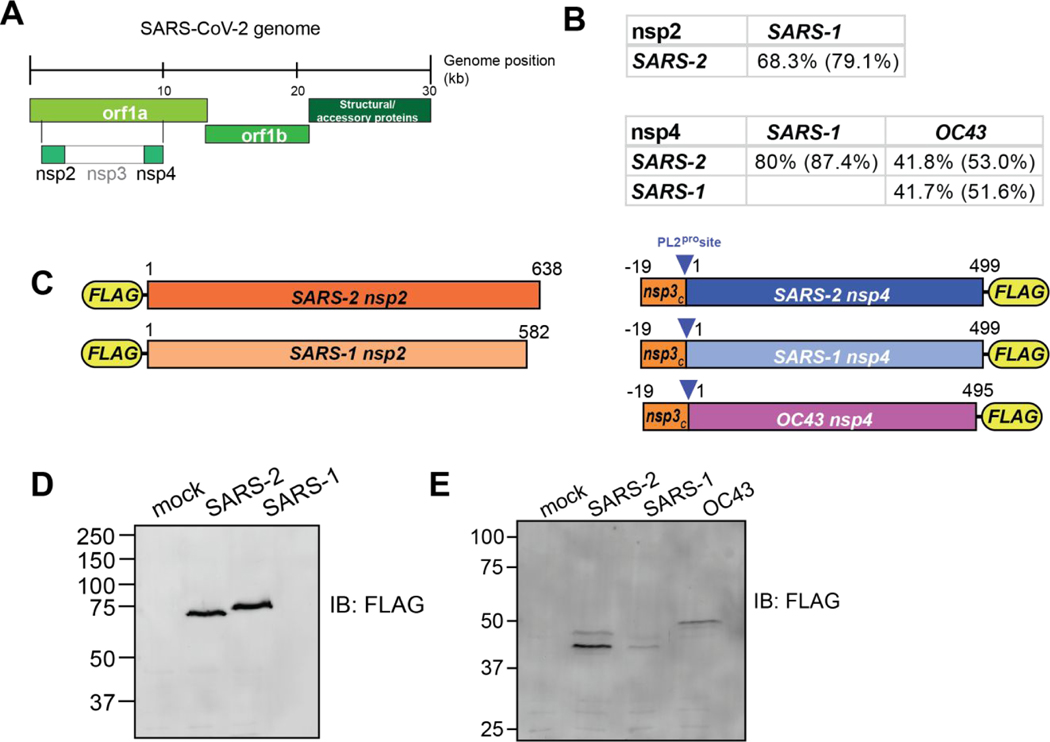
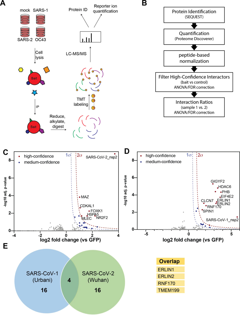
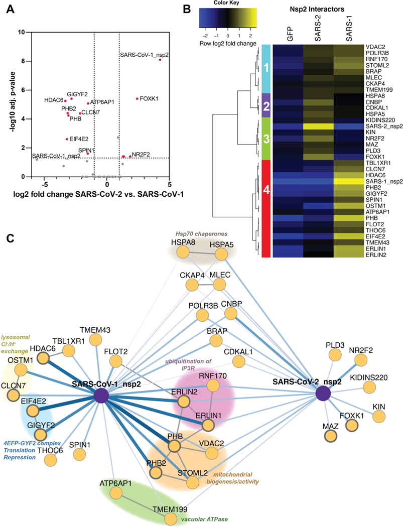
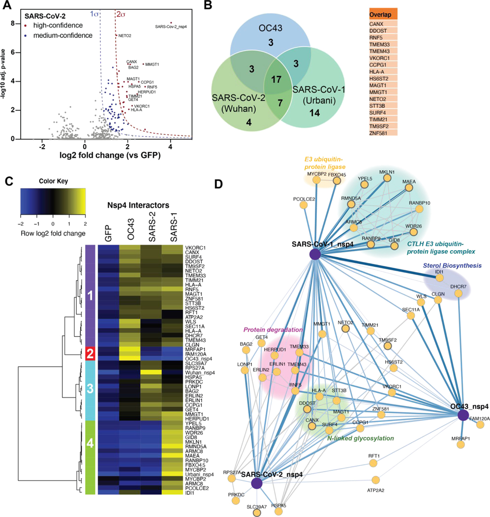
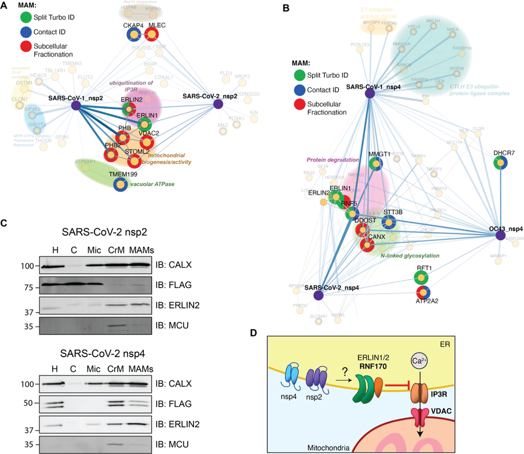
Human coronaviruses (hCoVs) have become a threat to global health and society, as evident from the SARS outbreak in 2002 caused by SARS-CoV-1 and the most recent COVID-19 pandemic caused by SARS-CoV-2. Despite a high sequence similarity between SARS-CoV-1 and -2, each strain has a distinctive virulence. A better understanding of the basic molecular mechanisms mediating changes in virulence is needed. Here, we profile the virus-host protein-protein interactions of two hCoV nonstructural proteins (nsps) that are critical for virus replication. We use tandem mass tag-multiplexed quantitative proteomics to sensitively compare and contrast the interactomes of nsp2 and nsp4 from three betacoronavirus strains: SARS-CoV-1, SARS-CoV-2, and hCoV-OC43-an endemic strain associated with the common cold. This approach enables the identification of both unique and shared host cell protein binding partners and the ability to further compare the enrichment of common interactions across homologues from related strains. We identify common nsp2 interactors involved in endoplasmic reticulum (ER) Ca2+ signaling and mitochondria biogenesis. We also identify nsp4 interactors unique to each strain, such as E3 ubiquitin ligase complexes for SARS-CoV-1 and ER homeostasis factors for SARS-CoV-2. Common nsp4 interactors include N-linked glycosylation machinery, unfolded protein response associated proteins, and antiviral innate immune signaling factors. Both nsp2 and nsp4 interactors are strongly enriched in proteins localized at mitochondria-associated ER membranes suggesting a new functional role for modulating host processes, such as calcium homeostasis, at these organelle contact sites. Our results shed light on the role these hCoV proteins play in the infection cycle, as well as host factors that may mediate the divergent pathogenesis of OC43 from SARS strains. Our mass spectrometry workflow enables rapid and robust comparisons of multiple bait proteins, which can be applied to additional viral proteins. Furthermore, the identified common interactions may present new targets for exploration by host-directed antiviral therapeutics.

Keywords: COVID-19; affinity purification-mass spectrometry; mitochondria-associated endoplasmic reticulum membrane; nsp2; nsp4; tandem mass tags.


Conflict of interest statement

CONFLICT OF INTEREST STATEMENT

The authors declare no conflicts of interest.

Figures

Figure 1.
Figure 1.. Design and validation of expression of CoV nsp2 and nsp4 constructs for affinity purification.
(A) Schematic of SARS-CoV-2 genome organization. (B) Amino acid sequence identity and similarity (in parentheses) for comparisons of nsp2 and nsp4 homologs. Sequence alignments are shown in Figure S1A–B. (C) Nsp2 and nsp4 FLAG-tagged construct designs. Nsp2 constructs contain an N-terminal FLAG-tag. Nsp4 constructs contain a 19 amino acid leader sequence from nsp3 at the N-terminus, including the PL2pro cleavage site, along with a C-terminal FLAG-tag. (D-E) Western blot of nsp2 and nsp4 homologs expressed in HEK293T cells. Cells were transiently transfected with FLAG-nsp2 (D) or nsp4-FLAG (E). Proteins were detected using an anti-FLAG antibody.
Figure 2.
Figure 2.. Affinity purification-mass spectrometry (AP-MS) identifies nsp2 interactors.
(A) General AP-MS workflow to quantitatively determine interactors of viral nsp homolog. HEK293T cells are transfected with FLAG-tagged expression constructs of nsps as bait or GFP (mock) and lysed. Bait proteins are immunoprecipitated (IP) along with interacting proteins, reduced, alkylated, and tryptic digested. Peptides are then tandem-mass tag (TMT) labeled, pooled, and analyzed by LC-MS/MS for identification and quantification. (B) Data processing workflow. Peptide spectra are identified and matched to corresponding proteins (SEQUEST HT), then quantified based on TMT reporter ion intensity (Proteome Discoverer 2.4). High confidence interactors are filtered by comparing bait vs control. Interaction ratios between bait homologs are determined (log2 fold change) and adjusted p-value calculated using ANOVA. (C-D) Volcano plot of SARS-CoV-2 nsp2 (C) and SARS-CoV-1 nsp2 (D) datasets to identify medium- and high-confidence interactors. Plotted are log2 TMT intensity fold changes for proteins between nsp2 bait channels and GFP mock transfections versus -log10 adjusted p-values. Curves for the variable cutoffs used to define high-confidence (red) or medium confidence (blue) interactors are shown. 1σ = 0.5 for (C), 1σ = 0.43 for (D). (E) Venn diagram comparing high-confidence interactors between nsp2 homologs. Sixteen unique proteins were identified each, while four proteins overlapped both data sets (listed in adjacent table).
Figure 3.
Figure 3.. Quantitative comparison of SARS-CoV-1 and SARS-CoV-2 nsp2 interactors.
(A) Volcano plot comparing interactions between nsp2 homolog from SARS-CoV-1 and SARS-CoV-2. Only high- and medium confidence interactors of nsp2 are shown. Highlighted proteins meet the filter criteria of adjusted p-value < 0.05 and | log2 fold change | >1. (B) Heatmap comparing the enrichment of SARS-CoV-1 and SARS-CoV-2 nsp2 interactors compared to GFP control. log2 fold change is color-coded and centered by row (blue low, yellow high enrichment). Hierarchical clustering using Ward’s method shown on the left was carried out on euclidean distances of log2 fold changes scaled by row. Clusters 1 and 2 corresponds to shared interactors of SARS-CoV-1 and −2 nsp2, while cluster 3 and 4 for are unique interactors for SARS-CoV-2 and SARS-CoV-1 nsp2, respectively. (C) Protein-protein interaction (PPI) network map of nsp2 homologs. Blue lines indicate viral-host PPIs, where line width corresponds to fold enrichment compared to the GFP control. Grey lines indicate annotated host-host PPIs in STRING (score > 0.75). Groups of interactors with a common functional role are highlighted.
Figure 4.
Figure 4.. Comparative profiling of nsp4 interactions.
(A) Volcano plot of the SARS-CoV-2 nsp4 datasets to identify medium- and high-confidence interactors. Plotted are log2 TMT intensity differences for proteins between nsp4 bait channels and GFP mock transfections versus -log10 adjusted p-values. Curves for the variable cutoffs used to define high-confidence (red) or medium confidence (blue) interactors are shown. 1σ = 0.66. Equivalent volcano plot for SARS-CoV-1 and OC43 nsp4 are shown in Figure S5B–C. (B) Venn diagram of interactors from nsp4 homologs. Overlapping nsp4 interactors between all strains are listed in the adjacent table. (C) Heatmap comparing the enrichment of interactors for the different nsp4 homologs. log2 fold change is color-coded and centered by row (blue low, yellow high enrichment). Hierarchical clustering using Ward’s method shown on the left was carried out on euclidean distances of log2 fold changes scaled by row. Clusters 1 corresponds to shared interactors of SARS-CoV-1, −2, and OC43 nsp4. Cluster 2, and 4 contain unique interactors for OC43 and SARS-CoV-1 nsp4, respectively, while cluster 3 contains shared interactors of SARS-CoV-1 and SARS-CoV-2. (D) Protein-protein interaction (PPI) network map of interactors of nsp4 homolog. Blue lines indicate measured viral-host PPIs, where line width corresponds to fold enrichment compared to the GFP control. Grey lines indicate annotated host-host PPIs in STRING (score > 0.75). Groups of interactors with a common functional role are highlighted.
Figure 5.
Figure 5.. Enrichment of mitochondria-associated membrane (MAM) proteins as nsp2 and nsp4 interactors.
(A–B) Interactors of nsp2 (A) and nsp4 (B) homolog annotated for MAM proteins. The lists of interactors were cross-referenced with previous publications profiling the MAM proteome (Split-Turbo ID(55), Contact-ID(56), and subcellular fractionation(57)). (C) Subcellular fractions of SARS-CoV-2 nsp2 or nsp4 transfected HEK293T cells to determine localization of viral proteins to MAMs. Homogenate (H), cytosol (C), microsome (Mic), crude mitochondria (CrM), and MAMs fractions were probed via Western blot for subcellular markers (CALX and ERLIN2 for MAMs; MCU for mitochondria) and viral proteins (FLAG). Subcellular fractionation was carried out in triplicate and representative blots shown. (D) Proposed model for how SARS-CoV nsp2 and nsp4 utilize ERLIN1/2 and interacting protein factors to regulate ER Ca2+ signaling at MAMs.

Update of

References

    1. Fehr AR; Perlman S. Coronaviruses: An Overview of Their Replication and Pathogenesis. Methods Mol. Biol 2015, 1282, 1. 10.1007/978-1-4939-2438-7_1. - DOI - PMC - PubMed
    1. Novel Coronavirus (2019-NCoV) Situation Report 1; 2020.
    1. Hoffmann M; Kleine-Weber H; Schroeder S; Mü MA; Drosten C; Pö S. SARS-CoV-2 Cell Entry Depends on ACE2 and TMPRSS2 and Is Blocked by a Clinically Proven Protease Inhibitor. Cell 2020, 181, 271–280. 10.1016/j.cell.2020.02.052. - DOI - PMC - PubMed
    1. Perlman S; Netland J. Coronaviruses Post-SARS: Update on Replication and Pathogenesis. Nature Reviews Microbiology. 2009, pp 439–450. 10.1038/nrmicro2147. - DOI - PMC - PubMed
    1. Blanco-Melo D; Nilsson-Payant BE; Liu W-C; Uhl S; Hoagland D; Møller R; Jordan TX; Oishi K; Panis M; Sachs D; Wang TT; Schwartz RE; Lim JK; Albrecht RA; TenOever BR Imbalanced Host Response to SARS-CoV-2 Drives Development of COVID-19. Cell 2020, 181 (5), 1036–1045.e9. 10.1016/j.cell.2020.04.026. - DOI - PMC - PubMed

Publication types

MeSH terms

Substances