

Secretin-Enhanced MRCP: How and Why- AJR Expert Panel Narrative Review
- PMID: 33263419
- PMCID: PMC8068672
- DOI: 10.2214/AJR.20.24857
Secretin-Enhanced MRCP: How and Why- AJR Expert Panel Narrative Review
Abstract
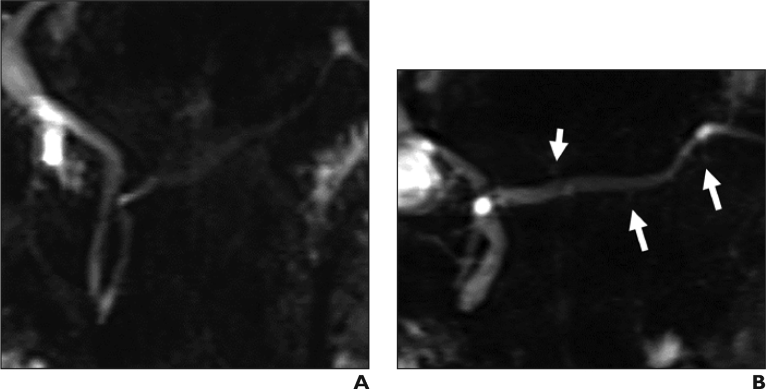
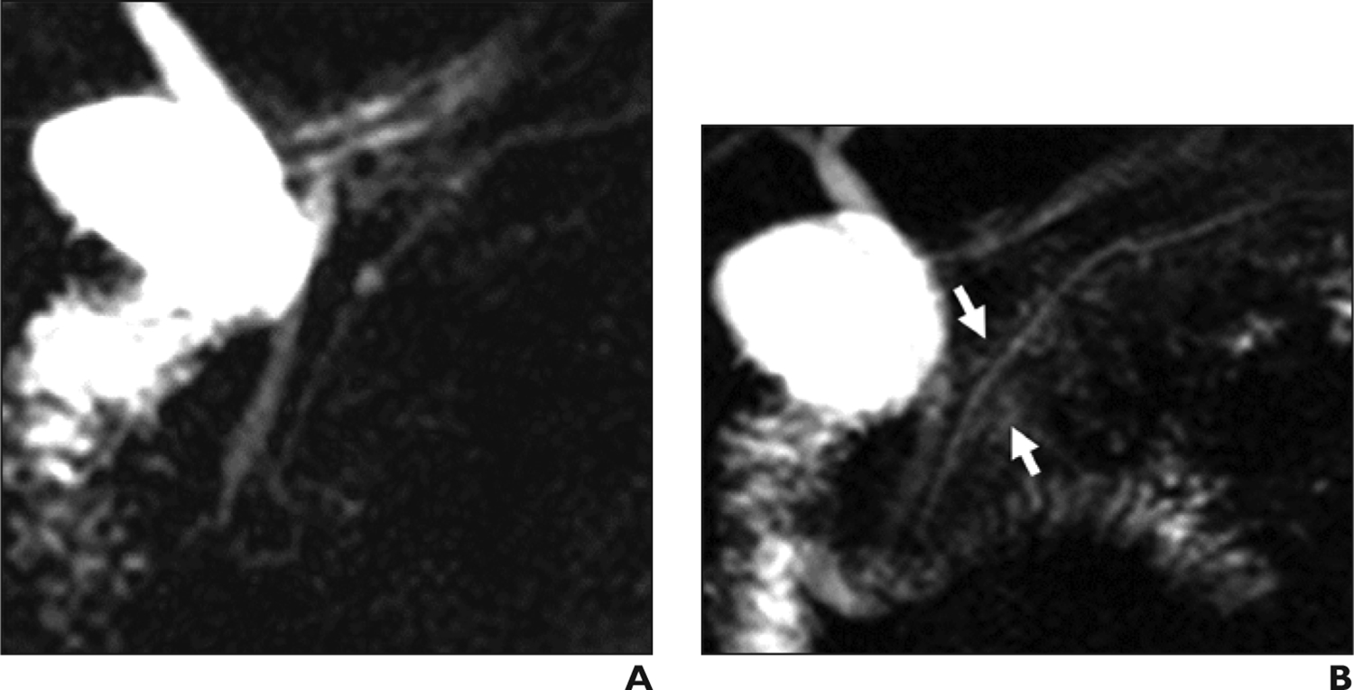
Secretin-enhanced MRCP (S-MRCP) has advantages over standard MRCP for imaging of the pancreaticobiliary tree. Through the use of secretin to induce fluid production from the pancreas and leveraging of fluid-sensitive MRCP sequences, S-MRCP facilitates visualization of ductal anatomy, and the findings provide insight into pancreatic function, allowing radiologists to provide additional insight into a range of pancreatic conditions. This narrative review provides detailed information on the practical implementation of S-MRCP, including patient preparation, logistics of secretin administration, and dynamic secretin-enhanced MRCP acquisition. Also discussed are radiologists' interpretation and reporting of S-MRCP examinations, including assessments of dynamic compliance of the main pancreatic duct and of duodenal fluid volume. Established indications for S-MRCP include pancreas divisum, anomalous pancreaticobiliary junction, Santorinicele, Wirsungocele, chronic pancreatitis, main pancreatic duct stenosis, and assessment of complex postoperative anatomy. Equivocal or controversial indications are also described along with an approach to such indications. These indications include acute and recurrent acute pancreatitis, pancreatic exocrine function, sphincter of Oddi dysfunction, and pancreatic neoplasms.
Keywords: MRCP; MRI; pancreas; secretin.
Figures
Similar articles
-
Secretin-enhanced MR cholangiopancreatography: spectrum of findings.Radiographics. 2013 Nov-Dec;33(7):1889-906. doi: 10.1148/rg.337125014. Radiographics. 2013. PMID: 24224585 Review.
-
Secretin Improves Visualization of Nondilated Pancreatic Ducts in Children Undergoing MRCP.AJR Am J Roentgenol. 2020 Apr;214(4):917-922. doi: 10.2214/AJR.19.21798. Epub 2020 Feb 5. AJR Am J Roentgenol. 2020. PMID: 32023119
-
Santorinicele: secretin-enhanced magnetic resonance cholangiopancreatography findings before and after minor papilla sphincterotomy.Eur Radiol. 2015 Aug;25(8):2437-44. doi: 10.1007/s00330-015-3644-0. Epub 2015 Feb 19. Eur Radiol. 2015. PMID: 25693665
-
The clinical value of secretin-enhanced MRCP in the functional and morphological assessment of pancreatic diseases.Br J Radiol. 2018 Apr;91(1084):20170677. doi: 10.1259/bjr.20170677. Epub 2018 Feb 5. Br J Radiol. 2018. PMID: 29206061 Free PMC article. Review.
-
Pancreatic ductal abnormalities documented by secretin-enhanced MRCP in asymptomatic subjects with chronic pancreatic hyperenzymemia.Am J Gastroenterol. 2009 Jul;104(7):1780-6. doi: 10.1038/ajg.2009.158. Epub 2009 May 12. Am J Gastroenterol. 2009. PMID: 19436288
Cited by
-
Risk Assessment and Radiomics Analysis in Magnetic Resonance Imaging of Pancreatic Intraductal Papillary Mucinous Neoplasms (IPMN).Cancer Control. 2024 Jan-Dec;31:10732748241263644. doi: 10.1177/10732748241263644. Cancer Control. 2024. PMID: 39293798 Free PMC article. Review.
-
AGA-PancreasFest Joint Symposium on Exocrine Pancreatic Insufficiency.Gastro Hep Adv. 2022 Nov 15;2(3):395-411. doi: 10.1016/j.gastha.2022.11.008. eCollection 2023. Gastro Hep Adv. 2022. PMID: 39132652 Free PMC article. Review.
-
Screening of pancreatic cancer: Target population, optimal timing and how?Ann Med Surg (Lond). 2022 Nov 5;84:104814. doi: 10.1016/j.amsu.2022.104814. eCollection 2022 Dec. Ann Med Surg (Lond). 2022. PMID: 36582884 Free PMC article. Review.
-
Intraductal papillary mucinous neoplasms of the pancreas: Uncommon imaging presentation, evolution and comparison of guidelines.Eur J Radiol Open. 2023 Oct 22;11:100531. doi: 10.1016/j.ejro.2023.100531. eCollection 2023 Dec. Eur J Radiol Open. 2023. PMID: 37920680 Free PMC article.
-
Wirsungocele as a Rare Cause of Recurrent Pancreatitis: Etiology and Therapeutic Insights.DEN Open. 2025 Jun 5;6(1):e70156. doi: 10.1002/deo2.70156. eCollection 2026 Apr. DEN Open. 2025. PMID: 40476145 Free PMC article.
References
-
- Tirkes T, Sandrasegaran K, Sanyal R, Sherman S, Schmidt CM, Cote GA, et al. Secretin-enhanced MR Cholangiopancreatography: Spectrum of Findings. RadioGraphics 2013; 33:1889–1906 - PubMed
-
- Carbognin G, Pinali L, Girardi V, Casarin A, Mansueto G, Mucelli RP. Collateral branches IPMTs: secretin-enhanced MRCP. Abdom Imaging 2007;32:374–380. - PubMed
-
- Akisik MF, Sandrasegaran K, Aisen AA, Maglinte DDT, Sherman S, Lehman GA. Dynamic secretin-enhanced MR cholangiopancreatography. RadioGraphics 2006;26(3):665–677. - PubMed
-
- Manfredi R, Mucelli RP. Secretin-enhanced MR imaging of the Pancreas. Radiology 2016;279(1):29–43 - PubMed
-
- Hoeffel C, Azizi L, Lewin M, Laurent V, Aubé C, Arrivé L et al. Normal and pathologic features of the postoperative biliary tract at 3D MR cholangiopancreatography and MR imaging. RadioGraphics 2006;26(6):1603–1620. - PubMed
Publication types
MeSH terms
Substances
Grants and funding
LinkOut - more resources
Full Text Sources
Other Literature Sources
Medical
