

White matter: A good reference for the signal intensity evaluation in magnetic resonance imaging for the diagnosis of uveal melanoma
- PMID: 33263498
- PMCID: PMC8041399
- DOI: 10.1177/1971400920973407
White matter: A good reference for the signal intensity evaluation in magnetic resonance imaging for the diagnosis of uveal melanoma
Abstract
Background: Comparing the signal intensity (SI) of an ocular mass to that of the vitreous body has been suggested. Most ocular lesions show a hyper-intense signal compared to the vitreous body on T1-weighted (T1w) images, and malignant melanomas have been almost always determined as 'cannot be excluded' in reports.
Purpose: This study aimed to determine the accuracy of magnetic resonance imaging (MRI) in the diagnosis of uveal melanoma by using normal white matter as reference tissue for SI evaluation on T1w images and vitreous body on T2w compared to the conventional method using the vitreous body as a reference on both T1w and T2w images.
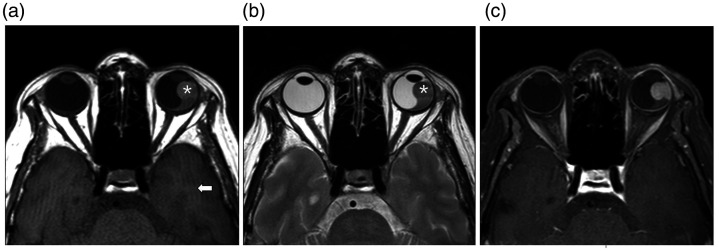
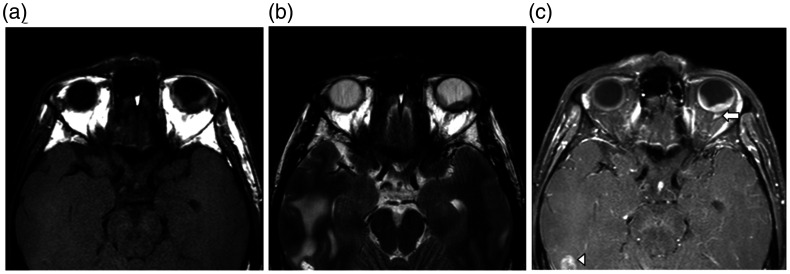
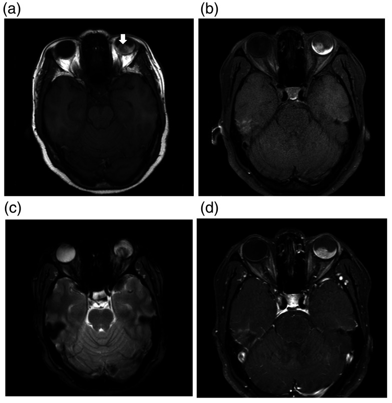
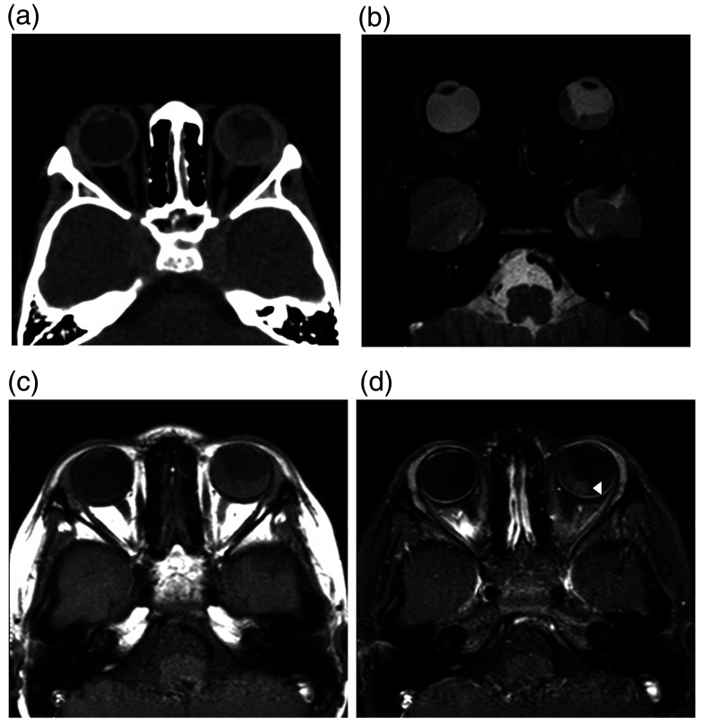
Methods: The MRIs of 43 patients (between August 2006 and July 2018) sent to rule out uveal melanoma were blindly reviewed by two radiologists. By using white matter as a reference for SI evaluation on T1w images and vitreous body as a reference on T2w images, uveal melanomas were suggested by hyper-intense signal on T1w and hypo-intense signal on T2w with homogeneous enhancement. The accuracy of diagnosis of uveal melanoma using white matter as a reference on T1w was compared to the conventional method using the vitreous body as a reference on both T1w and T2w images.
Results: The diagnosis of uveal melanoma using white matter as a reference gave a sensitivity of 92.31% (95% confidence interval (CI) 63.97-99.81) and specificity of 100.0% (95% CI 88.43-100.0). By using the vitreous body as a reference, sensitivity as high as 100.0% (95% CI 100.0-100.0) was obtained, but specificity was low at 53.33% (95% CI 34.33-71.66).
Conclusions: White matter is a good reference for the diagnosis of uveal melanoma, with high sensitivity and much higher specificity than conventional methods using the vitreous body as a reference.
Keywords: Uveal melanoma; magnetic resonance imaging (MRI); reference; white matter.
Figures
References
-
- Shields CL, Manalac J, Das C, et al. Choroidal melanoma: clinical features, classification, and top 10 pseudomelanomas. Curr Opin Ophthalmol 2014; 25: 177–185. - PubMed
-
- Smoker WR, Gentry LR, Yee NK, et al. Vascular lesions of the orbit: more than meets the eye. Radiographics 2008; 28: 185–204; quiz 325. - PubMed
-
- Enochs WS, Petherick P, Bogdanova A, et al. Paramagnetic metal scavenging by melanin: MR imaging. Radiology 1997; 204: 417–423. - PubMed
-
- Tailor TD, Gupta D, Dalley RW, et al. Orbital neoplasms in adults: clinical, radiologic, and pathologic review. Radiographics 2013; 33: 1739–1758. - PubMed
MeSH terms
Substances
LinkOut - more resources
Full Text Sources
Medical
