Review
. 2020 Dec 2;10(1):36.
doi: 10.1186/s12348-020-00228-w.

Current diagnostic tools and management modalities of Nocardia keratitis

Affiliations
Review

Current diagnostic tools and management modalities of Nocardia keratitis

Mohammad Soleimani et al. J Ophthalmic Inflamm Infect. .

Abstract

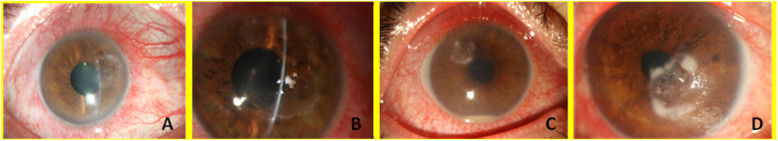
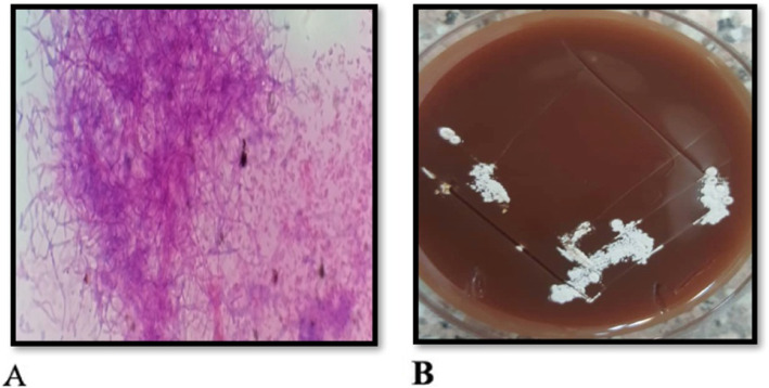
Nocardia species are an uncommon but important cause of keratitis. The purpose of this review is to discus previous published papers relation to the epidemiology, etiology, diagnosis and management of Nocardia keratitis. Nocardia asteroides is the most frequently reported from Nocardia keratitis. Pain, photophobia, blepharospasm and lid swelling are mainly clinical manifestations. Usual risk factors for Nocardia keratitis are trauma, surgery, corticosteroids, and contact lens wear. Several antibiotics were used for treatment of Nocardia infection but according to studies, topical amikacin is the drug of choice for Nocardia keratitis. Topical steroid should not prescribe in these patients. In conclusion, although Nocardia keratitis is rare, early diagnosis and treatment are essential to prevent any scar formation and preserve a good visual acuity.

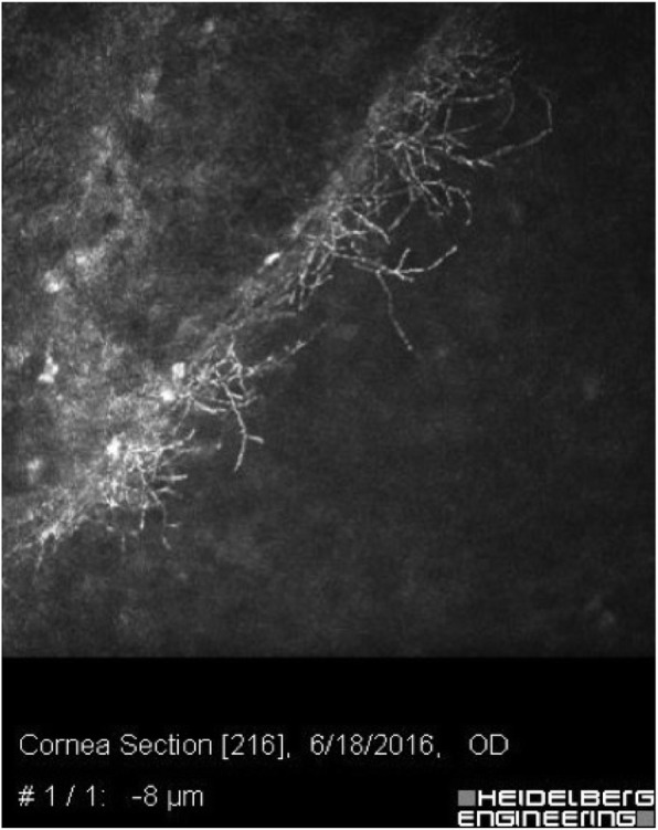
Keywords: Actinomycetes; Amikacin; Confocal microscopy; Corneal ulcer; Keratitis; Keratoplasty; Nocardia; Ocular trauma.


Conflict of interest statement

The authors declare that they have no conflicts of interest.

Figures

Fig. 1
Fig. 1
Nocardia keratitis (a, b, c and d). Typical superficial patchy, white and pin-head infiltrates in a wreath-like pattern. Active lesion margins are seen
Fig. 2
Fig. 2
A typical confocal scanning of an eye with Nocardia keratitis. Fine branching and interlocking elements are seen
Fig. 3
Fig. 3
a The modified Ziehl-Neelsen or Kinyoun stain method. Nocardia isolated from a corneal ulcer showing acid-fast filaments (magnification × 400). b Blood agar inoculated with corneal scrapings showing white, dry and chalky colonies

References

    1. Johansson B, Fagerholm P, Petranyi G, et al. Diagnostic and therapeutic challenges in a case of amikacin-resistant Nocardia keratitis. Acta Ophthalmol. 2017;95(1):103–105. doi: 10.1111/aos.13182. - DOI - PubMed
    1. Behaegel J, Dhubhghaill SN, Koppen C. Diagnostic challenges in Nocardia keratitis. Eye Contact Lens. 2018;44(1):370–372. doi: 10.1097/ICL.0000000000000462. - DOI - PubMed
    1. Lalitha P, Srinivasan M, Rajaraman R, et al. Nocardia keratitis: clinical course and effect of corticosteroids. Am J Ophthalmol. 2012;154(6):934–939. doi: 10.1016/j.ajo.2012.06.001. - DOI - PMC - PubMed
    1. Sridhar MS, Sharma S, Reddy MK, et al. Clinicomicrobiological review of Nocardia keratitis. Cornea. 1998;17(1):17–22. doi: 10.1097/00003226-199801000-00003. - DOI - PubMed
    1. Sridhar MS, Gopinathan U, Garg P, Sharma S, Rao GN. Ocular nocardia infections with special emphasis on the cornea. Surv Ophthalmol. 2001;45(5):361–378. doi: 10.1016/S0039-6257(00)00207-1. - DOI - PubMed

LinkOut - more resources