. 2020 Dec 2;15(12):e0239889.
doi: 10.1371/journal.pone.0239889. eCollection 2020.

Laminin N-terminus α31 protein distribution in adult human tissues

Affiliations

Laminin N-terminus α31 protein distribution in adult human tissues

Lee D Troughton et al. PLoS One. .

Abstract

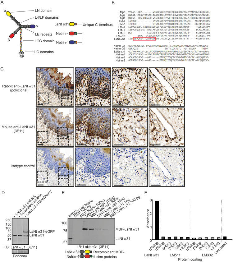
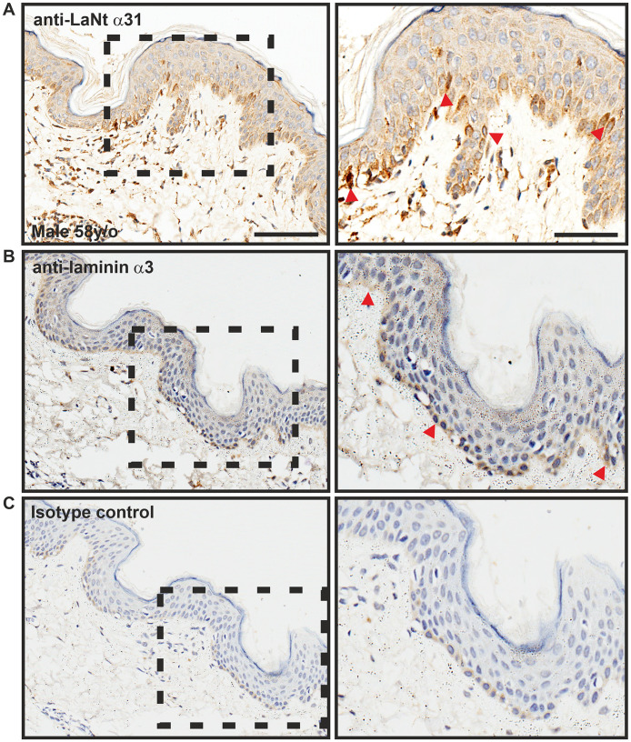
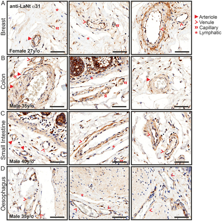
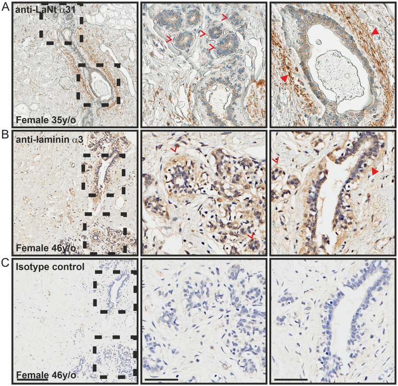
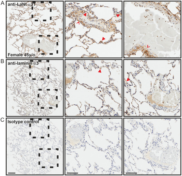
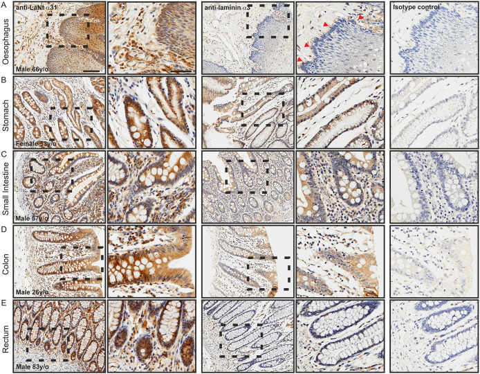
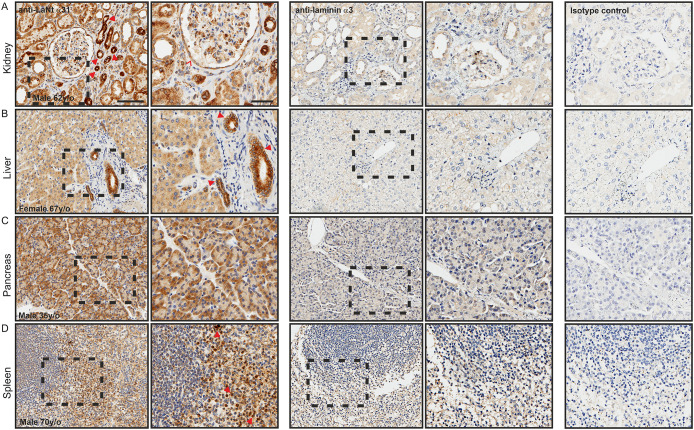
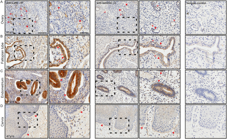
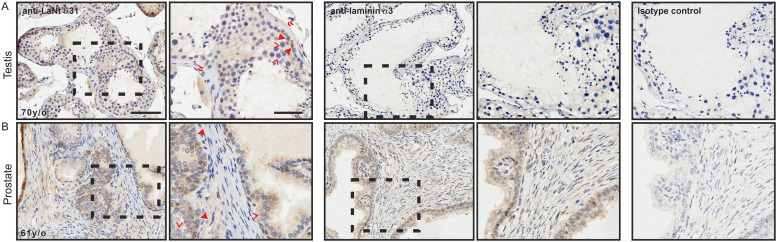
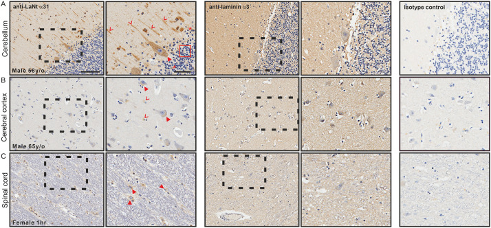
Laminin N-terminus α31 (LaNt α31) is a netrin-like protein derived from alternative splicing of the laminin α3 gene. Although LaNt α31 has been demonstrated to influence corneal and skin epithelial cell function, its expression has not been investigated beyond these tissues. In this study, we used immunohistochemistry to characterise the distribution of this protein in a wide-array of human tissue sections in comparison to laminin α3. The data revealed widespread LaNt α31 expression. In epithelial tissue, LaNt α31 was present in the basal layer of the epidermis, throughout the epithelium of the digestive tract, and in much of the epithelium of the reproductive system. LaNt α31 was also found throughout the vasculature of most tissues, with enrichment in reticular-like fibres in the extracellular matrix surrounding large vessels. A similar matrix pattern was observed around the terminal ducts in the breast and around the alveolar epithelium in the lung, where basement membrane staining was also evident. Specific enrichment of LaNt α31 was identified in sub-populations of cells of the kidney, liver, pancreas, and spleen, with variations in intensity between different cell types in the collecting ducts and glomeruli of the kidney. Intriguingly, LaNt α31 immunoreactivity was also evident in neurons of the central nervous system, in the cerebellum, cerebral cortex, and spinal cord. Together these findings suggest that LaNt α31 may be functionally relevant in a wider range of tissue contexts than previously anticipated, and the data provides a valuable basis for investigation into this interesting protein.


Conflict of interest statement

The authors have declared that no competing interests exist.

Figures

Fig 1
Fig 1. 3E11 mouse monoclonal antibodies against the unique C-terminus of LaNt α31 validation.
(A) Diagram of the laminin and laminin-related protein architecture. (B) Multiple sequence alignment of LaNt α31 C-terminus with equivalent regions of laminin N-terminal sequences. Peptide unique to LaNt α31 used to generate antibodies highlighted by red box. (C) Paraffin-embedded human tongue tissue sections processed for immunohistochemistry with rabbit polyclonal antibodies against LaNt α31 or mouse monoclonal antibodies against LaNt α31 (3E11). Scale bars represent 200 μm or 100 μm in magnified images. (D) Immunoblot of total protein from hTCEpi expressing either: shRNAs specific to LaNt α31, scrambled shRNA controls, or full-length LaNt α31-PAmCherry cDNA. Ponceau S stained membrane below. (E) Immunoblot of dilution series of recombinant MBP-LaNt α31, and MBP-netrin-4 probed with 3E11 antibodies. Diagram below shows the structure of the recombinant MBP-LaNt α31 and Netrin-4. (F) ELISA using recombinant MBP-LaNt α31, LM511, or LM332. Wells were coated overnight, blocked with BSA, and then probed with 3E11 antibodies. Bound 3E11 antibodies were detected with HRP-conjugated secondary antibodies, HRP-substrate added and absorbance read at 450 nm.
Fig 2
Fig 2. LaNt α31 is enriched in a sub-population of basal epidermal keratinocytes.
Paraffin-embedded human tissue microarray sections processed for immunohistochemistry with mouse monoclonal antibodies against LaNt α31 (3E11, A), laminin α3 (B), or mouse IgG (C). Scale bars represent 100 μm or 50 μm in magnified images. Arrowheads in (A) indicate localised enrichment of LaNt α31. Arrowheads in (B) indicate basement membrane reactivity for laminin α3.
Fig 3
Fig 3. LaNt α31 is expressed throughout the blood vasculature.
Paraffin-embedded human tissue microarray sections processed for immunohistochemistry with mouse monoclonal antibodies against LaNt α31 (3E11). Representative images taken from: (A) breast, (B) colon, (C) small intestine, (D) oesophagus. Scale bars represent 60 μm.
Fig 4
Fig 4. LaNt α31 is enriched in the ECM surround terminal ducts in breast tissue.
Paraffin-embedded human tissue microarray sections processed for immunohistochemistry with mouse monoclonal antibodies against LaNt α31 (3E11, A), laminin α3 (B), or mouse IgG (C). Arrowheads denote matrix immunoreactivity surrounding ducts, chevrons indicate lobular acini. Scale bars represent 200 μm or 100 μm in magnified images.
Fig 5
Fig 5. LaNt α31 localises to the alveolar basement membranes in lung tissue.
Paraffin-embedded human tissue microarray sections processed for immunohistochemistry with mouse monoclonal antibodies against LaNt α31 (3E11, A), laminin α3 (B), or mouse IgG (C). Arrowheads depicts basement membrane, chevrons indicate reticular fibre-like immunoreactivity. Scale bars represent 200 μm or 100 μm in magnified images.
Fig 6
Fig 6. LaNt α31 is expressed throughout the epithelium of the digestive tract.
Paraffin-embedded human tissue microarray sections processed for immunohistochemistry with mouse monoclonal antibodies against LaNt α31 (3E11), laminin α3, or mouse IgG. (A) oesophagus, (B) stomach, (C) small intestine, colon (D), rectum (E). Arrowheads in (A) indicate basement membrane immunoreactivity. Scale bars represent 100 μm or 50 μm in magnified images.
Fig 7
Fig 7. LaNt α31 expression in kidney, pancreas, liver, and spleen, is largely similar to that of LMα3.
Paraffin-embedded human tissue microarray sections processed for immunohistochemistry with mouse monoclonal antibodies against LaNt α31 (3E11), laminin α3, or mouse IgG. (A) kidney, (B) liver, (C) pancreas, (D) spleen. Arrowheads in (A) depict ducts displaying high immunoreactivity, chevrons indicate parietal epithelium of the Bowman’s capsule. Arrowheads in (B) indicate branches of the bile ducts. Arrowheads in (D) indicate splenic cords of Billroth. Scale bars represent 100 μm or 50 μm in magnified images.
Fig 8
Fig 8. LaNt α31 is expressed throughout the female reproductive system.
Paraffin-embedded human tissue microarray sections processed for immunohistochemistry with mouse monoclonal antibodies against LaNt α31 (3E11), laminin α3, or mouse IgG. (A) ovary, (B) fallopian tube, (C) endometrium, (D) cervix. Arrowheads in (A) indicate follicular cells of the primordial follicles, chevrons indicate primary follicles, and double chevrons indicate vasculature. Arrowheads in (B) indicate peg cells, chevrons indicate ciliated cells. Arrowheads in (C) indicates uterine glands of the endometrium. Arrowheads in (D) indicate reticular fibre-like immunoreactivity. Scale bars represent 100 μm or 50 μm in magnified images.
Fig 9
Fig 9. LaNt α31 is expressed in the testis and prostrate.
Paraffin-embedded human tissue microarray sections processed for immunohistochemistry with mouse monoclonal antibodies against LaNt α31 (3E11), laminin α3, or mouse IgG. (A) testis, (B) prostrate. Arrowheads in (A) indicate a sub-population of the spermatogonia, chevrons indicate Sertoli cells. Arrowheads in (B) indicate basal layer of the acini, chevron indicate acinar cells. Scale bars represent 100 μm or 50 μm in magnified images.
Fig 10
Fig 10. LaNt α31 is expressed by neurons in the central nervous system.
Paraffin-embedded human tissue microarray sections processed for immunohistochemistry with mouse monoclonal antibodies against LaNt α31 (3E11), laminin α3, or mouse IgG. (A) cerebellum, (B) cerebral cortex, (C) spinal cord. Arrowheads in (A) indicate Purkinje neurons in the ganglionic layer, chevrons indicate axons throughout granular layer. Arrowheads in (B) indicate cell bodies towards the axon hillock of pyramidal cells, chevrons indicate glial cells. Arrowheads in (C) indicate motor neurons. Scale bars represent 100 μm or 50 μm in magnified images.

Similar articles

Cited by

References

    1. Yurchenco PD, O’Rear JJ. Basement membrane assembly. Methods Enzymol. 1994;245:489–518. 10.1016/0076-6879(94)45025-0 - DOI - PubMed
    1. Hohenester E, Yurchenco PD. Laminins in basement membrane assembly. Cell Adh Migr. 2013;7(1):56–63. 10.4161/cam.21831 - DOI - PMC - PubMed
    1. Aumailley M. The laminin family. Cell Adh Migr. 2013;7(1):48–55. 10.4161/cam.22826 - DOI - PMC - PubMed
    1. Hamill KJ, Kligys K, Hopkinson SB, Jones JC. Laminin deposition in the extracellular matrix: a complex picture emerges. J Cell Sci. 2009;122(Pt 24):4409–17. 10.1242/jcs.041095 - DOI - PMC - PubMed
    1. Aumailley M, Bruckner-Tuderman L, Carter WG, Deutzmann R, Edgar D, Ekblom P, et al. A simplified laminin nomenclature. Matrix Biol. 2005;24(5):326–32. 10.1016/j.matbio.2005.05.006 - DOI - PubMed

Publication types