. 2020 Dec 2;15(12):e0243207.
doi: 10.1371/journal.pone.0243207. eCollection 2020.

Reperfused hemorrhagic myocardial infarction in rats

Affiliations

Reperfused hemorrhagic myocardial infarction in rats

Anand R Nair et al. PLoS One. .

Abstract

Background: Intramyocardial hemorrhage following reperfusion is strongly associated with major adverse cardiovascular events in myocardial infarction (MI) patients; yet the mechanisms contributing to these outcomes are not well understood. Large animal models have been used to investigate intramyocardial hemorrhage, but they are exorbitantly expensive and difficult to use for mechanistic studies. In contrast, rat models are widely used to investigate mechanistic aspects of cardiovascular physiology, but a rat model that consistently recapitulates the characteristics of an hemorrhagic MI does not exist. To bridge this gap, we investigated the physiological conditions of MI that would create intramyocardial hemorrhage in rats so that a reliable model of hemorrhagic MI would become available for basic research.

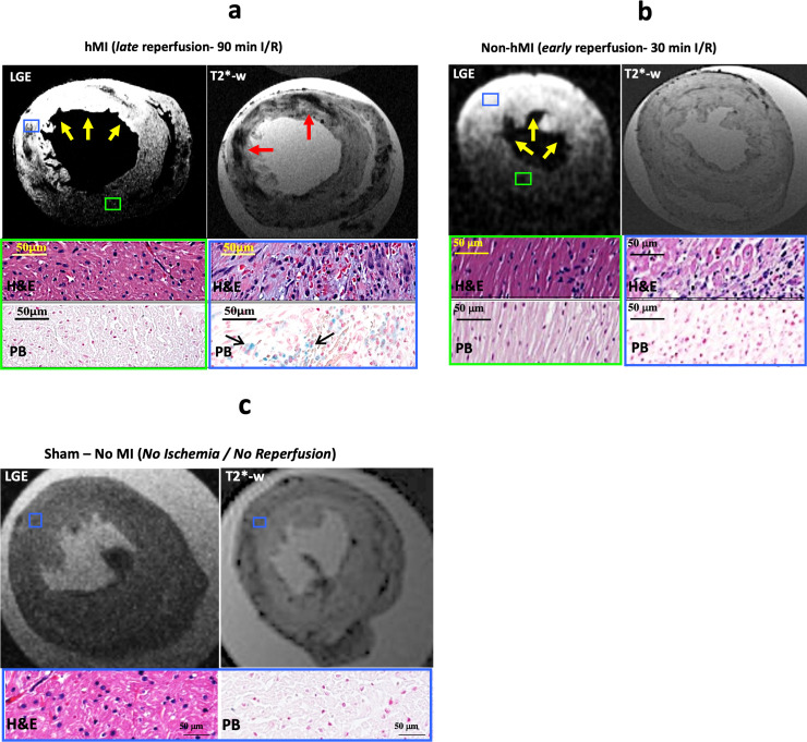
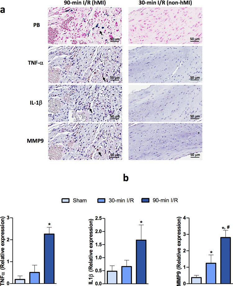
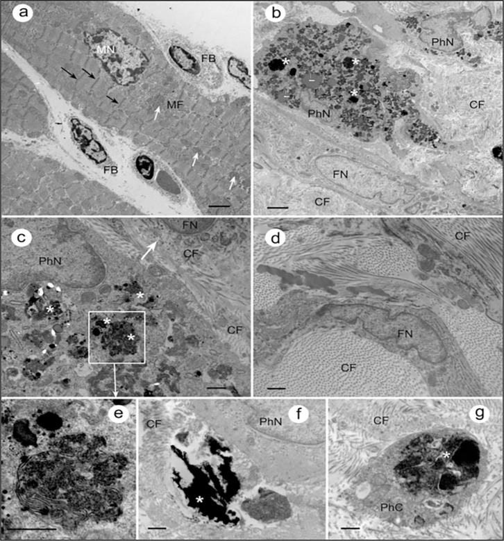
Methods & results: Sprague-Dawley rats underwent either a 90-minute (90-min) ischemia and then reperfusion (I/R) (n = 22) or 30-minute (30-min) I/R (n = 18) of the left anterior descending coronary artery. Sham rats (n = 12) were used as controls. 90-min I/R consistently yielded hemorrhagic MI, while 30-min I/R consistently yielded non-hemorrhagic MI. Twenty-four hours post-reperfusion, ex-vivo late-gadolinium-enhancement (LGE) and T2* cardiac MRI performed on excised hearts from 90-min I/R rats revealed colocalization of iron deposits within the scarred tissue; however, in 30-min I/R rats scar was evident on LGE but no evidence of iron was found on T2* CMR. Histological studies verified tissue damage (H&E) detected on LGE and the presence of iron (Perl's stain) observed on T2*-CMR. At week 4 post-reperfusion, gene and protein expression of proinflammatory markers (TNF-α, IL-1β and MMP-9) were increased in the 90-min I/R group when compared to 30-min I/R groups. Further, transmission electron microscopy performed on 90-min I/R myocardium that were positive for iron on T2* CMR and Perl's stain showed accumulation of granular iron particles within the phagosomes.

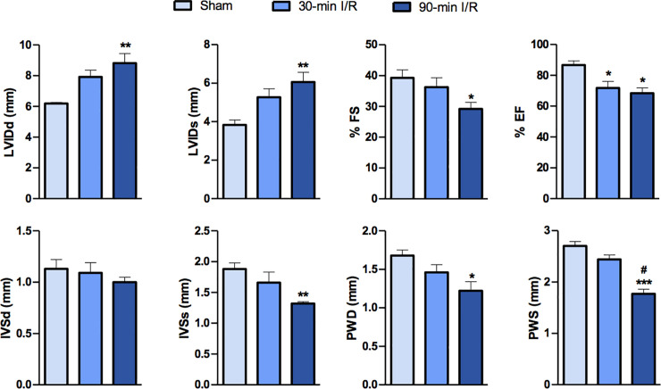
Conclusion: Ischemic time prior to reperfusion is a critical factor in determining whether a MI is hemorrhagic or non-hemorrhagic in rats. Specifically, a period of 90-min of ischemia prior to reperfusion can produce rat models of hemorrhagic MI, while 30-minutes of ischemia prior to reperfusion can ensure that the MIs are non-hemorrhagic. Hemorrhagic MIs in rats result in marked increase in iron deposition, proinflammatory burden and adverse left-ventricular remodeling compared to rats with non-hemorrhagic MIs.


Conflict of interest statement

The authors have declared that no competing interests exist.

Figures

Fig 1
Fig 1. Late reperfusion leads to development of hemorrhagic MI in rats.
Fig 1a-1c: Representative LGE and T2*-weighted images from ex-vivo cardiac MRI showing myocardial infarction (yellow arrows) and hemorrhage (red arrows) respectively from late-reperfusion (90-min I/R, panel a), early-reperfusion (30-min I/R, panel b) and sham (panel c) groups. Histological images with H&E staining from regions negative for MI (green box) and H&E and Perl’s stained images from regions positive for MI (blue box) are shown in late- and early-reperfusion groups. In the sham group, blue box represents region closer to the anterior descending wall.
Fig 2
Fig 2. Echocardiographic parameters show adverse LV remodeling in rats with hemorrhagic MI.
IVSd and IVSs (mm)—Intraventricular septal diameter at end diastole and at end systole, respectively; LVIDd and LVIDs (mm), left ventricular internal diameter at diastole and systole, respectively; PWD and PWS (mm), posterior wall thickness at end-diastole and end-systole, respectively; %FS, fractional shortening, %EF, ejection fraction (n = 7 per group). Data are presented as mean ± SEM. *P<0.05, **P<0.01 and ***P<0.001. # represents difference compared with 30-min I/R group (P<0.01).
Fig 3
Fig 3. Proinflammatory burden in chronic hemorrhagic MI territories in rats.
Panel a show hemorrhagic MI region with proinflammatory cytokines. Representative immunohistochemistry images of proinflammatory activation in hemorrhagic myocardial infarction are shown with immunoreactivity of inflammatory cytokines TNF-α and IL-1β, and fibrotic marker MMP-9 in myocardial tissue from 30-min I/R and 90-min I/R groups. Panel b shows gene expression of proinflammatory cytokines in rats with hemorrhagic MI. mRNA expression levels of the indicated genes in heart tissue from rats that underwent sham surgery, 30-min I/R and 90-min I/R (n = 7/group). Data are presented as mean ± SEM. *P<0.05 compared with sham, #P<0.05 compared with 30-min I/R group.
Fig 4
Fig 4. Granular traces of iron are evident in the ultrastructure of the hemorrhagic MI in rats.
a) Representative TEM image of a section through the myocardium of sham control rat. Black arrows indicate z-discs; white arrows show mitochondria. FB–fibroblast; MN–myocyte nucleus. Scale bar: 2um b) Section through the myocardium of rat with delayed reperfusion that was confirmed for iron within MI in T2* CMR and Perl’s staining. Organization of myocardial tissue is chaotic, no traces of regularly arranged myofibrils can be observed. Instead, the tissue is infiltrated with large fibroblast actively producing collagen fibers (CF). Accumulations of granular material within the phagosomes (white asterisks), are consistent with expected appearance of aggregated iron particles. FN, fibroblast nucleus, PhN, phagocyte nucleus. Scale bar: 2μm c) Section through another region of the myocardium that underwent delayed reperfusion post MI: a phagocyte with numerous phagosomes/secondary lysosomes loaded with aggregates of iron particles (asterisks), and a fibroblast (arrow) with intracellular and extracellular collagen fibers are in the view. Scale bar: 0.5μm d) Fibroblasts and collagen fibers filling the myocardial tissue after delayed-reperfusion. Scale bar: 0.5μm e) Fragment of panel (c) at higher magnification: phagosomes with iron particles inside (asterisk). Scale bar: 0.5μm f) Accumulation of iron (asterisk) outside the phagocytic cell. PhN, phagocyte nucleus. Scale bar: 0.5μm g) A phagosome with iron aggregates (asterisk) in the phagocyte cytoplasm (PhC) surrounded by CF. Scale bar: 0.5μm.

Similar articles

Cited by

References

    1. Kalogeris T, Baines Cp, Krenz M, Korthuis Rj. Cell Biology Of Ischemia/Reperfusion Injury. Int Rev Cell Mol Biol. 2012;298:229–317. 10.1016/B978-0-12-394309-5.00006-7 - DOI - PMC - PubMed
    1. Jm Torpy, Lynm C Glass Rm. Percutaneous Coronary Intervention. Jama. 2004;291(6):778–. 10.1001/jama.291.6.778 - DOI - PubMed
    1. Ir Degano, Subirana I, Fusco D, Tavazzi L, Kirchberger I, Farmakis D, et al. Percutaneous Coronary Intervention Reduces Mortality In Myocardial Infarction Patients With Comorbidities: Implications For Elderly Patients With Diabetes Or Kidney Disease. Int J Cardiol. 2017;249:83–9. 10.1016/j.ijcard.2017.07.054 - DOI - PubMed
    1. Rs Bricker, Ja Valle, Me Plomondon, Ej Armstrong, Sw Waldo. Causes Of Mortality After Percutaneous Coronary Intervention. Circ Cardiovasc Qual Outcomes. 2019;12(5):E005355 10.1161/CIRCOUTCOMES.118.005355 - DOI - PubMed
    1. Sezer M, Van Royen N, Umman B, Bugra Z, Bulluck H, Hausenloy Dj, et al. Coronary Microvascular Injury In Reperfused Acute Myocardial Infarction: A View From An Integrative Perspective. J Am Heart Assoc. 2018;7(21):E009949 10.1161/JAHA.118.009949 - DOI - PMC - PubMed

Publication types

MeSH terms