. 2020 Dec 2;16(12):e1009118.
doi: 10.1371/journal.ppat.1009118. eCollection 2020 Dec.

Auxin response factors (ARFs) differentially regulate rice antiviral immune response against rice dwarf virus

Affiliations

Auxin response factors (ARFs) differentially regulate rice antiviral immune response against rice dwarf virus

Qingqing Qin et al. PLoS Pathog. .

Abstract

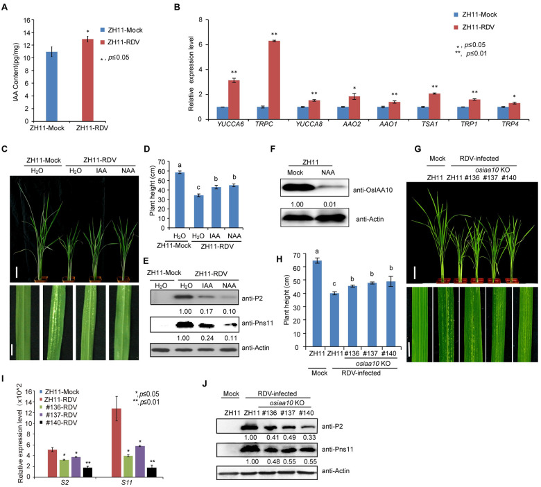
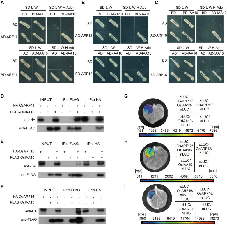
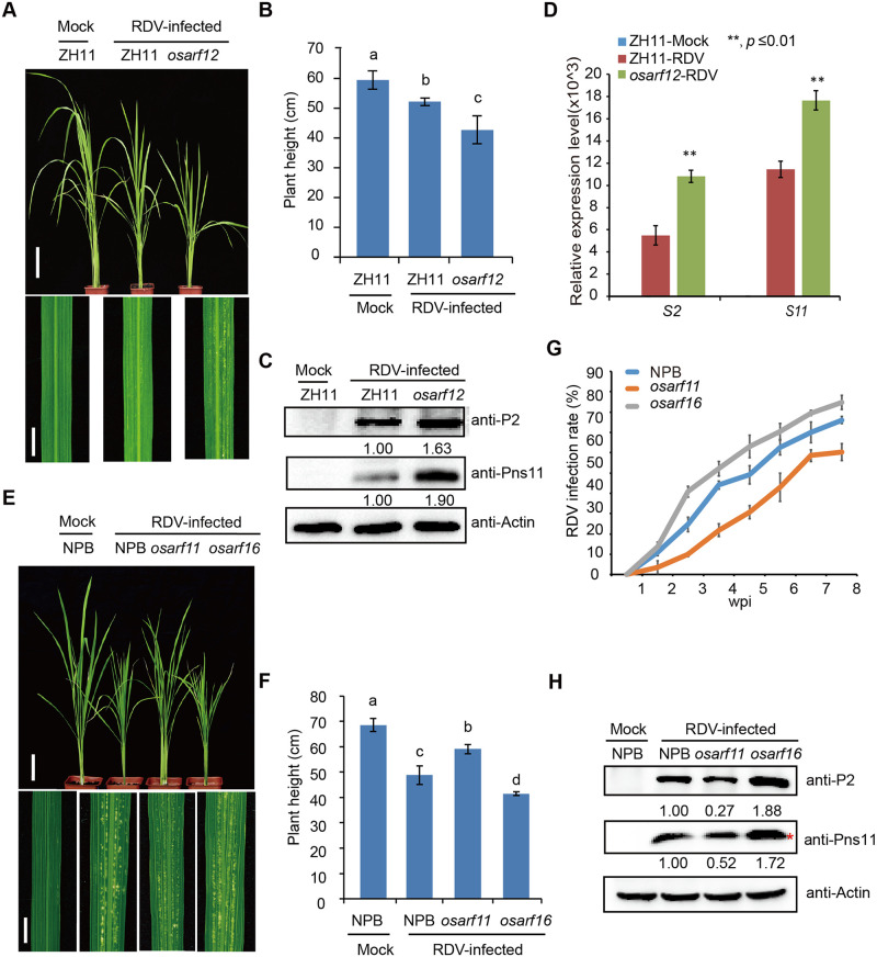
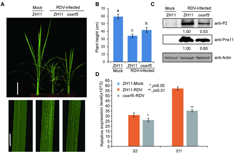
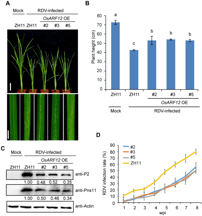
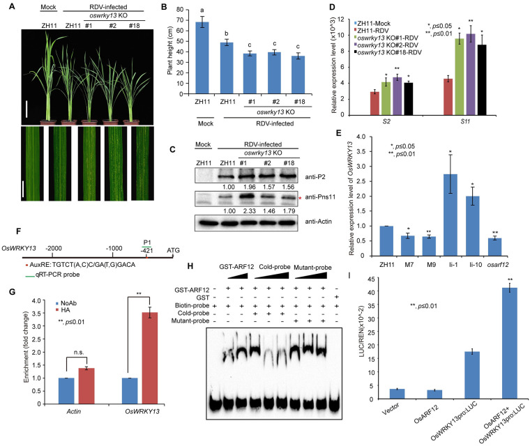
There are 25 auxin response factors (ARFs) in the rice genome, which play critical roles in regulating myriad aspects of plant development, but their role (s) in host antiviral immune defense and the underneath mechanism remain largely unknown. By using the rice-rice dwarf virus (RDV) model system, here we report that auxin signaling enhances rice defense against RDV infection. In turn, RDV infection triggers increased auxin biosynthesis and accumulation in rice, and that treatment with exogenous auxin reduces OsIAA10 protein level, thereby unleashing a group of OsIAA10-interacting OsARFs to mediate downstream antiviral responses. Strikingly, our genetic data showed that loss-of-function mutants of osarf12 or osarf16 exhibit reduced resistance whereas osarf11 mutants display enhanced resistance to RDV. In turn, OsARF12 activates the down-stream OsWRKY13 expression through direct binding to its promoter, loss-of-function mutants of oswrky13 exhibit reduced resistance. These results demonstrated that OsARF 11, 12 and 16 differentially regulate rice antiviral defense. Together with our previous discovery that the viral P2 protein stabilizes OsIAA10 protein via thwarting its interaction with OsTIR1 to enhance viral infection and pathogenesis, our results reveal a novel auxin-IAA10-ARFs-mediated signaling mechanism employed by rice and RDV for defense and counter defense responses.


Conflict of interest statement

The authors have declared that no competing interests exist.

Figures

Fig 1
Fig 1. Exogenous auxin treatment enhances rice tolerance to RDV infection via down-regulating OsIAA10.
(A) IAA content is higher in RDV-infected plants. ZH11-Mock, uninfected ZH11 plants, ZH11-RDV, RDV-infected ZH11. (B) Increased expression of some auxin biosynthesis genes in RDV-infected rice. ZH11-Mock, uninfected ZH11 plants, ZH11-RDV, RDV-infected ZH11. (C) Phenotypes of RDV-infected ZH11 rice plants pretreated with H2O, IAA or NAA, respectively. Photos were taken at four-week-post-inoculation (wpi). Scale bars, 10 cm (upper panel) and 1 cm (lower panel). (D) Schematic representation of plant height for the plants in (C). The average (±SD) values were obtained from three biological repeats. Different letters indicate significant difference (p< 0.05) based on the Tukey-Kramer HSD test. (E) Western blots showing the accumulation of RDV proteins in the corresponding rice lines shown in (C). Actin was used as a loading control. (F) Western blots showing the accumulation of OsIAA10 protein after auxin treatment and in the ZH11 (Mock). Actin was used as a loading control. (G) RDV-infected WT (ZH11) and osiaa10 KO rice plants. Photos were taken at 4 weeks after RDV-inoculation. The sizes of white specks on the leaves represent the degree of disease symptoms. Scale bars, 10 cm (upper panel) and 1 cm (lower panel). (H) Schematic representation of plant height for the plants in (G). The average (±SD) values were obtained from three biological repeats. Different letters indicate significant difference (p< 0.05) based on the Tukey-Kramer HSD test. (I) qRT-PCR showing the accumulation of RDV genomic RNAs in the corresponding rice lines shown in (G). (J) Western blots showing the accumulation of RDV proteins in the corresponding rice lines in (G). Actin was used as a loading control.
Fig 2
Fig 2. OsIAA10 interacts with OsARF11, 12, 16 in plant.
(A) Yeast two-hybrid assay confirming the OsARF11 and OsIAA10 interaction. (B) Yeast two-hybrid assay confirming the OsARF12 and OsIAA10 interaction. (C) Yeast two-hybrid assay confirming the OsARF16 and OsIAA10 interaction. (D) Co-immunoprecipitation confirmed the interaction between OsIAA10 and OsARF11. (E) Co-immunoprecipitation confirmed the interaction between OsIAA10 and OsARF12. (F) Co-immunoprecipitation confirmed the interaction between OsIAA10 and OsARF16. (G) LCI assay showed the interaction between OsIAA10 and OsARF11 in plants. (H) LCI assay showed the interaction between OsIAA10 and OsARF12 in plants. (I) LCI assay showed the interaction between OsIAA10 and OsARF16 in plants.
Fig 3
Fig 3. Loss-of-function mutants of osarf12 or osarf16 exhibit reduced resistance whereas osarf11 mutants display enhanced resistance to RDV.
(A) Phenotypes of RDV-infected WT (ZH11) and osarf12 mutants. Photos were taken at 4 weeks after RDV-inoculation. The sizes of white specks on the leaves represent the degree of disease symptoms. Scale bars, 10 cm (upper panel) and 1 cm (lower panel). (B) Schematic representation of plant height for the plants in (A). The average (±SD) values were obtained from three biological repeats. Different letters indicate significant difference (p< 0.05) based on the Tukey-Kramer HSD test. (C) Accumulation of RDV proteins in the corresponding lines. Actin was used as a loading control for proteins. (D) Accumulation of RDV RNAs in the corresponding lines. The average (±SD) values were obtained from three biological repeats. The error bars indicate SD. (E) Phenotypes of RDV-infected WT (NPB), osarf11 and osarf16 mutants. Photos were taken at 4 weeks after RDV-inoculation. The sizes of white specks on the leaves represent the degree of disease symptoms. Scale bars, 10 cm (upper panel) and 1 cm (lower panel). (F) Schematic representation of plant height for the plants in (E). The average (±SD) values were obtained from three biological repeats. Different letters indicate significant difference (p< 0.05) based on the Tukey-Kramer HSD test. (G) RDV infection rates of osarf11, osarf16 and WT (NPB) from one wpi to eight wpi. Inoculation assays were repeated three times. The error bars indicate SD. (H) Accumulation of RDV proteins in the corresponding lines. Actin was used as a loading control for proteins. “*” indicated the RDV Pns11 protein.
Fig 4
Fig 4. Loss-of-function osarf5 mutants are more resistant to RDV.
(A) Phenotypes of RDV-infected WT (ZH11) and osarf5 mutant. Photos were taken at 4 weeks after RDV-inoculation. The sizes of white specks on the leaves represent the degree of disease symptoms. Scale bars, 10 cm (upper panel) and 1 cm (lower panel). (B) Schematic representation of plant height for the plants in (A). The average (±SD) values were obtained from three biological repeats. Different letters indicate significant difference (p< 0.05) based on the Tukey-Kramer HSD test. (C) Accumulation of RDV proteins in the corresponding lines. Actin was used as a loading control for proteins. (D) Accumulation of RDV RNAs in the corresponding lines. The average (±SD) values were obtained from three biological repeats. The error bars indicate SD.
Fig 5
Fig 5. OsARF12 OE plants exhibit enhanced resistance to RDV infection.
(A) Phenotypes of RDV-infected WT (ZH11) and OsARF12 OE lines. Photos were taken at 4 weeks after RDV-inoculation. The sizes of white specks on the leaves represent the degree of disease symptoms. Scale bars, 10 cm (upper panel) and 1 cm (lower panel). (B) Schematic representation of plant height for the plants in (A). The average (±SD) values were obtained from three biological repeats. Different letters indicate significant difference (p< 0.05) based on the Tukey-Kramer HSD test. (C) Western blots showing the accumulation of RDV proteins in the corresponding lines. Actin was used as a loading control for proteins. (D) RDV infection rates of the corresponding lines from one wpi to eight wpi. Inoculation assays were repeated three times. The error bars indicate SD.
Fig 6
Fig 6. OsWRKY13 is a possible target of OsARF12, OsARF12 binds the promoter of OsWRKY13, oswrky13 KO are more susceptible to RDV infection.
(A) Symptoms of RDV-infected WT (ZH11) and oswrky13 KO lines. Photos were taken at 4 weeks after RDV-inoculation. The sizes of white specks on the leaves represent the degree of disease symptoms. Scale bars, 10 cm (upper panel) and 1 cm (lower panel). (B) Schematic representation of plant height for the plants in (A). The average (±SD) values were obtained from three biological repeats. Different letters indicate significant difference (p< 0.05) based on the Tukey-Kramer HSD test. (C) Accumulation of RDV proteins in the corresponding lines. Actin was used as a loading control for proteins. “*” indicated the product of RDV Pns11 protein. (D) Accumulation of RDV RNAs in the corresponding lines. The average (±SD) values were obtained from three biological repeats. The error bars indicate SD. (E) qRT-PCR analysis of OsWRKY13 expression in ZH11, M7, M9, Ii-1, Ii-10, and osarf12 mutant rice seedlings. M7 and M9 are transgenic rice lines overexpressing OsIAA10P116L. Ii-1 and Ii-10 are OsIAA10 RNAi transgenic rice lines. Expression levels were normalized against the values obtained for OsEF1a. The value obtained from WT (ZH11) plants was arbitrarily set at 1.0. The average (±SD) values were obtained from three biological repeats. The error bars indicate SD. (F) Sequence analysis of the auxin response element (AuxRE) in the OsWRKY13 promoter. AuxRE sequence: TGTCT (A, C) C/ GA (T, G) GACA indicated by red box. PCR primers are indicated by the green line. (G) ChIP-qPCR assay shows that OsARF12 binds to the OsWRKY13 promoter region containing the AuxRE element. NoAb, no antibody. HA, HA antibody. Actin as a negative control. (H) EMSA shows that the OsARF12 DNA binding domain binds to the AuxRE element of the OsWRKY13 promoter. The biotinylated probe containing the AuxRE sequence was incubated with GST-OsARF12 (121–247 aa), while the probes incubated with no protein or GST protein were used as negative controls. Non-labeled probes were used as the cold competitors. The probe sequence is TGGTTCGTGATTAAGGGTTTGGTTACACCGTGTCCCGCTCACGGATAGGCTGCTTAATTCTCTTT; Mutant probe: GTTACACCGAAAAAAGCTCACGGAT. (I) Ratio of firefly luciferase (LUC) to Renilla luciferase activity in rice protoplasts co-transformed with different reporter and effector construct combinations. Error bars represent standard deviations of three biological replicates run in triplicate. **P < 0.01 according to Student’s t-test.

References

    1. Wu J, Yang Z, Wang Y, Zheng L, Ye R, Ji Y, et al. Viral-inducible Argonaute18 confers broad-spectrum virus resistance in rice by sequestering a host microRNA. eLife. 2015; 4: e05733 10.7554/eLife.05733 - DOI - PMC - PubMed
    1. Wu J, Yang R, Yang Z, Yao S, Zhao S, Wang Y, et al. ROS accumulation and antiviral defence control by microRNA528 in rice. Nat Plants. 2017; 3: 16203 10.1038/nplants.2016.203 - DOI - PubMed
    1. Hibino H. Biology and epidemiology of rice viruses. Annu Rev Phytopathol. 1996; 34, 249–274. 10.1146/annurev.phyto.34.1.249 - DOI - PubMed
    1. Mandadi KK, Scholthof KB. Plant immune responses against viruses: how does a virus cause disease? Plant Cell. 2013; 25(5): 1489–505. 10.1105/tpc.113.111658 - DOI - PMC - PubMed
    1. Garcia-Ruiz H. Susceptibility Genes to Plant Viruses. Viruses. 2018; 10(9). 10.3390/v10090484 - DOI - PMC - PubMed

Publication types

MeSH terms

Supplementary concepts