. 2021 Sep;288(17):5010-5020.
doi: 10.1111/febs.15651. Epub 2020 Dec 14.

The structural basis of accelerated host cell entry by SARS-CoV-2†

Affiliations

The structural basis of accelerated host cell entry by SARS-CoV-2†

Murat Seyran et al. FEBS J. 2021 Sep.

Abstract

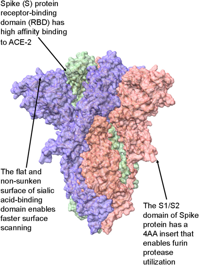
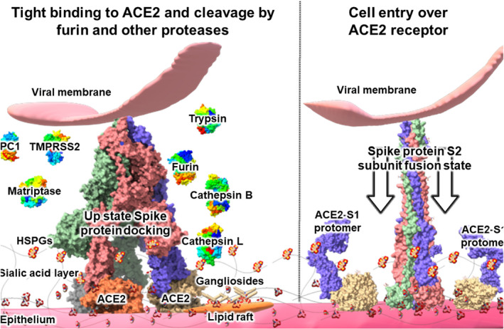
Severe acute respiratory syndrome coronavirus 2 (SARS-CoV-2) is the causative agent of the pandemic coronavirus disease 2019 (COVID-19) that exhibits an overwhelming contagious capacity over other human coronaviruses (HCoVs). This structural snapshot describes the structural bases underlying the pandemic capacity of SARS-CoV-2 and explains its fast motion over respiratory epithelia that allow its rapid cellular entry. Based on notable viral spike (S) protein features, we propose that the flat sialic acid-binding domain at the N-terminal domain (NTD) of the S1 subunit leads to more effective first contact and interaction with the sialic acid layer over the epithelium, and this, in turn, allows faster viral 'surfing' of the epithelium and receptor scanning by SARS-CoV-2. Angiotensin-converting enzyme 2 (ACE-2) protein on the epithelial surface is the primary entry receptor for SARS-CoV-2, and protein-protein interaction assays demonstrate high-affinity binding of the spike protein (S protein) to ACE-2. To date, no high-frequency mutations were detected at the C-terminal domain of the S1 subunit in the S protein, where the receptor-binding domain (RBD) is located. Tight binding to ACE-2 by a conserved viral RBD suggests the ACE2-RBD interaction is likely optimal. Moreover, the viral S subunit contains a cleavage site for furin and other proteases, which accelerates cell entry by SARS-CoV-2. The model proposed here describes a structural basis for the accelerated host cell entry by SARS-CoV-2 relative to other HCoVs and also discusses emerging hypotheses that are likely to contribute to the development of antiviral strategies to combat the pandemic capacity of SARS-CoV-2.

Keywords: COVID-19; SARS-CoV-2; furin protease; receptor-binding domain; sialic acid-binding domain.


Conflict of interest statement

MS has a utility model application with the file number of GM 63/2020 to the Österreichisches Patentamt.

Figures

Fig. 1
Fig. 1
Summary of the S protein structural features unique to SARS‐CoV‐2 over other HCoVs. (A) The binding affinity of the spike RBD to its primary cellular receptor ACE‐2 is more than 10‐fold higher than that of the SARS‐CoV spike RBD. (B) The flat, nonsunken sialic acid‐binding domain is in conflict with that of all other HCoVs, which are sunken in accord with the canyon hypothesis. (C) The S1/S2 domain of SARS‐CoV‐2 S protein contains a four‐amino acid long insert that constitutes a cleavage site for furin proteases, abundant in respiratory epithelia. See text for details. The protein model was S protein, and downstate (PDB ID 6X2C) was modeled with ChimeraX [41].
Fig. 2
Fig. 2
The flat sialic acid‐binding domain. The epithelium is covered with a sialic acid layer. The flat sialic acid‐binding domain of SARS‐CoV‐2 leads to a more effective first contact and interaction with the epithelium, which allows faster viral ‘surfing’ of the epithelial surface and entry receptor scanning. (A) The flat sialic acid, ganglioside, and other sugar‐binding domains (white spheres, left panel) are localized on the left side protomer of the S protein (purple oval, right panel). (B) Entry receptor ACE‐2‐binding domains (red spheres, left panel), C‐type lectin receptor (CLR, green), HSPG (heparan sulfate proteoglycan)‐binding domains based on charge interaction (blue), and HSPG‐binding domains based on GAG (glycosaminoglycan) motif (purple) are localized on the top of the peptomer adjacent to the sialic acid‐binding domain of the S protein (purple oval, right panel); (C) S1/S2 section of the S protein has a unique insert (purple spheres, left panel) that enables it to be cleaved by furin and other proteases, for example, TMPRSS2, PC1, trypsin, matriptase, cathepsin B, and cathepsin L. This domain is localized over the central section of the S protein (purple oval, right panel). Further, another region of the S2 subunit (purple spheres, left panel) has HSPG‐interaction domains (right side of left panel). In addition, the insert and downstream amino acids form a motif capable of binding NRP1 for cellular entry. See text for details. The protein model was S protein, and downstate (PDB ID 6X2C) was modeled with ChimeraX [41].
Fig. 3
Fig. 3
Cleavage of viral S protein by proteases. After viral surfing over the epithelium sialic acid layer, the SARS‐CoV‐2 S protein binds tightly to its entry receptor ACE‐2. A role of sialic acid‐rich gangliosides was also suggested. Unlike other HCoVs, the SARS‐CoV‐2 S1/S2 protein subdomain contains a four‐amino acid insert that constitutes an enzymatic cleavage site for furin and other proteases, which are abundant on respiratory epithelia. Compared to SARS‐CoV, a wider variety of proteases (displayed in left side of Fig. 3) is capable of cleaving the S1/S2 subunit domains of SARS‐CoV‐2, which is believed to form screw‐like S2 fusion conformations composed of spiral trimeric protomers (see right side of Fig. 4) that facilitate host cell entry by SARS‐CoV‐2. See text for details. The protein models were S protein, S protein‐ACE2 complex (PDB ID 7A98), S protein S1 subunit with ACE2 (PDB ID 7A92), S2 subunit postfusion state (PDB ID 6M3W), furin (PDB ID 5JXG), trypsin (PDB ID 3MI4), matriptase (PDB ID 4R0I), cysteine proteases cathepsin B (PDB ID 3MOR), and cathepsin L (PDB ID 3OF9). The models of TMPRSS2 (UniProtKB—O15393), PC1 (GenBank NP_000430.3), were constructed using Swiss‐model (https://swissmodel.expasy.org/). The structures were modeled with ChimeraX and visualized over Microsoft Paint 3D [41].
Fig. 4
Fig. 4
Hypothetical roles for HSPGs, gangliosides, CLRs, NRP1, and lipid rafts in viral S protein –epithelial cell interactions. In a proposed cell entry mechanism based on CLRs, the S protein moves over epithelium with its flat sialic acid‐binding domain (at left of Fig. 4), interacting with free state sialic acids, gangliosides, and various motifs on HSPGs, primarily though relatively weak interactions such as hydrogen bonding. CLRs localized either over the plasma membrane or over lipid rafts (at center of Fig. 4) interact with the S protein and may bypass ACE‐2 and promote virion entry into the cell. Some viruses are known to use CLRs for cell entry to avoid degradation by phagocytes, a process sometimes referred to as the ‘viral escape’ mechanism. Lipid rafts are known to contain many of these components, and thus may also be involved in viral entry. After protease action on the S2 subunit, NRP1 localized either over the plasma membrane or over lipid rafts (at right of Fig. 4) may interact with the S protein and help bypass ACE‐2 to promote virion entry into the cell. See text for details. The protein models were S protein, and downstate (PDB ID 6X2C), S protein S1 subunit (PDB ID 7CHF), S2 subunit postfusion state (PDB ID 6M3W), CLRs (PDB IDs 1XAR with 3JQH), furin (PDB ID 5JXG), and Neuropilin‐1 (PDB ID 2QQM) were modeled with ChimeraX and visualized over Microsoft Paint 3D [41].

References

    1. Seyran M, Pizzol D, Adadi P, El‐Aziz TMA, Hassan SS, Soares A, Kandimalla R, Lundstrom K, Tambuwala M, Aljabali AA et al. (2020) Questions concerning the proximal origin of SARS‐CoV‐2. J Med Virol. 10.1002/jmv.26478. - DOI - PMC - PubMed
    1. Elrashdy F, Redwan EM & Uversky VN (2020) Why COVID‐19 transmission is more efficient and aggressive than viral transmission in previous coronavirus epidemics? Biomolecules 10, 1312. - PMC - PubMed
    1. Yao H, Song Y, Chen Y, Wu N, Xu J, Sun C, Zhang J, Weng T, Zhang Z & Wu Z (2020) Molecular architecture of the SARS‐CoV‐2 virus. Cell 183, 730–738 e13. - PMC - PubMed
    1. Casalino L, Gaieb Z, Goldsmith JA, Hjorth CK, Dommer AC, Harbison AM, Fogarty CA, Barros EP, Taylor BC & McLellan JS (2020) Beyond shielding: the roles of glycans in the SARS‐CoV‐2 spike protein. ACS Cent Sci 6, 1722–1734. - PMC - PubMed
    1. Shang J, Ye G, Shi K, Wan Y, Luo C, Aihara H, Geng Q, Auerbach A & Li F (2020) Structural basis of receptor recognition by SARS‐CoV‐2. Nature 581, 221–224. - PMC - PubMed

Publication types

MeSH terms

Associated data