. 2020 Dec 1;33(9):108456.
doi: 10.1016/j.celrep.2020.108456.

Rare Variant Burden Analysis within Enhancers Identifies CAV1 as an ALS Risk Gene

Collaborators, Affiliations

Rare Variant Burden Analysis within Enhancers Identifies CAV1 as an ALS Risk Gene

Johnathan Cooper-Knock et al. Cell Rep. .

Erratum in

  • Rare variant burden analysis within enhancers identifies CAV1 as an ALS risk gene.
    Cooper-Knock J, Zhang S, Kenna KP, Moll T, Franklin JP, Allen S, Nezhad HG, Iacoangeli A, Yacovzada NY, Eitan C, Hornstein E, Elhaik E, Celadova P, Bose D, Farhan S, Fishilevich S, Lancet D, Morrison KE, Shaw CE, Al-Chalabi A; Project MinE ALS Sequencing Consortium; Veldink JH, Kirby J, Snyder MP, Shaw PJ. Cooper-Knock J, et al. Cell Rep. 2021 Feb 2;34(5):108730. doi: 10.1016/j.celrep.2021.108730. Cell Rep. 2021. PMID: 33535055 Free PMC article. No abstract available.

Abstract

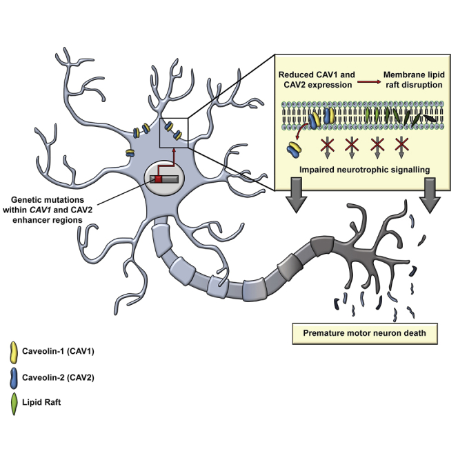
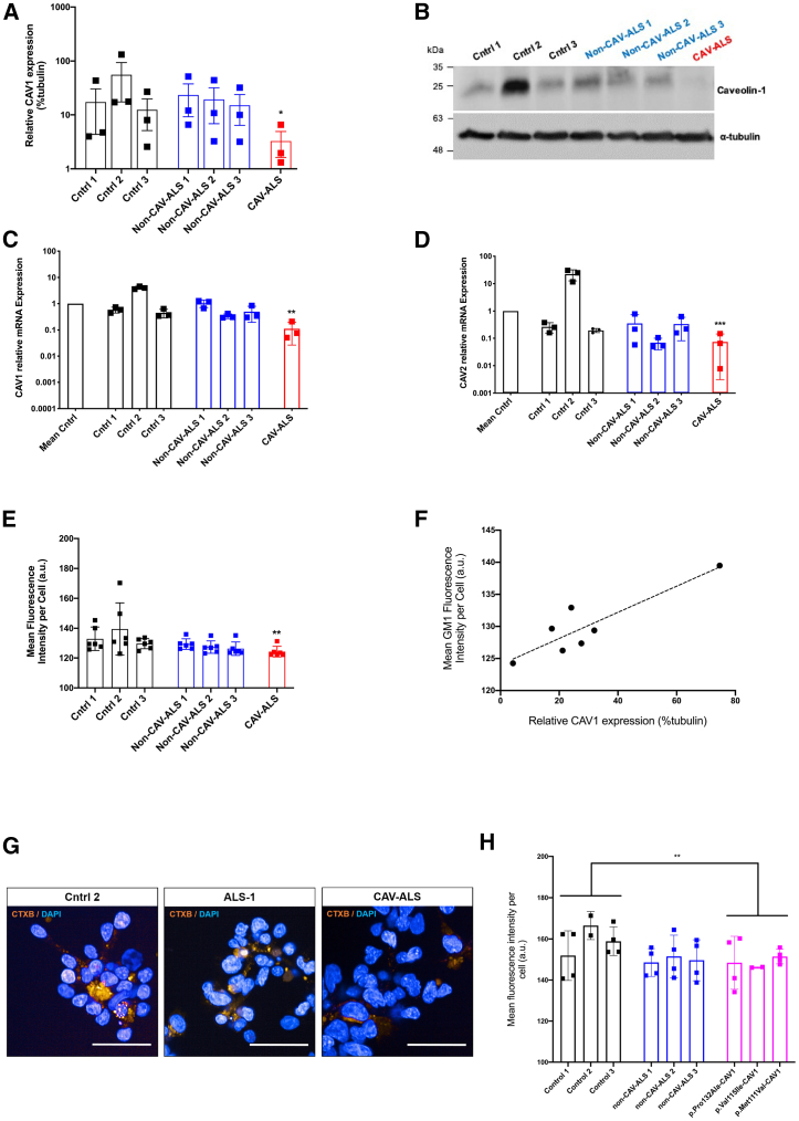
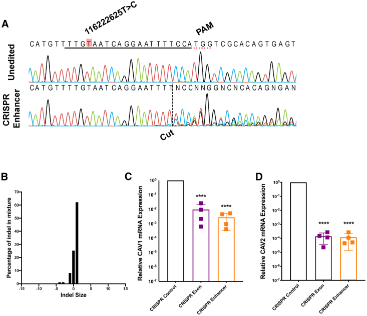
Amyotrophic lateral sclerosis (ALS) is an incurable neurodegenerative disease. CAV1 and CAV2 organize membrane lipid rafts (MLRs) important for cell signaling and neuronal survival, and overexpression of CAV1 ameliorates ALS phenotypes in vivo. Genome-wide association studies localize a large proportion of ALS risk variants within the non-coding genome, but further characterization has been limited by lack of appropriate tools. By designing and applying a pipeline to identify pathogenic genetic variation within enhancer elements responsible for regulating gene expression, we identify disease-associated variation within CAV1/CAV2 enhancers, which replicate in an independent cohort. Discovered enhancer mutations reduce CAV1/CAV2 expression and disrupt MLRs in patient-derived cells, and CRISPR-Cas9 perturbation proximate to a patient mutation is sufficient to reduce CAV1/CAV2 expression in neurons. Additional enrichment of ALS-associated mutations within CAV1 exons positions CAV1 as an ALS risk gene. We propose CAV1/CAV2 overexpression as a personalized medicine target for ALS.

Keywords: CAV1; CAV2; amyotrophic lateral sclerosis; gene enhancers; membrane lipid rafts; non-coding DNA; whole-genome sequencing.


Conflict of interest statement

Declaration of Interests The authors declare no competing interests.

Figures

None
Graphical abstract
Figure 1
Figure 1
Significant Enrichment of ALS Genetic Risk within Enhancers and Coding Regions Linked to CAV1 and CAV2 (A) Pipeline for variant filtering and burden testing; enhancers are first associated with genes based on epigenetic and transcriptome data (Fishilevich et al., 2017); enhancer variants are prioritized for further analysis if they are rare (MAF < 0.01; Lek et al., 2016) and evolutionary conserved (LINSIGHT score > 0.8; Huang et al., 2017). (B) Q-Q plot depicting on x axis the −log 10 of expected p value versus the actually measured p value for 3,761 enhancer groups using whole-genome sequencing (WGS) data from 4,495 ALS cases and 1,925 controls. CAV1/CAV2 enhancers deviate from the null distribution (diagonal), revealing that the burden of variants measured in CAV1/CAV2 enhancers is significantly associated with risk of ALS even after correction for multiple testing. (C) Quantity of genetic material (bp) relative to CNS enhancers derived from Hi-C data (Rhie et al., 2018); CNS enhancers = 100. Upper two bars denote total genetic material; lower two bars denote ALS-associated genetic variants only. Gray shading denotes material unique to CNS or tissue-agnostic enhancers versus material shared by both (white). (D and E) CAV1-coding variants passing filtering criteria are depicted in the table and figure. This analysis utilized WGS data from 4,495 ALS cases and 1,925 controls. One variant is present at higher frequency in controls (orange arrow), and one variant is present is multiple ALS patients (bold arrow); all other variants were discovered in a single ALS patient and zero controls.
Figure 2
Figure 2
Patient-Derived Lymphoblastoid Cell Lines Carrying an ALS-Associated CAV-Enhancer/Coding Variants Have Reduced Expression of CAV1/CAV2 and Disrupted MLR Lymphoblastoid cells were derived from neurologically normal controls (n = 3, black), ALS patients without CAV-enhancer variants (n = 3, blue), ALS patients carrying CAV1-coding mutations (n = 3, magenta), and cells carrying a disease-associated chr7:116222625T>C-CAV1/CAV2 enhancer variant (red). (A and B) Immunoblotting measurement of CAV1 protein expression relative to α-tubulin loading control with an example blot. (C and D) qPCR measurement of CAV1 and CAV2 mRNA expression relative to mean expression in normal controls; expression normalized relative to loading control. (E, G, and H) Measurement of MLR integrity by live-cell imaging and GM1 labeling with CTxB. CTxB fluorescence is plotted with example images. Scale bar, 50 μm. (F) CAV1 protein expression is plotted versus MLR integrity as measured by CTxB fluorescence in the same cell line, with regression line (dotted). Data presented as mean ± 1 SD. p < 0.05; ∗∗p < 0.01; ∗∗∗p < 0.001.
Figure 3
Figure 3
CRISPR-Directed Perturbation of a CAV-Enhancer Region Proximate to a Patient Mutation Reduces CAV1/CAV2 Expression in a Differentiated SH-SY5Y Neuronal Cell (A) Sanger sequencing traces demonstrating spCas9 cut site adjacent to PAM and subsequent waveform decomposition in enhancer edited cells. Position of chr7:116222625T>C mutation is indicated. Black line indicates gRNA sequence. (B) Indel distribution within CAV-enhancer region in CRISPR-edited SH-SY5Y cells. (C and D) qPCR measurement of CAV1 mRNA and CAV2 mRNA reveals reduced expression in CAV-enhancer and CAV1-exon CRISPR-edited neurons compared to CRISPR editing of HPRT; expression normalized relative to loading control. Data presented as mean ± 1 SD. ∗∗∗∗p < 0.0001.

References

    1. Al-Chalabi A., Calvo A., Chio A., Colville S., Ellis C.M., Hardiman O., Heverin M., Howard R.S., Huisman M.H.B., Keren N. Analysis of amyotrophic lateral sclerosis as a multistep process: a population-based modelling study. Lancet Neurol. 2014;13:1108–1113. - PMC - PubMed
    1. Aman A.T., Fraser S., Merritt E.A., Rodigherio C., Kenny M., Ahn M., Hol W.G.J., Williams N.A., Lencer W.I., Hirst T.R. A mutant cholera toxin B subunit that binds GM1- ganglioside but lacks immunomodulatory or toxic activity. Proc. Natl. Acad. Sci. USA. 2001;98:8536–8541. - PMC - PubMed
    1. Arganda-Carreras I., Fernández-González R., Muñoz-Barrutia A., Ortiz-De-Solorzano C. 3D reconstruction of histological sections: Application to mammary gland tissue. Microsc. Res. Tech. 2010;73:1019–1029. - PubMed
    1. Basu S., Pan W. Comparison of statistical tests for disease association with rare variants. Genet. Epidemiol. 2011;35:606–619. - PMC - PubMed
    1. Bemelmans A.-P., Husson I., Jaquet M., Mallet J., Kosofsky B.E., Gressens P. Lentiviral-mediated gene transfer of brain-derived neurotrophic factor is neuroprotective in a mouse model of neonatal excitotoxic challenge. J. Neurosci Res. 2006;83:50–60. - PubMed

Publication types