. 2021 Mar;16(1):70-77.
doi: 10.5469/neuroint.2020.00255. Epub 2020 Dec 3.

Peri-Aneurysmal Brain Edema in Native and Treated Aneurysms: The Role of Thrombosis

Affiliations

Peri-Aneurysmal Brain Edema in Native and Treated Aneurysms: The Role of Thrombosis

Valeria Onofrj et al. Neurointervention. 2021 Mar.

Erratum in

Abstract

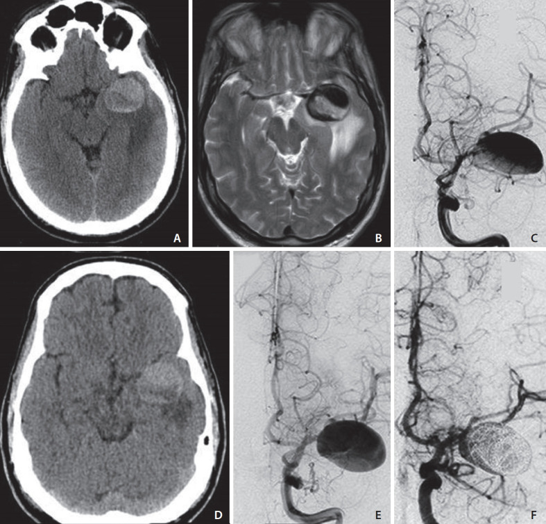
Cerebral peri-aneurysmal edema (PE) is typically associated with giant partially-thrombosed aneurysms and less frequently with smaller aneurysms treated with endovascular embolization. An understanding of the pathophysiologic mechanism of PE is still limited. We report 3 cases of cerebral aneurysms associated with PE. We describe 2 cases of giant partially thrombosed aneurysms surrounded by vasogenic edema with apposition of an intramural and juxtamural thrombus. Our third case is a smaller aneurysm inciting vasogenic edema several years after coil embolization. Vessel-wall magnetic resonance imaging (MRI) showed avid wall enhancement and an enhancing thrombus embedded within the coils, reflecting inflammation of the aneurysm wall and proliferation of the vasa vasorum. Thrombosis within the aneurysmal sac and walls, both in native and treated aneurysms, may promote inflammatory changes and sustain the occurrence of PE. Vessel-wall MRI has a potential role in the evaluation process of this subgroup of aneurysms.

Keywords: Cerebral aneurysms; Inflammation; Magnetic resonance vessel-wall imaging; Perianeurysmal edema; Thrombosis.


Conflict of interest statement

Conflicts of Interest

The authors have no conflicts to disclose.

Figures

Fig. 1.
Fig. 1.
Case 1. A 55 years-old male presenting with generalized seizures. On admission, non contrast computed tomography (NCCT) suggests the presence of a giant partially-thrombosed aneurysm of the left middle cerebral artery (MCA) inciting vasogenic edema in the surrounding brain parenchyma (A). A significant amount of peri-aneurysmal edema (PE) is confirmed on a T2-weighted axial image (B) in the white matter of the left temporal lobe resulting in mass effect on the ipsilateral cerebral peduncle. The aneurysm lumen shows signal flow-void in keeping with patency of the lumen. Catheter digital subtraction angiography (DSA) demonstrates the left MCA aneurysm (C) and its mild increase in size in a follow-up study (E) regardless of the interval decrease of PE on NCCT (D). Early post-treatment DSA after flow-diverter implant and loose intrasaccular coiling shows residual blood-flow in the proximal lumen (F).
Fig. 2.
Fig. 2.
Case 2. A 23 years-old female with right hemiparesis. Head non contrast computed tomography (NCCT) (A) and CT angiography (CTA) (B) after intravenous (i.v.) administration of iodinated contrast medium show a partially-thrombosed giant aneurysm of the left posterior cerebral artery (P3 tract) with a small patent lumen (B) confirmed at catheter digital subtraction angiography (DSA) (C). Magnetic resonance (MR) reveals a significant amount of PE in the left cerebral peduncle and thalamo-capsular region (D) and demonstrates the heterogeneous signal intensity of the intraluminal clot suggesting repetitive intra-aneurysmal hemorrhage (E). Subtle contrast-enhancement is noted along the aneurysmal walls after i.v. administration of gadolinium (F). A 1-month follow-up MR study after endovascular treatment reveals the complete resolution of peri-aneurysmal edema and visible shrinkage of the thrombus (G, H). No signs of recanalization of the treated aneurysm are observed on post-treatment DSA (I).
Fig. 3.
Fig. 3.
Case 3. A 79 years old female with a sudden episode of seizure. Magnetic resonance imaging and magnetic resonance angiography (MRA) before endovascular treatment (2007) show a 10 mm left middle cerebral artery (MCA) aneurysm (white arrow in A) with minimal peri-aneurysmal edema (PE) surrounding the aneurysmal sac (black arrows in B) and a small T1 hyperintense intra-luminal clot (white arrowhead in C). MRA 2 days after coils embolization shows no intraluminal filling of the treated aneurysm (D). Follow-up MR studies respectively 4 (2011), 8 (2015) and 12 (2019) years after initial treatment reveal a progressive and significant increase in PE (E, G, J) despite no interval recanalization of the left MCA aneurysm on 3D-time of flight (TOF) images (F, H, M). A small T1 hyperintense spot is observed within the aneurysm in the 8-year post-treatment study on TOF and T1W images respectively (white arrows in H and I), indicating new apposition of the intraluminal clot. Axial T1-space images before and after i.v administration of gadolinium (K, L) demonstrate vivid contrast-enhancement of the aneurysmal walls and sac suggestive of ongoing inflammatory changes.

Similar articles

Cited by

References

    1. Bose B, Northrup B, Osterholm J. Giant basilar artery aneurysm presenting as a third ventricular tumor. Neurosurgery. 1983;13:699–702. - PubMed
    1. Heros RC, Kolluri S. Giant intracranial aneurysms presenting with massive cerebral edema. Neurosurgery. 1984;15:572–577. - PubMed
    1. Craven I, Patel UJ, Gibson A, Coley SC. Symptomatic perianeurysmal edema following bare platinum embolization of a small unruptured cerebral aneurysm. AJNR Am J Neuroradiol. 2009;30:1998–2000. - PMC - PubMed
    1. Lanzino G. Inflammation after embolization of intracranial aneurysms. J Neurosurg. 2008;108:1071–1073. - PubMed
    1. Lehman VT, Brinjikji W, Kallmes DF, Huston J Rd, Lanzino G, Rabinstein AA, et al. Clinical interpretation of high-resolution vessel wall MRI of intracranial arterial diseases. Br J Radiol. 2016;89:20160496. - PMC - PubMed

LinkOut - more resources