. 2020 Nov 30;21(23):9096.
doi: 10.3390/ijms21239096.

Inhibition of EphA2 by Dasatinib Suppresses Radiation-Induced Intestinal Injury

Affiliations

Inhibition of EphA2 by Dasatinib Suppresses Radiation-Induced Intestinal Injury

Areumnuri Kim et al. Int J Mol Sci. .

Abstract

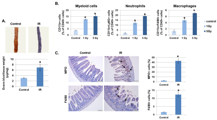
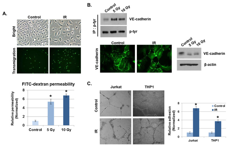
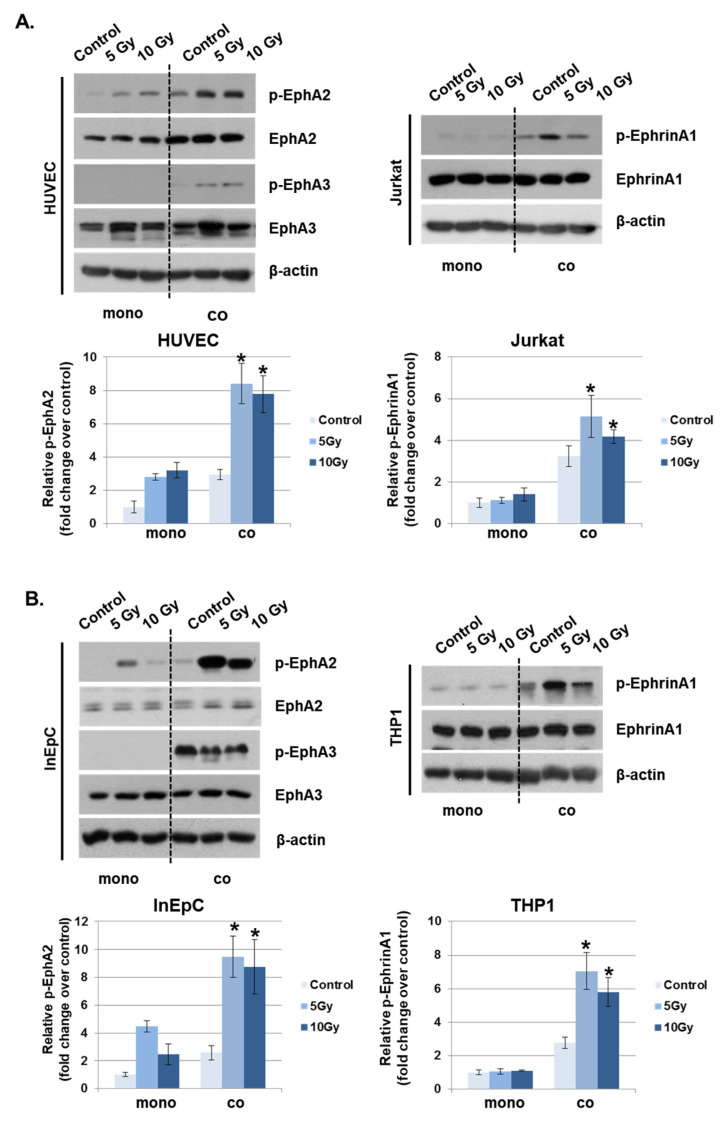
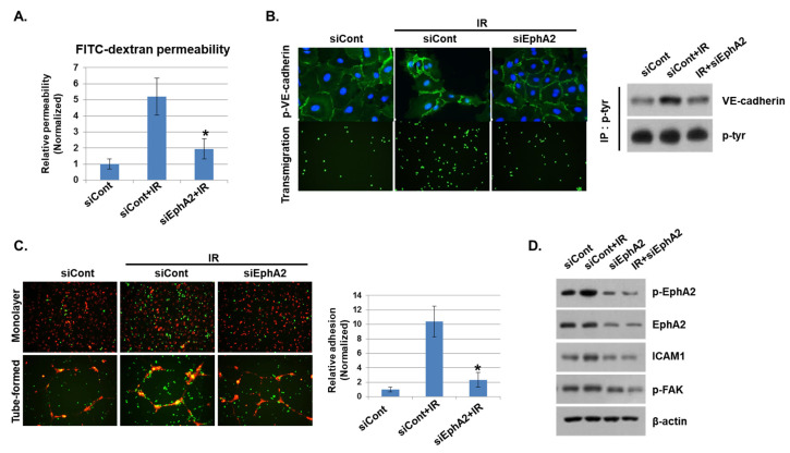
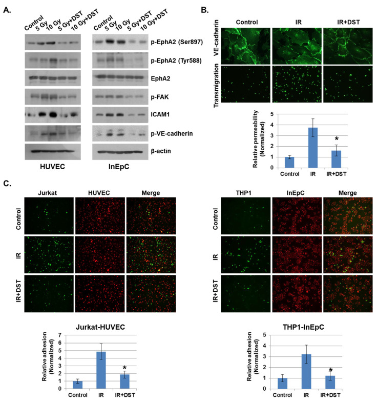
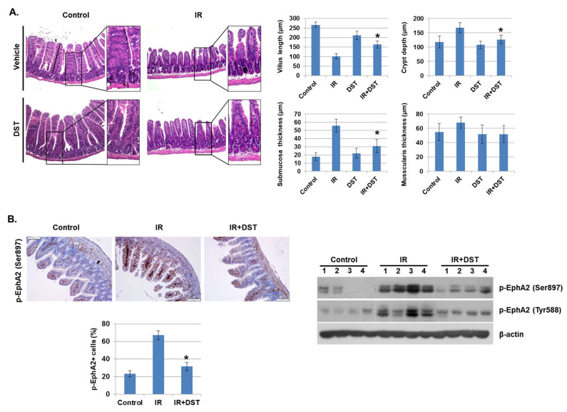
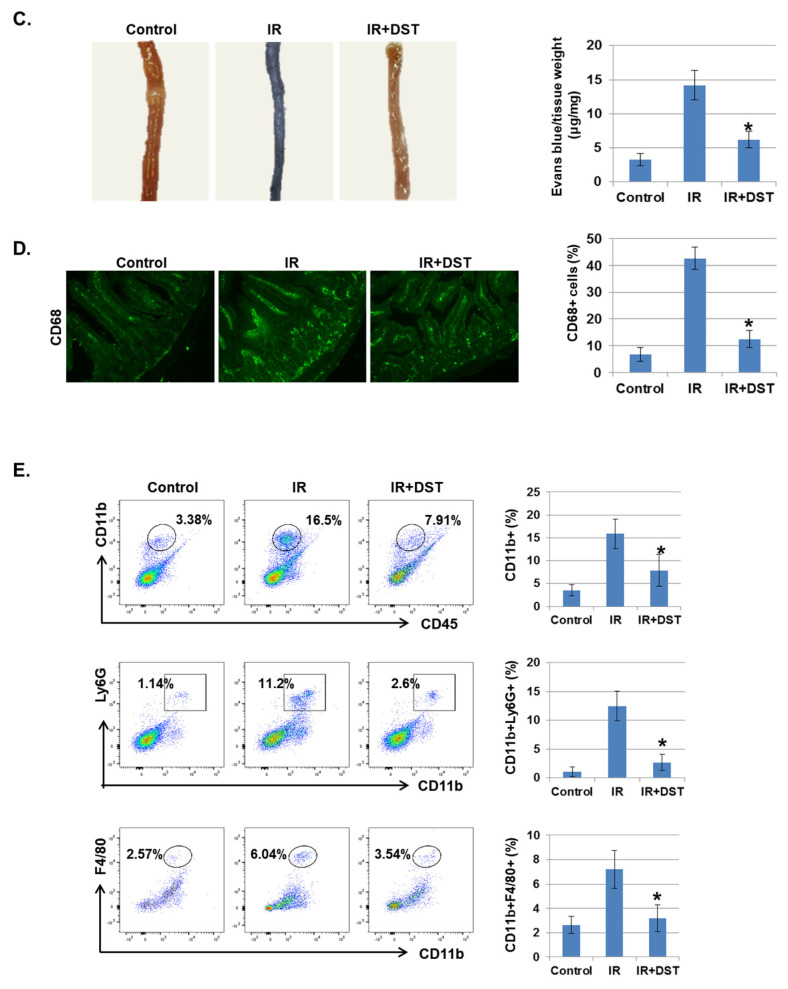
Radiation-induced multiorgan dysfunction is thought to result primarily from damage to the endothelial system, leading to a systemic inflammatory response that is mediated by the recruitment of leukocytes. The Eph-ephrin signaling pathway in the vascular system participates in various disease developmental processes, including cancer and inflammation. In this study, we demonstrate that radiation exposure increased intestinal inflammation via endothelial dysfunction, caused by the radiation-induced activation of EphA2, an Eph receptor tyrosine kinase, and its ligand ephrinA1. Barrier dysfunction in endothelial and epithelial cells was aggravated by vascular endothelial-cadherin disruption and leukocyte adhesion in radiation-induced inflammation both in vitro and in vivo. Among all Eph receptors and their ligands, EphA2 and ephrinA1 were required for barrier destabilization and leukocyte adhesion. Knockdown of EphA2 in endothelial cells reduced radiation-induced endothelial dysfunction. Furthermore, pharmacological inhibition of EphA2-ephrinA1 by the tyrosine kinase inhibitor dasatinib attenuated the loss of vascular integrity and leukocyte adhesion in vitro. Mice administered dasatinib exhibited resistance to radiation injury characterized by reduced barrier leakage and decreased leukocyte infiltration into the intestine. Taken together, these data suggest that dasatinib therapy represents a potential approach for the protection of radiation-mediated intestinal damage by targeting the EphA2-ephrinA1 complex.

Keywords: EphA2; dasatinib; intestinal injury; radiation.


Conflict of interest statement

The authors declare no conflict of interest.

Figures

Figure 1
Figure 1
Irradiation (IR) increases intestinal permeability and leukocyte infiltration. (A) Mice were subjected to whole body IR (5 Gy). After 3 days, mice were injected with Evans blue solution (100 µL of 30 mg/mL) via the tail vein and sacrificed after 30 min. The intestines were collected and photographed. The amount of extracted Evans blue dye was quantified. Results were normalized by tissue weight (mean ± SD); n = 8 control and 8 irradiated mice, * p < 0.01. (B) Three days following IR, intestinal lamina propria cells were isolated and stained for myeloid cells (CD11b+), neutrophils (CD11b+Ly6G+), and macrophages (CD11b+F4/80+) and analyzed using flow cytometry. Graphs show the percent of the CD45+ cell population (mean ± SD); n = 5. * p < 0.01. (C) Representative results of the immunohistochemical staining of control and IR-treated intestines for myeloperoxidase (MPO) and F4/80 3 days after IR (7 Gy). Arrows, stained cells. Graphs show the MPO or F4/80 positive cell population (mean ± SD); n = 5. * p < 0.01.
Figure 2
Figure 2
IR enhances vascular barrier permeability and leukocyte adhesion. (A) Representative images show the morphology of human umbilical vein endothelial cells (HUVECs) and transmigration of THP1 cells co-cultured with HUVECs 24 h after IR. The graph shows the amount of FITC–dextran leakage. Results were normalized to those in control cells (mean ± SD); * p < 0.01. (B) Twenty-four hours after IR, immunofluorescence staining for vascular endothelial (VE)–cadherin in IR-treated HUVECs was performed. VE–cadherin and phosphorylated VE–cadherin levels were measured by Western blotting. β-actin and p-tyr were used as protein loading controls. (C) HUVECs were seeded onto Matrigel, subjected to IR (5 Gy), and co-cultured with leukocytes (THP1 and Jurkat cells) for 6 h. The graph shows the relative adhesion rate of leukocytes to HUVECs (mean ± SD); * p < 0.01.
Figure 3
Figure 3
Phosphorylated EphA2 and ephrinA1 expression levels are upregulated by radiation exposure. Twenty-four hours after IR, p-EphA2, EphA2, p-EphA3, EphA3, p-ephrinA1, and ephrinA1 expression levels were measured in whole-cell lysates by Western blotting. (A) HUVECs were exposed to IR and either co-cultured with Jurkat cells (co) or singly cultured (monoculture; mono). (B) Human intestinal epithelial cells (InEpCs) were exposed to IR and either co-cultured with THP1 cells (co) or singly cultured (monoculture; mono). β-actin was used as the protein loading control. Bars represent mean ± SD of 3 independent experiments. * p < 0.01.
Figure 4
Figure 4
Depletion of EphA2 with siRNAs blocks IR-induced endothelial cell damage. (A) HUVECs were transfected for 24 h with EphA2 siRNAs, which was followed by IR (5 Gy) exposure for an additional 24 h. Endothelial permeability is represented by the amount of FITC–dextran staining. * p < 0.05. (B) Twenty-four hours after IR, siCont or siEphA2-treated HUVECs were stained with FITC-conjugated p-VE–cadherin and visualized using immunofluorescence (upper panel). Carboxyfluorescein succinimidyl ester (CFSE)-labeled THP1 cell transmigration when co-cultured with siCont or siEphA2-treated HUVECs (lower panel). Western blotting results show levels of p-VE–cadherin in siCont or siEphA2-treated HUVECs after IR. P-tyr was used as a protein loading control. (C) Monolayers or tubes of PKH26-labeled HUVECs (red) were exposed to IR and then co-cultured with CFSE-labeled THP1 cells (green) for 24 h. The graph shows the relative adhesion rate. * p < 0.05. (D) Expression levels of p-EphA2, EphA2, ICAM1, and p-FAK in HUVECs were determined using Western blotting. β-actin was used as a protein loading control.
Figure 5
Figure 5
Downregulation of EphA2 by dasatinib reduces IR-induced permeability and adhesion. (A) HUVECs and InEpCs were treated with dasatinib (500 nM) and then exposed to IR for 24 h. Expression levels of p-EphA2 (Ser897 and Tyr588), EphA2, p-FAK, ICAM1 and p-VE–cadherin were measured by Western blotting (left panel). (B) Twenty-four hours after IR (5 Gy), VE–cadherin expression and transmigration were analyzed in HUVECs with or without dasatinib (500 nM). HUVEC permeability is represented by the amount of FITC–dextran staining. * p < 0.05. (C) PKH26-labeled HUVECs (red) were exposed to IR (5 Gy) with or without dasatinib (500 nM) for 12 h, followed by co-culture with CFSE-labeled Jurkat cells (green). PKH26-labeled InEpCs (red) were exposed to IR with or without dasatinib for 12 h, followed by co-culture with CFSE-labeled THP1 cells (green). The adhesion rate was measured at 6 h. * p < 0.05.
Figure 6
Figure 6
Dasatinib suppresses intestinal injury by radiation exposure in mice. Mice were exposed to IR (5 Gy) followed by treatment with dasatinib. Twenty-seven days after IR, mice were sacrificed. (A) Representative images show hematoxylin and eosin (H&E) staining from intestine of control, IR, dasatinib (DST) and IR+DST animals. The sections were analyzed for villus length (1), crypt depth (2), submucosa thickness (3) and muscularis thickness (4) (original magnification ×40, insert ×100). Each bar represents mean ±SD of 7–9 analyzed sections per animal (n = 5). * p < 0.05. (B) Representative images show EphA2 (Ser897) expression in mouse intestine using immunohistochemical staining. The graph indicates the percentage of p-EphA2-positive cells. Expression of p-EphA2 (Ser897 and Tyr588) was measured in mouse intestinal tissues by Western blotting; n = 4. β-actin was used as a protein loading control.; mean ± SD, * p < 0.05. (C) Intestinal permeability was assessed using the Evans blue assay. Representative images show mouse intestine. The graph shows the measurement of Evans blue dye leakage into the intestine; mean ± SD, * p < 0.05. (D) Immunofluorescence staining of CD68 in mouse intestine. The graph shows the percentage of CD68-positive cells; mean ± SD, * p < 0.05. (E) Mouse intestinal lamina propria cells were stained for CD45/CD11b/Ly6G/F480 and analyzed by flow cytometry. Representative images show CD11b+ (myeloid cells), CD11b+Ly6G+ (neutrophils), and CD11b+F4/80+ (macrophages); mean ± SD, n = 5, * p < 0.05.
Figure 6
Figure 6
Dasatinib suppresses intestinal injury by radiation exposure in mice. Mice were exposed to IR (5 Gy) followed by treatment with dasatinib. Twenty-seven days after IR, mice were sacrificed. (A) Representative images show hematoxylin and eosin (H&E) staining from intestine of control, IR, dasatinib (DST) and IR+DST animals. The sections were analyzed for villus length (1), crypt depth (2), submucosa thickness (3) and muscularis thickness (4) (original magnification ×40, insert ×100). Each bar represents mean ±SD of 7–9 analyzed sections per animal (n = 5). * p < 0.05. (B) Representative images show EphA2 (Ser897) expression in mouse intestine using immunohistochemical staining. The graph indicates the percentage of p-EphA2-positive cells. Expression of p-EphA2 (Ser897 and Tyr588) was measured in mouse intestinal tissues by Western blotting; n = 4. β-actin was used as a protein loading control.; mean ± SD, * p < 0.05. (C) Intestinal permeability was assessed using the Evans blue assay. Representative images show mouse intestine. The graph shows the measurement of Evans blue dye leakage into the intestine; mean ± SD, * p < 0.05. (D) Immunofluorescence staining of CD68 in mouse intestine. The graph shows the percentage of CD68-positive cells; mean ± SD, * p < 0.05. (E) Mouse intestinal lamina propria cells were stained for CD45/CD11b/Ly6G/F480 and analyzed by flow cytometry. Representative images show CD11b+ (myeloid cells), CD11b+Ly6G+ (neutrophils), and CD11b+F4/80+ (macrophages); mean ± SD, n = 5, * p < 0.05.

References

    1. Dimitrievich G.S., Fischer-Dzoga K., Griem M.L. Radiosensitivity of vascular tissue. I. Differential radiosensitivity of capillaries: A quantitative in vivo study. Radiat. Res. 1984;99:511–535. doi: 10.2307/3576327. - DOI - PubMed
    1. Georgakilas A.G., Pavlopoulou A., Louka M., Nikitaki Z., Vorgias C.E., Bagos P.G., Michalopoulos I. Emerging molecular networks common in ionizing radiation, immune and inflammatory responses by employing bioinformatics approaches. Cancer Lett. 2015;368:164–172. doi: 10.1016/j.canlet.2015.03.021. - DOI - PubMed
    1. Gourmelon P., Benderitter M., Bertho J.M., Huet C., Gorin N.C., De Revel P. European consensus on the medical management of acute radiation syndrome and analysis of the radiation accidents in Belgium and Senegal. Health Phys. 2010;98:825–832. doi: 10.1097/HP.0b013e3181ce64d4. - DOI - PubMed
    1. Fliedner T.M., Chao N.J., Bader J.L., Boettger A., Case C., Jr., Chute J., Confer D.L., Ganser A., Gorin N.C., Gourmelon P., et al. Stem cells, multiorgan failure in radiation emergency medical preparedness: A U.S./European Consultation Workshop. Stem Cells. 2009;27:1205–1211. doi: 10.1002/stem.16. - DOI - PMC - PubMed
    1. Fajardo L.F. Is the pathology of radiation injury different in small vs large blood vessels? Cardiovasc. Radiat. Med. 1999;1:108–110. - PubMed

MeSH terms

LinkOut - more resources