. 2020 Nov 30;9(12):1202.
doi: 10.3390/antiox9121202.

Comparative Proteomics Unveils LRRFIP1 as a New Player in the DAPK1 Interactome of Neurons Exposed to Oxygen and Glucose Deprivation

Affiliations

Comparative Proteomics Unveils LRRFIP1 as a New Player in the DAPK1 Interactome of Neurons Exposed to Oxygen and Glucose Deprivation

Núria DeGregorio-Rocasolano et al. Antioxidants (Basel). .

Abstract

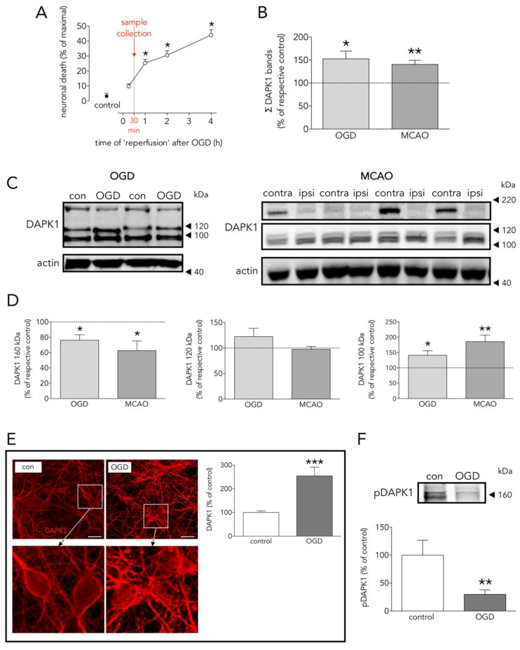
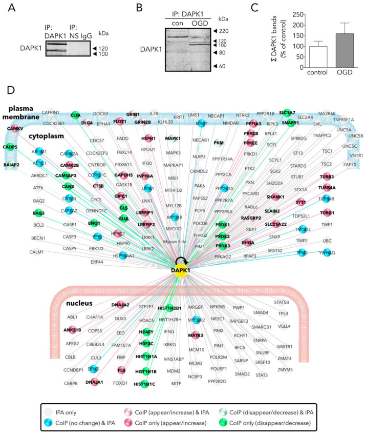
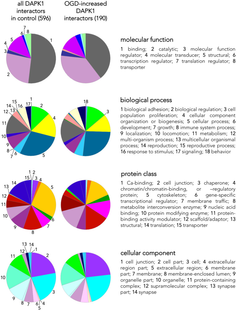
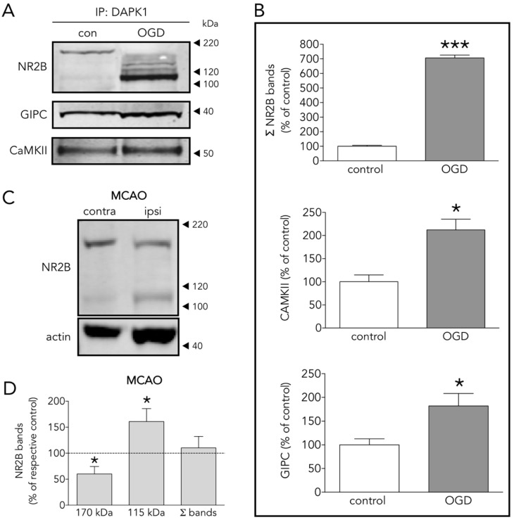
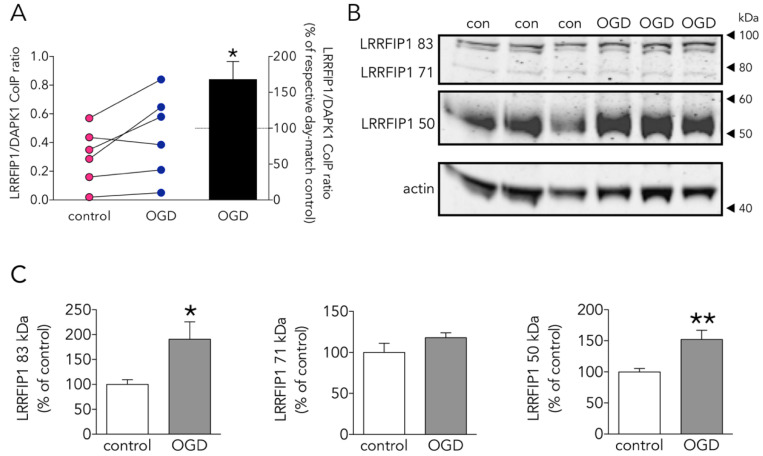
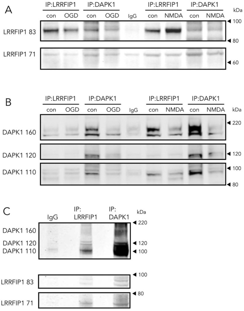
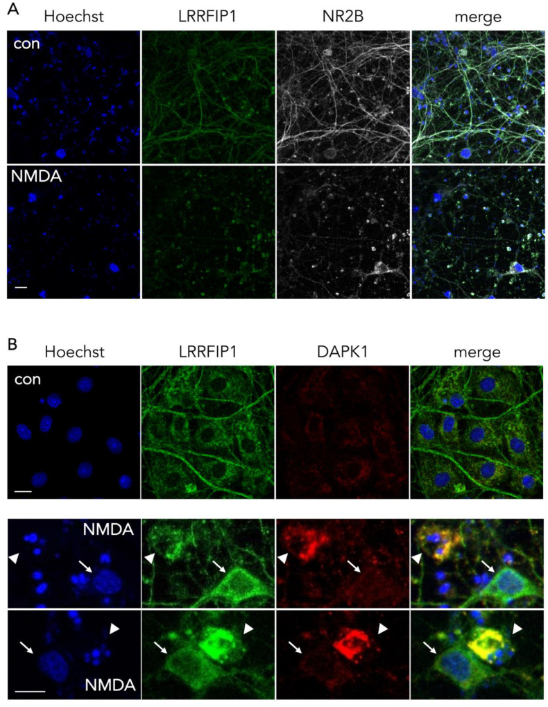
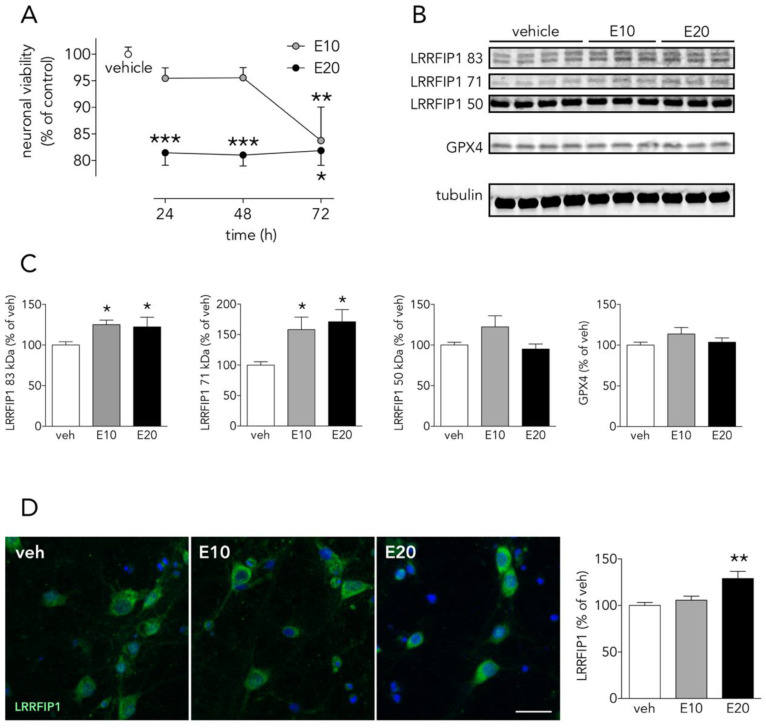
Death-associated protein kinase 1 (DAPK1) is a pleiotropic hub of a number of networked distributed intracellular processes. Among them, DAPK1 is known to interact with the excitotoxicity driver NMDA receptor (NMDAR), and in sudden pathophysiological conditions of the brain, e.g., stroke, several lines of evidence link DAPK1 with the transduction of glutamate-induced events that determine neuronal fate. In turn, DAPK1 expression and activity are known to be affected by the redox status of the cell. To delineate specific and differential neuronal DAPK1 interactors in stroke-like conditions in vitro, we exposed primary cultures of rat cortical neurons to oxygen/glucose deprivation (OGD), a condition that increases reactive oxygen species (ROS) and lipid peroxides. OGD or control samples were co-immunoprecipitated separately, trypsin-digested, and proteins in the interactome identified by high-resolution LC-MS/MS. Data were processed and curated using bioinformatics tools. OGD increased total DAPK1 protein levels, cleavage into shorter isoforms, and dephosphorylation to render the active DAPK1 form. The DAPK1 interactome comprises some 600 proteins, mostly involving binding, catalytic and structural molecular functions. OGD up-regulated 190 and down-regulated 192 candidate DAPK1-interacting proteins. Some differentially up-regulated interactors related to NMDAR were validated by WB. In addition, a novel differential DAPK1 partner, LRRFIP1, was further confirmed by reverse Co-IP. Furthermore, LRRFIP1 levels were increased by pro-oxidant conditions such as ODG or the ferroptosis inducer erastin. The present study identifies novel partners of DAPK1, such as LRRFIP1, which are suitable as targets for neuroprotection.

Keywords: DAPK1; LRRFIP1; MCAO; NMDA; OGD; ROS; ferroptosis; neuron.


Conflict of interest statement

The authors declare no conflict of interest.

Figures

Figure 1
Figure 1
Effects of OGD on neuronal death, and DAPK1 expression, cleavage, and dephosphorylation. (A) Effect of time of ‘reperfusion’ after OGD on neuronal death; as depicted, experimental samples were taken at 30 min for further processing (one-way ANOVA plus SNK, n = at least 5 independent primary neuron culture preparations, with at least 3 technical replicates each). (B) Effect of OGD and MCAO on total DAPK1 levels (OGD: t test, n = 3 independent primary neuron culture preparations, with 3 technical replicates each; MCAO: paired t test, n = 9 rats, comparing ipsilateral (ipsi, ischemic) brain hemisphere with the contralateral (contra, control) one). (C) WB showing the effect of OGD or MCAO on DAPK1 bands. (D) Quantification of the effect of OGD or MCAO on DAPK1 cleavage and levels of the resulting bands (OGD: t test, n = 3 independent primary neuron culture preparations, with 3 technical replicates each; MCAO: paired t test, n = 9 rats, comparing ipsilateral (ipsi, ischemic) brain hemisphere with the contralateral (contra, control) one). (E) Effect of OGD on DAPK1 levels as measured by immunocytochemistry; insets in the upper pictures are shown magnified in the bottom pictures (t test, n = 3 independent primary neuron culture preparations, with 5 technical replicates each). (F) WB and quantification of the effect of OGD on pDAPK1 levels (t test, n = 3 independent primary neuron culture preparations, with 3 technical replicates each). Results are shown as the mean and SEM. * p < 0.05, ** p < 0.01, *** p < 0.005 vs. respective control in all graphs. Scale bar: 30 µm. Note that in (B), total DAPK1 levels represent protein expression.
Figure 2
Figure 2
Main effects of OGD on the neuronal DAPK1 interactome. (A) WB shows that DAPK1 immunoprecipitates using a specific anti-DAPK1 antibody (IP: DAPK1, left lane), but it does not when using a non-specific antibody (IP: NS IgG, right lane). (B) WB showing the effect of OGD on the DAPK1 bands obtained by immunoprecipitation. (C) Graph showing the amount of DAPK1 obtained after immunoprecipitation of control and OGD samples (t test, n = 3 independent primary neuron culture preparations, with 3 technical replicates each). Note that, in this graph, differently than in WB in Figure 1B, DAPK1 data represent protein co-immunoprecipitated from neurons. (D) Drawing depicting some of the DAPK1 interactors found in the present study and those present in the IPA database; color codes of proteins are self-explained in the legend within the drawing. Results are shown as the mean and SEM.
Figure 3
Figure 3
Classes and categorization of proteins in the DAPK1 interactome. Panther™ v15.0 (pantherdb.org) was used to classify proteins of the DAPK1 interactome in control neurons and those interactors found increased in OGD-exposed neurons (they are shown in a pie chart on the left). Four main classes are defined, each encompassing a number of different categories encoded by colors and explained on the right side of each class.
Figure 4
Figure 4
Several DAPK1 interactors are CTD-NR2B binding partners. (A) WB and (B) quantification of the effect of OGD on the co-immunoprecipitation of DAPK1 with the well-known DAPK1 interactors NR2B, CAMKII and GIPC (t test, n = 4–5 co-immunoprecipitations from independent primary neuron culture preparations). (C) WB showing the effect of MCAO on the NR2B band levels. (D) Effect of MCAO on 170 kDa, 115 kDa and total NR2B levels (paired t test, n = 9 rats, comparing ipsilateral (ipsi, ischemic) brain hemisphere with the contralateral (contra, control) one). The results are shown as the mean and SEM. * p < 0.05, *** p < 0.005 vs. respective control in all graphs.
Figure 5
Figure 5
Effect of OGD on the new DAPK1-interacting candidate protein LRRFIP1. (A) Effect of OGD on the LRRFIP1/DAPK1 co-immunoprecipitation ratio (t test, n = 6 co-immunoprecipitations from independent primary neuron culture preparations). (B) WB showing the effect of OGD on the expression of different LRRFIP1 transcripts, and (C) graphs showing the quantification of this effect (83 and 50 kDa graphs: t test, and 71 kDa graph: Mann-Whitney U test, n = 3 independent primary neuron culture preparations, with 3 technical replicates each). Results in A (left panel) are shown as individual co-immunoprecipitation values, and in A (right panel) and C as the mean and SEM. * p < 0.05, ** p < 0.01 vs. respective control in all graphs.
Figure 6
Figure 6
Direct and reverse co-immunoprecipitation of LRRFIP1 during ischemia in vivo and excitotoxicity in vitro. (A,B) WB showing the effect of OGD or NMDA on direct and reverse co-immunoprecipitation of LRRFIP1 and DAPK1 from rat cultured neurons. (C) WB showing direct and reverse co-immunoprecipitation of DAPK1 and LRRFIP1 from ischemic rat brain homogenates.
Figure 7
Figure 7
Representative ICC images showing the colocalization pattern of NR2B and DAPK1 with LRRFIP1. (A) Images showing the main cellular location of LRRFIP1 and NR2B in control and NMDA-treated neurons in vitro; scale bar: 30 µm. (B) Images showing the colocalization pattern of LRRFIP1 and DAPK1 in control neurons (upper panel, con), and in neurons with normal (arrows) or pyknotic (arrowheads) nucleus shortly after NMDA treatment (lower panels, NMDA); scale bars: 10 µm.
Figure 8
Figure 8
The ferroptosis inducer erastin increases neuronal LRRFIP1 levels. (A) Time-course effect of erastin on neuronal viability in vitro (one-way ANOVAs plus SNK, n = 3 independent primary neuron culture preparations, with 3 technical replicates each). (B) WB showing the effect of erastin on LRRFIP1 and GXP4 levels at 24 h, and (C) graphs depicting the quantification of this effect (one-way ANOVA plus SNK, n = 3 independent primary neuron culture preparations, with 3 technical replicates each). (D) Representative ICC images showing LRRFIP1 expression in neurons exposed to vehicle (veh) or erastin for 24 h, and a graph showing the quantification of this effect. Ten µM erastin: filled grey circles/bars or E10; 20 µM erastin: filled black circles/bars or E20. Results are shown as the mean and SEM. * p < 0.05, ** p < 0.01, *** p < 0.001 vs. vehicle in all graphs. Scale bar: 20 µm.

References

    1. Zhou Q., Song W., Xiao W. Dioscin induces demethylation of DAPK-1 and RASSF-1α genes via the antioxidant capacity, resulting in apoptosis of bladder cancer T24 cells. EXCLI J. 2017;16:101–112. doi: 10.17179/excli2016-571. - DOI - PMC - PubMed
    1. Tsai Y.T., Chuang M.J., Tang S.H., Wu S.T., Chen Y.C., Sun G.H., Hsiao P.W., Huang S.M., Lee H.J., Yu C.P., et al. Novel cancer therapeutics with allosteric modulation of the mitochondrial C-Raf-DAPK complex by Raf inhibitor combination therapy. Cancer Res. 2015;75:3568–3582. doi: 10.1158/0008-5472.CAN-14-3264. - DOI - PubMed
    1. Yu S., Zhai J., Yu J., Yang Q., Yang J. miR-98-5p protects against cerebral ischemia/reperfusion injury through anti-apoptosis and anti-oxidative stress in mice. J. Biochem. 2020 doi: 10.1093/jb/mvaa099. - DOI - PubMed
    1. Martel M.A., Ryan T.J., Bell K.F.S., Fowler J.H., McMahon A., Al-Mubarak B., Komiyama N.H., Horsburgh K., Kind P.C., Grant S.G.N., et al. The subtype of GluN2 C-terminal domain determines the response to excitotoxic insults. Neuron. 2012;74:543–556. doi: 10.1016/j.neuron.2012.03.021. - DOI - PMC - PubMed
    1. Tang N., Wu J., Zhu H., Yan H., Guo Y., Cai Y., Yan H., Shi Y., Shu S., Pei L., et al. Genetic mutation of GluN2B protects brain cells against stroke damages. Mol. Neurobiol. 2018;55:2979–2990. doi: 10.1007/s12035-017-0562-y. - DOI - PubMed

LinkOut - more resources