

EcDBS1R4, an Antimicrobial Peptide Effective against Escherichia coli with In Vitro Fusogenic Ability
- PMID: 33265989
- PMCID: PMC7730630
- DOI: 10.3390/ijms21239104
EcDBS1R4, an Antimicrobial Peptide Effective against Escherichia coli with In Vitro Fusogenic Ability
Abstract
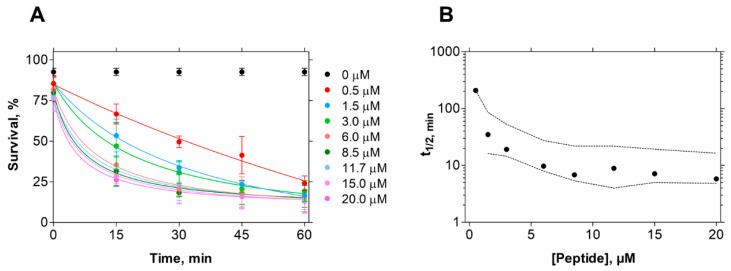
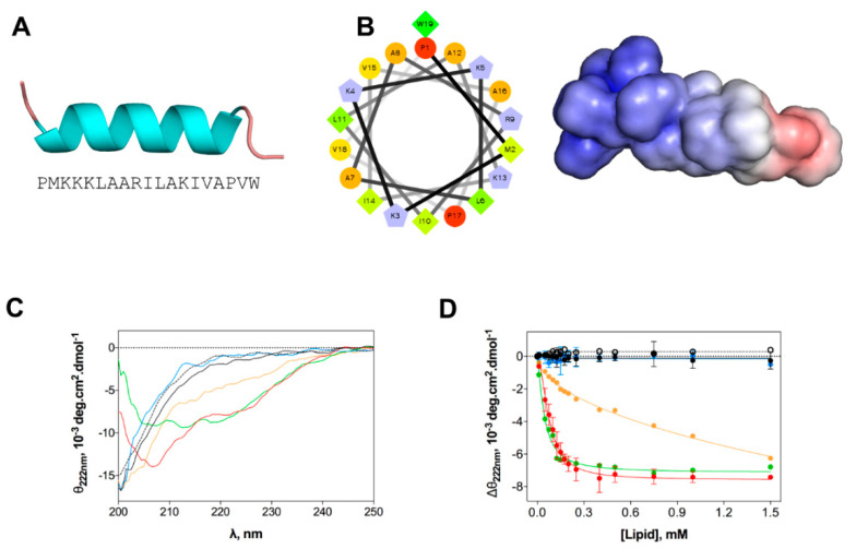
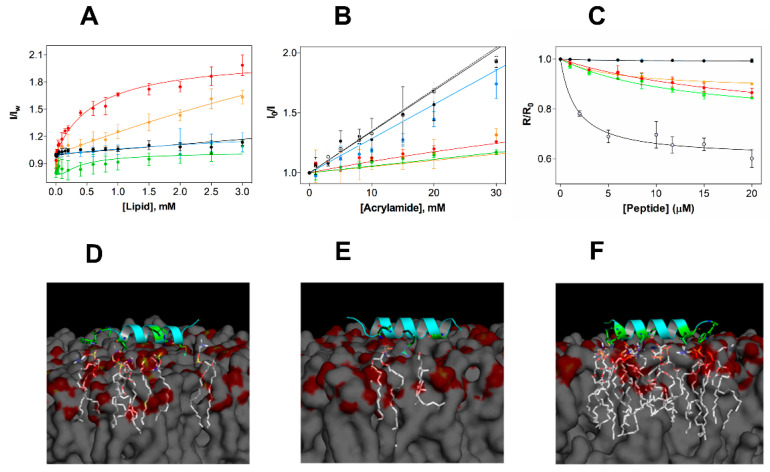
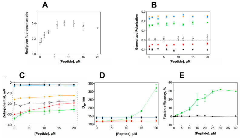
Discovering antibiotic molecules able to hold the growing spread of antimicrobial resistance is one of the most urgent endeavors that public health must tackle. The case of Gram-negative bacterial pathogens is of special concern, as they are intrinsically resistant to many antibiotics, due to an outer membrane that constitutes an effective permeability barrier. Antimicrobial peptides (AMPs) have been pointed out as potential alternatives to conventional antibiotics, as their main mechanism of action is membrane disruption, arguably less prone to elicit resistance in pathogens. Here, we investigate the in vitro activity and selectivity of EcDBS1R4, a bioinspired AMP. To this purpose, we have used bacterial cells and model membrane systems mimicking both the inner and the outer membranes of Escherichia coli, and a variety of optical spectroscopic methodologies. EcDBS1R4 is effective against the Gram-negative E. coli, ineffective against the Gram-positive Staphylococcus aureus and noncytotoxic for human cells. EcDBS1R4 does not form stable pores in E. coli, as the peptide does not dissipate its membrane potential, suggesting an unusual mechanism of action. Interestingly, EcDBS1R4 promotes a hemi-fusion of vesicles mimicking the inner membrane of E. coli. This fusogenic ability of EcDBS1R4 requires the presence of phospholipids with a negative curvature and a negative charge. This finding suggests that EcDBS1R4 promotes a large lipid spatial reorganization able to reshape membrane curvature, with interesting biological implications herein discussed.
Keywords: EcDBS1R4; Escherichia coli; Gram-negative bacteria; antimicrobial peptide; cardiolipin; hemifusion; hyperpolarization.
Conflict of interest statement
The authors declare no conflict of interest.
Figures
Similar articles
-
Activity modulation of the Escherichia coli F1FO ATP synthase by a designed antimicrobial peptide via cardiolipin sequestering.iScience. 2023 May 30;26(7):107004. doi: 10.1016/j.isci.2023.107004. eCollection 2023 Jul 21. iScience. 2023. PMID: 37416464 Free PMC article.
-
Differential interactions of the antimicrobial peptide, RQ18, with phospholipids and cholesterol modulate its selectivity for microorganism membranes.Biochim Biophys Acta Gen Subj. 2021 Sep;1865(9):129937. doi: 10.1016/j.bbagen.2021.129937. Epub 2021 May 27. Biochim Biophys Acta Gen Subj. 2021. PMID: 34052310
-
The introduction of L-phenylalanine into antimicrobial peptide protonectin enhances the selective antibacterial activity of its derivative phe-Prt against Gram-positive bacteria.Amino Acids. 2021 Jan;53(1):23-32. doi: 10.1007/s00726-020-02919-z. Epub 2020 Nov 24. Amino Acids. 2021. PMID: 33236256
-
Recent Applications of Aggregation Induced Emission Probes for Antimicrobial Peptide Studies.Chem Asian J. 2021 May 3;16(9):1027-1040. doi: 10.1002/asia.202100102. Epub 2021 Mar 25. Chem Asian J. 2021. PMID: 33723926 Review.
-
Anti-bacterial activity of inorganic nanomaterials and their antimicrobial peptide conjugates against resistant and non-resistant pathogens.Int J Pharm. 2020 Aug 30;586:119531. doi: 10.1016/j.ijpharm.2020.119531. Epub 2020 Jun 12. Int J Pharm. 2020. PMID: 32540348 Review.
Cited by
-
Novel antibiotics against Staphylococcus aureus without detectable resistance by targeting proton motive force and FtsH.MedComm (2020). 2025 Jan 8;6(1):e70046. doi: 10.1002/mco2.70046. eCollection 2025 Jan. MedComm (2020). 2025. PMID: 39781293 Free PMC article.
-
Expression of the Antimicrobial Peptide SE-33-A2P, a Modified Analog of Cathelicidin, and an Analysis of Its Properties.Antibiotics (Basel). 2024 Feb 16;13(2):190. doi: 10.3390/antibiotics13020190. Antibiotics (Basel). 2024. PMID: 38391576 Free PMC article.
-
Activity modulation of the Escherichia coli F1FO ATP synthase by a designed antimicrobial peptide via cardiolipin sequestering.iScience. 2023 May 30;26(7):107004. doi: 10.1016/j.isci.2023.107004. eCollection 2023 Jul 21. iScience. 2023. PMID: 37416464 Free PMC article.
-
Recent advances in the development of antimicrobial peptides against ESKAPE pathogens.Heliyon. 2024 May 24;10(11):e31958. doi: 10.1016/j.heliyon.2024.e31958. eCollection 2024 Jun 15. Heliyon. 2024. PMID: 38868046 Free PMC article. Review.
-
Creation of New Antimicrobial Peptides.Int J Mol Sci. 2023 May 29;24(11):9451. doi: 10.3390/ijms24119451. Int J Mol Sci. 2023. PMID: 37298402 Free PMC article.
References
-
- Tackling Drug-Resistant Infections Globally: Final Report and Recommendations. [(accessed on 17 March 2020)]; Available online: http://amr-review.org/sites/default/files/160525_Final paper_with cover.pdf.
MeSH terms
Substances
Grants and funding
- NA/Fundação para a Ciência e a Tecnologia - Ministério da Ciência, Tecnologia e Ensino Superior (FCT-MCTES, Portugal)
- grant agreement 644167/Marie Skłodowska-Curie Research and Innovation Staff Exchange (MSCA-RISE, European Union) project INPACT
- NA/Conselho Nacional de Desenvolvimento Científico e Tecnológico (CNPq, Brazil), Coordenação de Aperfeiçoamento de Pessoal de Nível Superior (CAPES, Brazil), Fundação de Amparo a Pesquisa do Distrito Federal (FAPDF, Brazil)
- NA/Fundação de Apoio ao Desenvolvimento do Ensino, Ciência e Tecnologia do Estado de Mato Grosso do Sul (FUNDECT, Brazil)
- SPRH/BD/128290/2017 and SPRH/BD/100517/2014/Fundação para a Ciência e a Tecnologia - Ministério da Ciência, Tecnologia e Ensino Superior (FCT-MCTES, Portugal)
LinkOut - more resources
Full Text Sources
