Review
. 2020 Nov 30;8(12):1899.
doi: 10.3390/microorganisms8121899.

The Role of APOBECs in Viral Replication

Affiliations
Review

The Role of APOBECs in Viral Replication

Wendy Kaichun Xu et al. Microorganisms. .

Abstract

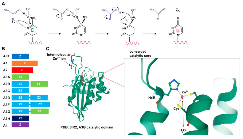
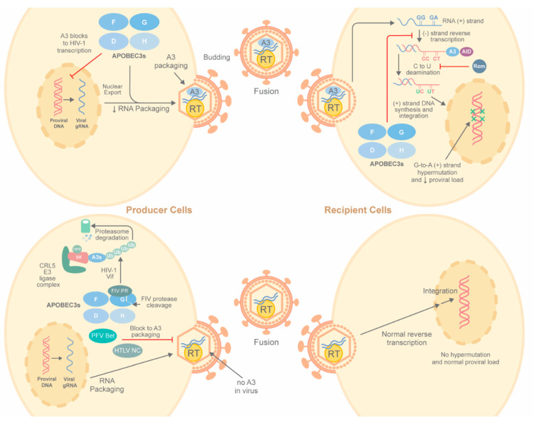
Apolipoprotein B mRNA-editing enzyme catalytic polypeptide-like (APOBEC) proteins are a diverse and evolutionarily conserved family of cytidine deaminases that provide a variety of functions from tissue-specific gene expression and immunoglobulin diversity to control of viruses and retrotransposons. APOBEC family expansion has been documented among mammalian species, suggesting a powerful selection for their activity. Enzymes with a duplicated zinc-binding domain often have catalytically active and inactive domains, yet both have antiviral function. Although APOBEC antiviral function was discovered through hypermutation of HIV-1 genomes lacking an active Vif protein, much evidence indicates that APOBECs also inhibit virus replication through mechanisms other than mutagenesis. Multiple steps of the viral replication cycle may be affected, although nucleic acid replication is a primary target. Packaging of APOBECs into virions was first noted with HIV-1, yet is not a prerequisite for viral inhibition. APOBEC antagonism may occur in viral producer and recipient cells. Signatures of APOBEC activity include G-to-A and C-to-T mutations in a particular sequence context. The importance of APOBEC activity for viral inhibition is reflected in the identification of numerous viral factors, including HIV-1 Vif, which are dedicated to antagonism of these deaminases. Such viral antagonists often are only partially successful, leading to APOBEC selection for viral variants that enhance replication or avoid immune elimination.

Keywords: AID; APOBEC; DNA virus replication; G-to-A mutations; HIV-1 Vif; MMTV Rem; RNA virus replication; genome hypermutation; viral variants.


Conflict of interest statement

The authors declare no conflict of interest.

Figures

Figure 1
Figure 1
Structural characteristics and activity of APOBECs. (A) Reaction of APOBEC-mediated cytidine deamination (see text for details). (B) Different APOBECs are composed of distinct zinc-coordinating (Z) domains. In the mammalian A3 family, Z domains are categorized into three distinct phylogenetic groups, Z1, Z2, and Z3. Members of the human APOBEC genes and the relative lengths of their Z domains are shown. (C) A ribbon diagram of the A3G catalytic domain shows its globular structure and an expanded view of the zinc-coordinating active site (from PDB 3IR2) [10]. Zinc ions are depicted in gray. The expanded view shows the interactions of the histidine ring and cysteine side chains with zinc. The red dot near the zinc ion represents a water molecule.
Figure 2
Figure 2
Deamination-dependent restriction of viruses by APOBECs. In the absence of an antagonist, such as HIV-1 Vif, virus-producing cells (top left) often package A3 proteins, including A3D, A3F, A3G and A3H, (abbreviated as D, F, G, and H) into virions together with reverse transcriptase and two copies of genomic RNA. A3 enzymes (specifically A3G) also may block HIV-1 transcription, lowering the amount of viral RNA available for packaging. Subsequent infection of susceptible recipient cells (upper right) leads to reverse transcription of viral plus-strand RNA. During minus-strand DNA synthesis, C-to-U deamination occurs, leading to G-to-A hypermutation on the viral DNA plus strands. Blocks to reverse transcription also occur, resulting in integrated viral DNA with increased G-to-A transition mutations and reduced proviral loads. Virus-producing cells (lower left) that make an antagonist, such as Prototype Foamy Virus (PFV) Bet or Human T-Cell Leukemia Virus type 1 (HTLV-1) nucleocapsid (NC), fail to package A3. Other A3 antagonists, such as HIV-1 Vif, recruit an E3 ligase complex to A3 enzymes to mediate their ubiquitylation and degradation, whereas the Feline Immunodeficiency Virus (FIV) protease (PR) reportedly cleaves A3 proteins. In either case, A3 levels are reduced sufficiently to prevent virion incorporation. A3-free virions then mediate successful reverse transcription without hypermutation in recipient cells (lower right). Mouse Mammary Tumor Virus regulator of export of MMTV mRNA (MMTV Rem) expression leads to AID proteasomal degradation (not shown), but AID is not incorporated into virions in the presence or absence of Rem. RT = reverse transcriptase; gRNA = genomic RNA; CRL = cullin–RING ubiquitin ligase.
Figure 3
Figure 3
Deamination-independent inhibition of viral replication. In virus-producing cells, A3 and AID proteins are present in cytoplasmic complexes (left). AID is a shuttling protein and its nuclear activity may block cell proliferation and the number of infected cells. Alternatively, multiple APOBEC proteins may inhibit viral RNA translation. In the case of murine leukemia viruses, several different virally specified inhibitors, such as p50 and glycoGag (gGag) are produced in infected cells. The p50 protein prevents A3 incorporation into virions, whereas gGag is incorporated into virus particles to promote their stability, allowing normal proviral DNA synthesis in recipient cells (right). If gGag is present in viral particles, A3 may be incorporated into virions, leading to reduced proviral DNA. If virion RNA is packaged in the absence of gGag, viral cores are unstable and allow A3 enzymes in recipient cells to block reverse transcription. RT = reverse transcriptase.

References

    1. Harris R.S., Dudley J.P. APOBECs and virus restriction. Virology. 2015;479–480:131–145. doi: 10.1016/j.virol.2015.03.012. - DOI - PMC - PubMed
    1. Olson M.E., Harris R.S., Harki D.A. APOBEC Enzymes as Targets for Virus and Cancer Therapy. Cell Chem. Biol. 2018;25:36–49. doi: 10.1016/j.chembiol.2017.10.007. - DOI - PMC - PubMed
    1. Conticello S.G., Thomas C.J., Petersen-Mahrt S.K., Neuberger M.S. Evolution of the AID/APOBEC family of polynucleotide (deoxy)cytidine deaminases. Mol. Biol. Evol. 2005;22:367–377. doi: 10.1093/molbev/msi026. - DOI - PubMed
    1. Krzysiak T.C., Jung J., Thompson J., Baker D., Gronenborn A.M. APOBEC2 is a monomer in solution: Implications for APOBEC3G models. Biochemistry. 2012;51:2008–2017. doi: 10.1021/bi300021s. - DOI - PMC - PubMed
    1. Ko T.P., Lin J.J., Hu C.Y., Hsu Y.H., Wang A.H., Liaw S.H. Crystal structure of yeast cytosine deaminase. Insights into enzyme mechanism and evolution. J. Biol. Chem. 2003;278:19111–19117. doi: 10.1074/jbc.M300874200. - DOI - PubMed

LinkOut - more resources