

The Role of Extracellular Vesicles in Demyelination of the Central Nervous System
- PMID: 33266211
- PMCID: PMC7729475
- DOI: 10.3390/ijms21239111
The Role of Extracellular Vesicles in Demyelination of the Central Nervous System
Abstract
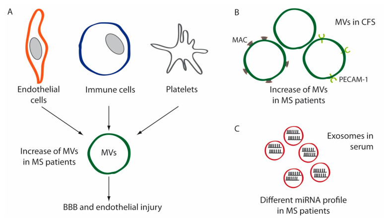
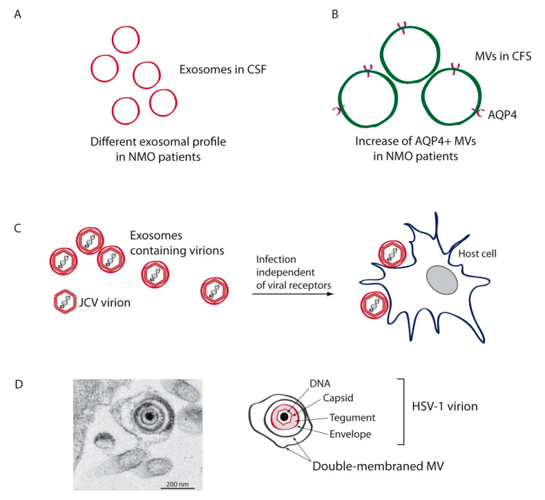
It is being increasingly demonstrated that extracellular vesicles (EVs) are deeply involved in the physiology of the central nervous system (CNS). Processes such as synaptic activity, neuron-glia communication, myelination and immune response are modulated by EVs. Likewise, these vesicles may participate in many pathological processes, both as triggers of disease or, on the contrary, as mechanisms of repair. EVs play relevant roles in neurodegenerative disorders such as Alzheimer's or Parkinson's diseases, in viral infections of the CNS and in demyelinating pathologies such as multiple sclerosis (MS). This review describes the involvement of these membrane vesicles in major demyelinating diseases, including MS, neuromyelitis optica, progressive multifocal leukoencephalopathy and demyelination associated to herpesviruses.
Keywords: central nervous system; demyelination; extracellular vesicles; herpesviruses; multiple sclerosis; neuromyelitis optica; progressive multifocal leukoencephalopathy.
Conflict of interest statement
The authors declare no conflict of interest.
Figures

Similar articles
-
HSV-1 and Endogenous Retroviruses as Risk Factors in Demyelination.Int J Mol Sci. 2021 May 27;22(11):5738. doi: 10.3390/ijms22115738. Int J Mol Sci. 2021. PMID: 34072259 Free PMC article. Review.
-
Pediatric central nervous system inflammatory demyelination: acute disseminated encephalomyelitis, clinically isolated syndromes, neuromyelitis optica, and multiple sclerosis.Curr Opin Neurol. 2009 Jun;22(3):233-40. doi: 10.1097/wco.0b013e32832b4c47. Curr Opin Neurol. 2009. PMID: 19434783 Review.
-
Brain incoming call from glia during neuroinflammation: Roles of extracellular vesicles.Neurobiol Dis. 2024 Oct 15;201:106663. doi: 10.1016/j.nbd.2024.106663. Epub 2024 Sep 7. Neurobiol Dis. 2024. PMID: 39251030 Review.
-
Central nervous system-derived extracellular vesicles: the next generation of neural circulating biomarkers?Transl Neurodegener. 2024 Jun 19;13(1):32. doi: 10.1186/s40035-024-00418-9. Transl Neurodegener. 2024. PMID: 38898538 Free PMC article. Review.
-
Potential Roles of Extracellular Vesicles as Biomarkers and a Novel Treatment Approach in Multiple Sclerosis.Int J Mol Sci. 2021 Aug 20;22(16):9011. doi: 10.3390/ijms22169011. Int J Mol Sci. 2021. PMID: 34445717 Free PMC article. Review.
Cited by
-
Role of Extracellular Vesicles in Glia-Neuron Intercellular Communication.Front Mol Neurosci. 2022 Apr 13;15:844194. doi: 10.3389/fnmol.2022.844194. eCollection 2022. Front Mol Neurosci. 2022. PMID: 35493327 Free PMC article. Review.
-
Motor skill learning modulates striatal extracellular vesicles' content in a mouse model of Huntington's disease.Cell Commun Signal. 2024 Jun 11;22(1):321. doi: 10.1186/s12964-024-01693-9. Cell Commun Signal. 2024. PMID: 38863004 Free PMC article.
-
Delivery Platforms for CRISPR/Cas9 Genome Editing of Glial Cells in the Central Nervous System.Front Genome Ed. 2021 Mar 3;3:644319. doi: 10.3389/fgeed.2021.644319. eCollection 2021. Front Genome Ed. 2021. PMID: 34713256 Free PMC article. Review.
-
Extracellular Vesicles: The Next Generation of Biomarkers and Treatment for Central Nervous System Diseases.Int J Mol Sci. 2024 Jul 5;25(13):7371. doi: 10.3390/ijms25137371. Int J Mol Sci. 2024. PMID: 39000479 Free PMC article. Review.
-
Human Glial Cells as Innovative Targets for the Therapy of Central Nervous System Pathologies.Cells. 2024 Mar 30;13(7):606. doi: 10.3390/cells13070606. Cells. 2024. PMID: 38607045 Free PMC article. Review.
