Review
. 2020 Nov 30;21(23):9133.
doi: 10.3390/ijms21239133.

Cross Talk at the Cytoskeleton-Plasma Membrane Interface: Impact on Neuronal Morphology and Functions

Affiliations
Review

Cross Talk at the Cytoskeleton-Plasma Membrane Interface: Impact on Neuronal Morphology and Functions

Rossella Di Giaimo et al. Int J Mol Sci. .

Abstract

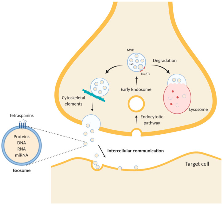
The cytoskeleton and its associated proteins present at the plasma membrane not only determine the cell shape but also modulate important aspects of cell physiology such as intracellular transport including secretory and endocytic pathways. Continuous remodeling of the cell structure and intense communication with extracellular environment heavily depend on interactions between cytoskeletal elements and plasma membrane. This review focuses on the plasma membrane-cytoskeleton interface in neurons, with a special emphasis on the axon and nerve endings. We discuss the interaction between the cytoskeleton and membrane mainly in two emerging topics of neurobiology: (i) production and release of extracellular vesicles and (ii) local synthesis of new proteins at the synapses upon signaling cues. Both of these events contribute to synaptic plasticity. Our review provides new insights into the physiological and pathological significance of the cytoskeleton-membrane interface in the nervous system.

Keywords: cystatin B; cytoskeleton; exosomes; local protein synthesis; plasma membrane; synaptic plasticity.


Conflict of interest statement

The authors declare no conflict of interest.

Figures

Figure 1
Figure 1
Schematic diagram of exosome trafficking in a neuron. While multivesicular bodies (MVBs) undergo a maturing process, the composition of the intraluminal proteins is gradually changed. The endosomal sorting complex required for transport (ESCRT) remodels the inner membrane of MVB to generate the intraluminal vesicles (ILVs), which represent exosomes inside the MVBs. After intracellular translocation mediated by cytoskeleton elements, such as F-actin, and microtubules, MVBs merge with the plasma membrane and undergo exocytosis. Alternatively, MVBs can fuse with lysosomes to degrade themselves. Exosomes contain in their lumen free proteins, RNA, microRNA, and DNA and are characterized by transmembrane proteins of the tetraspanins family.
Figure 2
Figure 2
Schematic diagrams of the subplasmalemmal regions in specialized neuronal compartments. (a) the axonal plasma membrane is associated with “periaxoplasmic plaques”, which contain ribosomes and therefore serve as a center for intra-axonal protein synthesis. (b) The plasma membrane of the growth cones contains receptors for various guiding cues. When netrin-1 receptor DCC (Deleted in Colorectal Carcinoma) is bound by netrin, the sequestered ribosomes are released from the membrane and move to the unspecified cytoskeleton, which may serve as a scaffold for protein synthesis. Translation of β-actin mRNA increases local concentration of β-actin, which leads to enhanced F-actin polymerization. Binding of a repulsive guidance cue slit-2 has the opposite effect because of the increased local translation of cofilin, an actin-binding protein. (c) Stimulation of the serotonin receptor 7 (5-HT7R), localized in the neuronal plasma membrane, promotes de novo protein synthesis and activation of several signaling pathways such as ERK, Cdk5, Cdc42, and mTOR. These intracellular pathways converge to promote reorganization of the neuronal cytoskeleton, including microtubules and actin filaments.

References

    1. Ishizuka N., Cowan W.M., Amaral D.G. A quantitative analysis of the dendritic organization of pyramidal cells in the rat hippocampus. J. Comp. Neurol. 1995;362:17–45. doi: 10.1002/cne.903620103. - DOI - PubMed
    1. Holt C.E., Martin K.C., Schuman E.M. Local translation in neurons: Visualization and function. Nat. Struct. Mol. Biol. 2019;26:557–566. doi: 10.1038/s41594-019-0263-5. - DOI - PubMed
    1. Arimura N., Kaibuchi K. Neuronal polarity: From extracellular signals to intracellular mechanisms. Nat. Rev. Neurosci. 2007;8:194–205. doi: 10.1038/nrn2056. - DOI - PubMed
    1. Leterrier C., Dubey P., Roy S. The nano-architecture of the axonal cytoskeleton. Nat. Rev. Neurosci. 2017;18:713–726. doi: 10.1038/nrn.2017.129. - DOI - PubMed
    1. Conde C., Cáceres A. Microtubule assembly, organization and dynamics in axons and dendrites. Nat. Rev. Neurosci. 2009;10:319–332. doi: 10.1038/nrn2631. - DOI - PubMed