. 2020 Nov 30;21(23):9135.
doi: 10.3390/ijms21239135.

Functional Identification of Serine Hydroxymethyltransferase as a Key Gene Involved in Lysostaphin Resistance and Virulence Potential of Staphylococcus aureus Strains

Affiliations

Functional Identification of Serine Hydroxymethyltransferase as a Key Gene Involved in Lysostaphin Resistance and Virulence Potential of Staphylococcus aureus Strains

Nayab Batool et al. Int J Mol Sci. .

Abstract

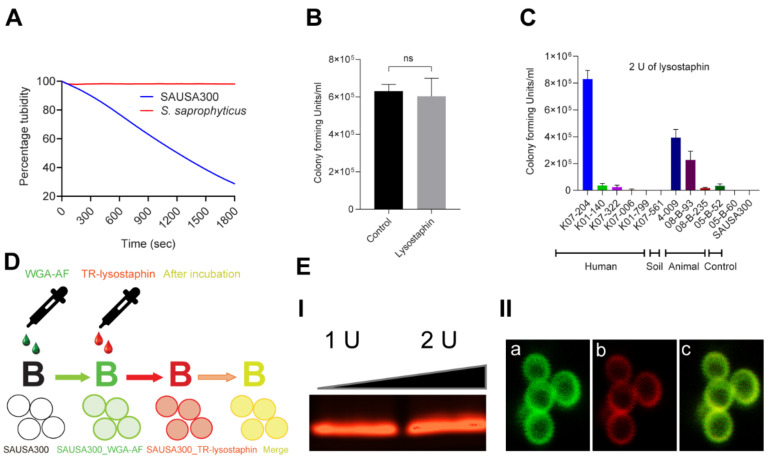
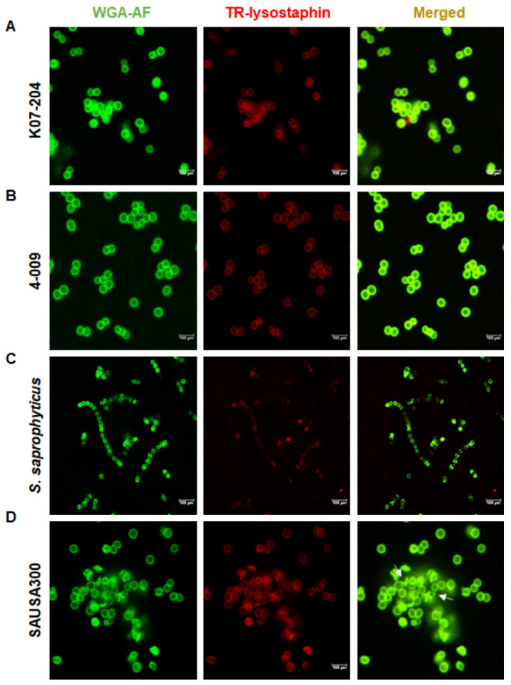
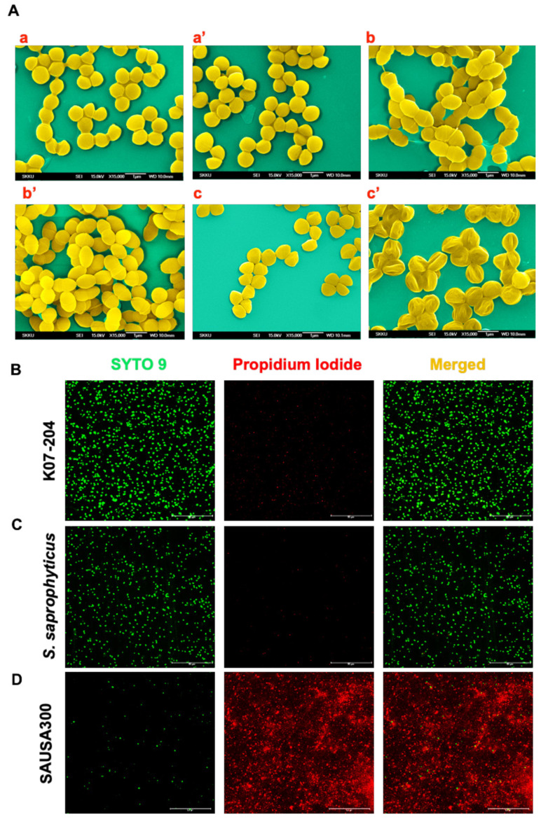
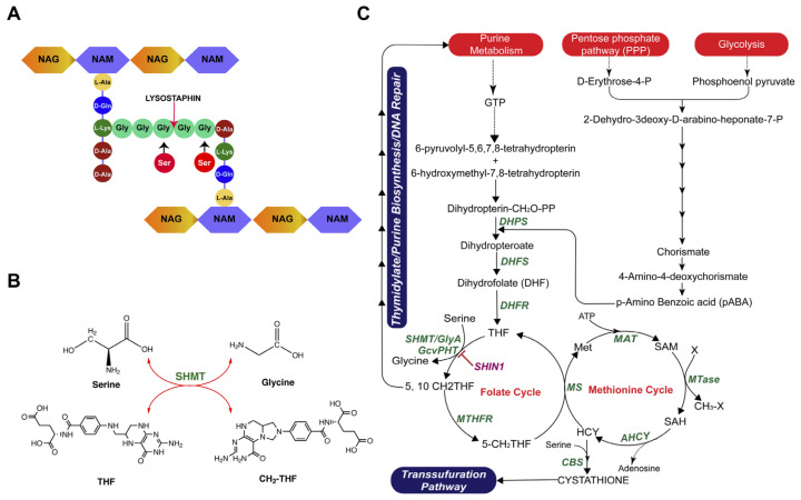
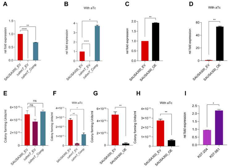
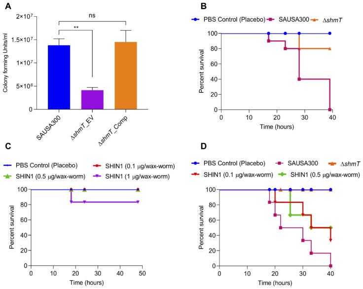
Gaining an insight into the mechanism underlying antimicrobial-resistance development in Staphylococcus aureus is crucial for identifying effective antimicrobials. We isolated S. aureus sequence type 72 from a patient in whom the S. aureus infection was highly resistant to various antibiotics and lysostaphin, but no known resistance mechanisms could explain the mechanism of lysostaphin resistance. Genome-sequencing followed by subtractive and functional genomics revealed that serine hydroxymethyltransferase (glyA or shmT gene) plays a key role in lysostaphin resistance. Serine hydroxymethyltransferase (SHMT) is indispensable for the one-carbon metabolism of serine/glycine interconversion and is linked to folate metabolism. Functional studies revealed the involvement of SHMT in lysostaphin resistance, as ΔshmT was susceptible to the lysostaphin, while complementation of the knockout expressing shmT restored resistance against lysostaphin. In addition, the ΔshmT showed reduced virulence under in vitro (mammalian cell lines infection) and in vivo (wax-worm infection) models. The SHMT inhibitor, serine hydroxymethyltransferase inhibitor 1 (SHIN1), protected the 50% of the wax-worm infected with wild type S. aureus. These results suggest SHMT is relevant to the extreme susceptibility to lysostaphin and the host immune system. Thus, the current study established that SHMT plays a key role in lysostaphin resistance development and in determining the virulence potential of multiple drug-resistant S. aureus.

Keywords: SHMT; SHMT inhibitor; ST72; Staphylococcus aureus; folate cycle; lysostaphin resistance; serine hydroxymethyltransferase; virulence factor.


Conflict of interest statement

The authors declare no conflict of interest.

Figures

Figure 1
Figure 1
Lysostaphin resistance pattern in ST72 isolates and the efficiency of lysostaphin binding to the cell wall of Staphylococcus aureus. (A) Lysostaphin mediated killing kinetics of lysostaphin susceptible S. aureus USA300 (lyss) in comparison to the lysostaphin-resistant Staphylococcus saprophyticus (lysr) using the cell turbidity reduction assay. S. aureus USA300 (lyss) showed 70% reduction of cell turbidity compared to S. saprophyticus (lysr); (B) The lysostaphin resistance of S. saprophyticus (lysr) was further confirmed by colony forming unit (CFU) counting without (control) and with lysostaphin treatment, showing no significant difference in CFU counts; (C) Differential resistance pattern in 11 isolates of S. aureus ST72 against 2 U of lysostaphin upon 5 min of incubation wherein K07-204 (human), 4-009 (soil), and 08-B-93 (animal) showed lysostaphin resistance compared to lysostaphin-susceptible S. aureus USA300 (control); (D) Schematic diagram displays lysostaphin binding to the cell wall labeled with wheat germ agglutinin Alexa Fluor 488 (WGA-AF) (green fluorescence) with colocalized Texas Red labeled lysostaphin (red fluorescence of TR-lysostaphin); and (E) (I) Texas Red-labeled lysostaphin on agarose gel showing the red fluorescent protein band, (II) Colocalization of TR-lysostaphin on WGA-AF labeled green fluorescent cell wall of lysostaphin resistant human isolate of ST72 K07-204 wherein (a) the green channel of confocal photomicrograph shows WGA-AF labeled green fluorescent cell wall boundary of staphylococcal cell, (b) the red channel of the confocal photomicrograph shows the red fluorescent cell wall upon TR-lysostaphin binding, and (c) the merged channel of green (a) and red (b) shows the yellow fluorescent cell boundary, confirming the efficient binding of lysostaphin with the staphylococcal cell wall.
Figure 2
Figure 2
Phenotypic assessment of the lysostaphin-binding and catalytic cleavage activity of lysostaphin resistant isolates of ST72. (AD) Confocal microscopic images of lysostaphin-resistant ST72 human isolate K07-204 (A); lysostaphin-resistant ST72 soil isolate 4-009 (B); lysostaphin-resistant control S. saprophyticus (C); and lysostaphin-susceptible control S. aureus USA300 (D) upon treatment with TR-lysostaphin wherein white arrows indicate the broken cells after lysostaphin treatment. TR-lysostaphin binds efficiently with both ST72 isolates and S. aureus USA300, while TR-lysostaphin showed the least binding with S. saprophyticus.
Figure 3
Figure 3
Confirmation of lysostaphin-mediated alteration in the cell wall and consequent cell death. (A) Scanning electron microscopy (SEM) to visualize the alteration in the cellular morphology showing staphylococcal cells without lysostaphin treatment wherein K07-204 (a); S. saprophyticus (b); and SAUSA300 (c); and after lysostaphin treatment K07-204 (a’); S. saprophyticus (b’); and SAUSA300 (c’). Both K07-204 and S. saprophyticus (lysr) did not show any alteration, while the SAUSA300 (lyss) cells were shrunk upon lysostaphin treatment due to the catalytic cleavage activity of lysostaphin; (BD) Confocal microscopy images of live/dead staining of ST72 resistant isolates and its comparison with SAUSA300 to assess the consequent proportion of live/dead staphylococcal cells, wherein SYTO9 stains the total cells (green), whereas propidium iodide (PI) exclusively stains dead cells (red). Both K07-204 (B) and S. saprophyticus (C) showed a lower number of dead (red) cells compared to SAUSA300 (D) upon 2 U of lysostaphin treatment.
Figure 4
Figure 4
Novel mechanism of lysostaphin resistance in ST72 isolate and its associated metabolic pathway. After screening all the possible genes and mutation(s) known for lysostaphin resistance, no existing mechanisms (genes, mutations) of lysostaphin resistance was found to be functioned in ST72. (A) The schematic diagram shows the fundamental reason of lysostaphin resistance in staphylococcal cells due to the modification of glycine residues of pentaglycine bridge to serine; (B) Serine hydroxymethyltransferase (SHMT) is an indispensable enzyme for the one-carbon metabolism of serine/glycine interconversion and is linked to folate/methionine cycle. Therefore, glyA/shmT gene was hypothesized to be involved in lysostaphin resistance. (C) The metabolic pathway showing the interdependence of folate/methionine cycle and the key role of shmT serine/glycine homeostasis. One-carbon metabolism is responsible for the transfer of methyl group to various substrate and cofactors in folate, methionine cycle, and transsulfuration pathways. Various enzymes are denoted in green font while the substrates are depicted in regular font. The abbreviation used in the pathway wherein enzymes are DHPS (Dihydropteroate synthase); DHFS (Dihydrofolate synthase); DHFR (Dihydrofolate reductase); SHMT (Serine hydroxymethyltransferase); GcvPHT (glycine cleavage system); MTHFR (Methylene tetrahydrofolate reductase); MS (Methionine synthase); MAT (Methionine adenosyltransferase); MTases (Methyl transferases); AHCY (S-adenosylhomocysteine hydrolase), and CBS (Cystathionine beta-synthase) and substrates are THF (Tetrahydrofolate); 5, 10 CH2-THF (5, 10 methylene tetrahydrofolate); 5-CH2-THF (5-methylene tetrahydrofolate); Met (Methionine); SAM (S-adenosyl methionine); SAH (S-adenosyl homocysteine), and HCY (Homocysteine). One-carbon metabolism is important in cellular homeostasis by maintaining cellular seine/glycine through folate cycle, methionine cycle (protein synthesis), DNA synthesis and repair, redox balance, and various methylation reactions.
Figure 5
Figure 5
Assessment of shmT expression and functional genomics to establish the role of shmT in lysostaphin resistance. (AB) Gene expression of shmT in SAUSA300 recombinant strains including SAUSA300_EV, ΔshmT_EV and ΔshmT_Comp without induction (A), and with anhydrotetracycline (aTc) induction (B) wherein no transcript was detected in ΔshmT knockout with empty vector (ΔshmT_EV) as compared to the wild type S. aureus USA300 with empty vector (SAUSA300_EV) and ΔshmT complemented strain harboring pRMC2_shmTshmT_Comp). The shmT expression in (ΔshmT_Comp) strain was found to be moderate without aTc induction while it was significantly enhanced (3.5-fold) upon aTc induction. (CD) Gene expression of shmT in SAUSA300 recombinant strains SAUSA300_EV and SAUSA300_OE constructed by expressing shmT in trans (plasmid: pRMC2_shmT) under tetracycline inducible promoter in the wild type S. aureus USA300. Expression of shmT in SAUSA300_OE versus SAUSA300_EV without (C) and with aTc induction (D) was found to be 2 and 53-fold higher, respectively, than control. (EF) Assessment of colony forming unit in SAUSA300 recombinant strains SAUSA300_EV, ΔshmT_EV and ΔshmT_Comp showing the relative susceptibility of ΔshmT_EV strain compared to SAUSA300_EV and ΔshmT_Comp strains (E) whereas the susceptibility of ΔshmT_Comp strain was enhanced upon higher expression of shmT using aTc induction (F). The susceptibility of ΔshmT_EV showed the plausible involvement of shmT in lysostaphin resistance. (GH) The SAUSA300_OE strain without induction (G), and with aTc induction (H) showed extreme susceptibility towards lysostaphin as compared to SAUSA300_EV. Both ΔshmT_Comp and SAUSA300_OE showed higher susceptibility to lysostaphin as compared to empty vector control SAUSA300_EV upon aTc induction and resultant overexpression of shmT; and (I) Expression of shmT in ST72 isolates K07-204 versus K07-561, wherein the K07-561 showed overexpression of shmT, which is the plausible reason of why K07-561 was susceptible to lysostaphin compared to K07-204.
Figure 6
Figure 6
Validating the role of shmT in virulence potential of SAUSA300 using in vitro mammalian cells, and in vivo wax-worm infection model. (A) Assessment of internalization (invasion/phagocytosis) and survival potential of SAUSA300, ΔshmT knockout and ΔshmT complemented strains under in vitro mammalian cell culture conditions using murine macrophage, RAW264.7 cells. The ΔshmT knockout showed significantly reduced survival inside the macrophage as compared to wild type SAUSA300 and ΔshmT complemented strains; (B) Survival graph for wax-worms infected by SAUSA300 and ΔshmT knockout (2.0 × 10⁵ bacterial cells). The number of wax-worms in each group was 10 (n =10). (C) Assessment of toxicity of the SHMT inhibitor (SHIN1) for wax-worms (n = 10). Varying concentrations of SHIN1 (0.1 μg, 0.5 μg and 1μg in 20 μL solution) were injected into the wax-worms and the survival of the worms was observed for up to 80 h along with 20 μL placebo PBS control. The SHIN1 did not show any toxicity to the worms up to 0.5 μg; and (D) The treatment of SHIN1 inhibitor protected 50% wax-worm infected by the wild type SAUSA300, indicating that SHIN1 inhibits the pathogenesis of the wild type SAUSA300.

References

    1. Nahrgang S., Nolte E., Rechel B. The role of public health organizations in addressing antimicrobial resistance in Europe. Eur. J. Public Health. 2018;28:169–170. doi: 10.1093/eurpub/cky212.497. - DOI - PubMed
    1. Monserrat-Martinez A., Gambin Y., Sierecki E. Thinking outside the bug: Molecular targets and strategies to overcome antibiotic resistance. Int. J. Mol. Sci. 2019;20:1255. doi: 10.3390/ijms20061255. - DOI - PMC - PubMed
    1. Tong S.Y.C., Davis J.S., Eichenberger E., Holland T.L., Fowler V.G. Staphylococcus aureus infections: Epidemiology, pathophysiology, clinical manifestations, and management. Clin. Microbiol. Rev. 2015;28:603–661. doi: 10.1128/CMR.00134-14. - DOI - PMC - PubMed
    1. Bouvet C., Gjoni S., Zenelaj B., Lipsky B.A., Hakko E., Uckay I. Staphylococcus aureus soft tissue infection may increase the risk of subsequent staphylococcal soft tissue infections. Int. J. Infect. Dis. 2017;60:44–48. doi: 10.1016/j.ijid.2017.05.002. - DOI - PubMed
    1. Ryu S., Song P.I., Seo C.H., Cheong H., Park Y. Colonization and infection of the skin by S. aureus: Immune system evasion and the response to cationic antimicrobial peptides. Int. J. Mol. Sci. 2014;15:8753–8772. - PMC - PubMed

MeSH terms