. 2020 Nov 30;21(23):9140.
doi: 10.3390/ijms21239140.

Procathepsin V Is Secreted in a TSH Regulated Manner from Human Thyroid Epithelial Cells and Is Accessible to an Activity-Based Probe

Affiliations

Procathepsin V Is Secreted in a TSH Regulated Manner from Human Thyroid Epithelial Cells and Is Accessible to an Activity-Based Probe

Alaa Al-Hashimi et al. Int J Mol Sci. .

Abstract

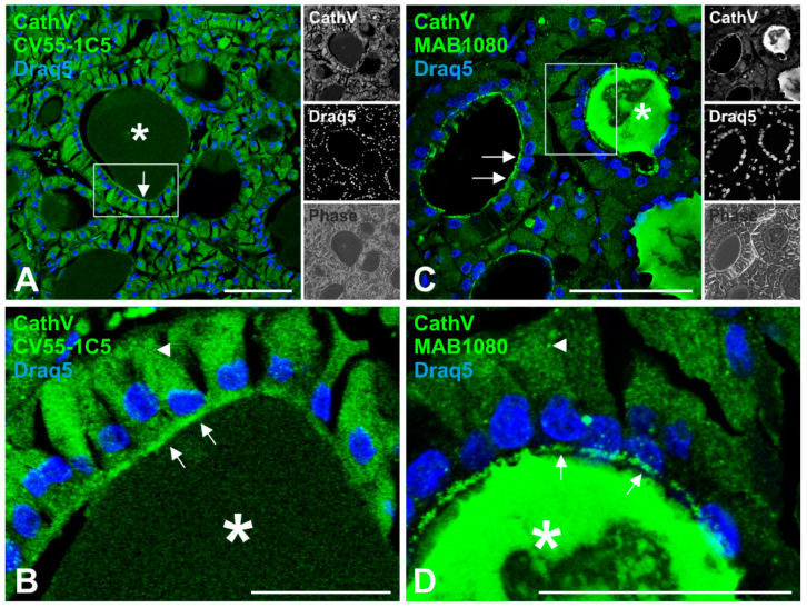
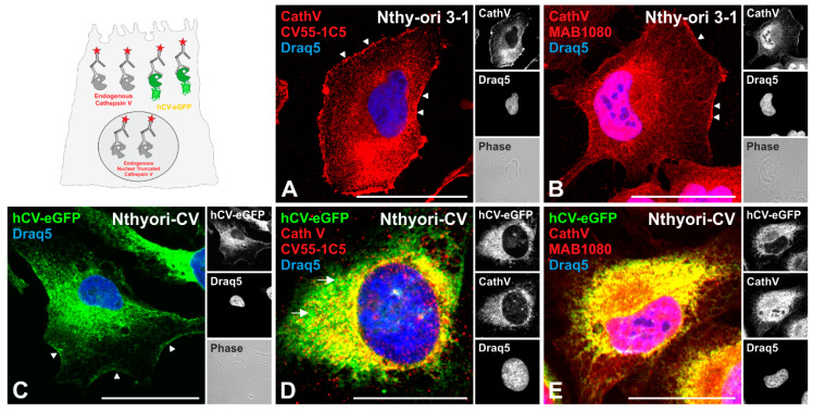
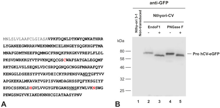
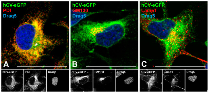
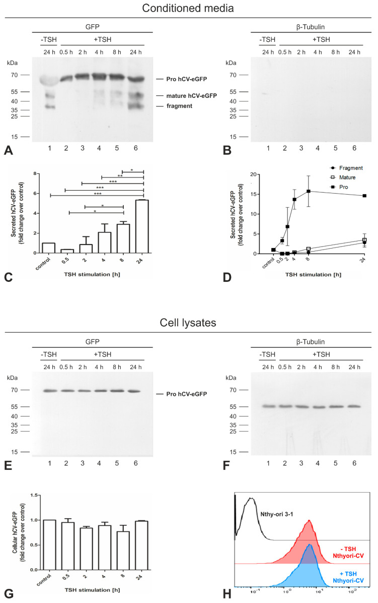
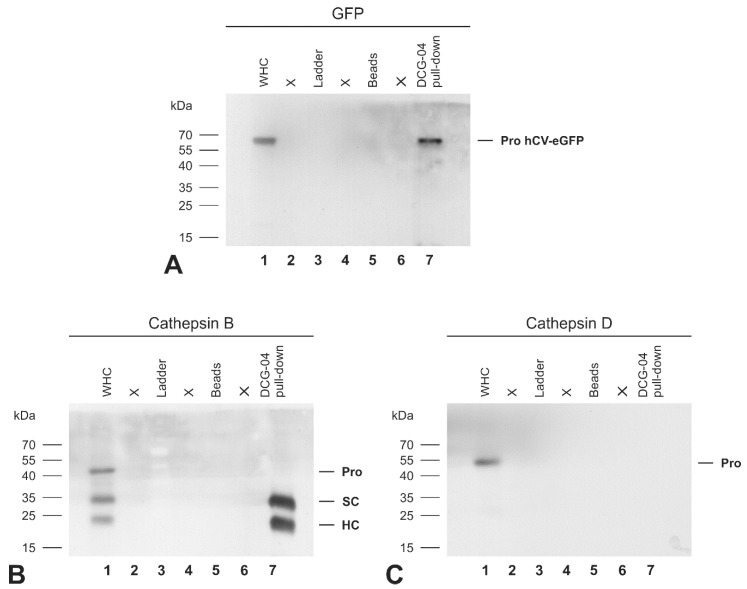
The significance of cysteine cathepsins for the liberation of thyroid hormones from the precursor thyroglobulin was previously shown by in vivo and in vitro studies. Cathepsin L is most important for thyroglobulin processing in mice. The present study aims at specifying the possible contribution of its closest relative, cysteine cathepsin L2/V, to thyroid function. Immunofluorescence analysis on normal human thyroid tissue revealed its predominant localization at the apical plasma membrane of thyrocytes and within the follicle lumen, indicating the secretion of cathepsin V and extracellular tasks rather than its acting within endo-lysosomes. To explore the trafficking pathways of cathepsin V in more detail, a chimeric protein consisting of human cathepsin V tagged with green fluorescent protein (GFP) was stably expressed in the Nthy-ori 3-1 thyroid epithelial cell line. Colocalization studies with compartment-specific markers and analyses of post-translational modifications revealed that the chimeric protein was sorted into the lumen of the endoplasmic reticulum and subsequently transported to the Golgi apparatus, while being N-glycosylated. Immunoblotting showed that the chimeric protein reached endo-lysosomes and it became secreted from the transduced cells. Astonishingly, thyroid stimulating hormone (TSH)-induced secretion of GFP-tagged cathepsin V occurred as the proform, suggesting that TSH upregulates its transport to the plasma membrane before it reaches endo-lysosomes for maturation. The proform of cathepsin V was found to be reactive with the activity-based probe DCG-04, suggesting that it possesses catalytic activity. We propose that TSH-stimulated secretion of procathepsin V is the default pathway in the thyroid to enable its contribution to thyroglobulin processing by extracellular means.

Keywords: cysteine cathepsins; green fluorescent protein tagging; protein trafficking; secretion; thyroid epithelial cells; thyroid stimulating hormone.


Conflict of interest statement

The authors declare no conflict of interest. The funders had no role in the design of the study; in the collection, analyses, or interpretation of data; in the writing of the manuscript, or in the decision to publish the results.

Figures

Figure 1
Figure 1
Immunohistochemistry of cysteine cathepsin V in human thyroid tissue. Human thyroid tissue sections were immunolabeled with different cathepsin V-specific antibodies (green signals), namely, anti-human cathepsin V CV55 1C5 (A,B) that exclusively immunoreacts with the proform of cathepsin V, and anti-human cathepsin V MAB1080 (C,D) recognizing both the pro- and mature cathepsin V forms. Nuclei were counter-stained with Draq5™ (blue signals). Single-channel fluorescence and corresponding phase contrast micrographs are depicted as indicated. Cathepsin V-immunopositive signals were detected within the cytoplasm (arrowheads), at the apical plasma membrane (arrows), and dispersed within the follicle lumen (asterisks). Rectangular boxes in A and C denote regions magnified in B and D, respectively. Scale bars represent 50 μm.
Figure 2
Figure 2
Subcellular localization of endogenous cathepsin V and eGFP-tagged full-length cathepsin V chimeric protein in thyroid epithelial cells. Non-transduced Nthy-ori 3-1 (A,B) and transduced Nthyori-CV cells (CE) were immunostained with different cathepsin V antibodies (red signals), i.e., anti-human cathepsin V CV55-1C5 that exclusively immunodetects the proform of cathepsin V (A,D) and anti-human cathepsin V MAB1080 recognizing both, the pro- and mature cathepsin V forms (B,E). Green channels (CE) represent fluorescence signals of hCV-eGFP. Yellow signals are indicative of colocalizing signals from cathepsin V antibodies and hCV-eGFP. The sketch (upper left panel) outlines this approach schematically. Nuclei were counter-stained with Draq5™ (blue signals). Pink signals are indicative of colocalizing signals from cathepsin V antibodies and nuclear Draq5™ staining. Single-channel fluorescence and corresponding phase contrast micrographs are depicted as indicated. Arrowheads denote immunoreactive and hCV-eGFP-derived signals at the cell surface, while arrows point to vesicular hCV-eGFP signals in Nthyori-CV cells. Scale bars represent 50 μm.
Figure 3
Figure 3
N-glycosylation state of hCV-eGFP chimeric protein in thyroid epithelial cell lines. (A) The amino acid sequence of human cathepsin V (UniProt accession number O60911) contains two potential N-glycosylation sites of the motif “N-X-S/T-NOT P” at positions Asn-221 and Asn-292 (underlined). The active site residues Cys-138, His-277 and Asn-301 indicative of cysteine peptidases are highlighted in red. The propeptide is indicated in gray font. (B) Immunoblots of lysates prepared from Nthy-ori 3-1 control (lane 1) or Nthyori-CV cells, treated without (lanes 2 and 4) or with (lanes 3 and 5) EndoF1 or PNGase F, were probed with anti-GFP antibodies. Molecular mass markers are indicated in the left margin. Faster migration due to glycosidase-mediated reduction in the molecular mass of the proform of hCV-eGFP chimeras was indicative of the removal of N-linked glycans.
Figure 4
Figure 4
Trafficking of eGFP-tagged full-length cathepsin V in thyroid epithelial cells. Confocal laser scanning micrographs of Nthyori-CV cells expressing hCV-eGFP chimeras ((AC), green signals) after immunolabeling with antibodies against PDI (A), GM130 (B) and Lamp1 (C) proteins residing in the ER, at the cytosolic face of Golgi cisternae and vesicles, and in endo-lysosomes, respectively (red signals). Yellow signals are indicative of co-localization. Nuclei were counter-stained with Draq5™ (blue signals). Single-channel fluorescence micrographs are depicted in the bottom panels as indicated. Scale bars represent 50 μm.
Figure 5
Figure 5
Presence of mature hCV-eGFP chimeric protein within endo-lysosomal fractions. Immunoblots of lysates prepared from whole cells (WHC, lanes 1, respectively) or endo-lysosome enriched fractions (Lyso, lanes 2, respectively) of Nthyori-CV cells were probed with anti-GFP (A), anti-cathepsin D (B), and anti-PCNA antibodies (C), respectively. Molecular mass markers are indicated in the left margins (AC). Only the proforms (pro) of hCV-eGFP and cathepsin D were detected in the whole cell lysates of Nthyori-CV cells ((A,B), lanes 1, respectively). In addition to the proform, the endo-lysosomal fractions of Nthyori-CV cells contained the processed mature form of hCV-eGFP and its degradation fragment ((A), lane 2). The molecular forms of cathepsin D, i.e., proform (Pro), intermediate, and heavy chain of two-chain cathepsin D (HC), were detected in the endo-lysosomal fractions ((B), lane 2). Immunoblotting with anti-PCNA antibodies confirmed the purity of the endo-lysosomal fractions (C).
Figure 6
Figure 6
Secretion of hCV-eGFP chimeric protein from thyroid epithelial cells. TCA precipitated proteins of 24 h conditioned media (AD) collected from confluent Nthy-ori 3-1 (lanes 1) or Nthyori-CV cell cultures (lanes 2) and the corresponding cell lysates (EH) were immunoblotted with anti-GFP (A,E), anti-cathepsin B (B,F), anti-cathepsin L (C,G), and anti-β-tubulin (D,H) antibodies. The conditioned media of Nthyori-CV cells contained mainly the processed mature form of hCV-eGFP in addition to its proform and a degradation fragment of approximately 37 kDa ((A), lane 2). Cathepsin L was found extracellularly only as proform ((C), lanes 1 and 2). hCV-eGFP was present predominantly in its proform in the lysate of Nthyori-CV cells ((E), lane 2). However, the expected molecular forms of cathepsins B and L, namely the proform (pro), single-chain (SC), and the heavy chain (HC) of the two-chain forms were detected in the lysates of both Nthy-ori 3-1 and Nthyori-CV cells ((F,G), respectively). Molecular mass markers are indicated in the left margins. No anti-GFP bands were observed in conditioned media or lysates of non-transduced Nthy-ori 3-1 cells ((A,E), lane 1), confirming specificity of the GFP-specific antibodies. Immunoblots with anti-β-tubulin antibodies (D,H) confirmed that conditioned media were not contaminated by cell debris, and that the TCA precipitates represented only the proteins secreted from the investigated cells.
Figure 7
Figure 7
TSH regulates the secretion of the proform of hCV-eGFP chimeric protein from thyroid epithelial cells. Proteins precipitated with TCA from the conditioned media of confluent Nthyori-CV cells stimulated with 100 µU/mL TSH for different time intervals, as indicated ((A,B), lanes 2–6), and corresponding cell lysates ((E,F), lanes 2–6) were separated by SDS-PAGE. The 24 h conditioned media collected from confluent Nthyori-CV cells and its respective cell lysate represented the non-stimulated control (lanes 1). After blotting, proteins were immunolabeled with anti-GFP (A,E). To verify that TCA-precipitated proteins were not contaminated with cellular debris, the same membrane was stripped and reblotted with anti-β-tubulin antibodies (B,F). No bands corresponding to β-tubulin were seen in any of the conditioned media precipitates, verifying that the secreted proteins of Nthyori CV cells exclusively were analyzed. Molecular mass markers are indicated in the left margins. The total amounts (C) and the amounts of proform and processed forms, i.e., the mature form and the derived fragment (D), of secreted hCV-eGFP chimeric protein were quantified by densitometry. The amounts of intracellular hCV-eGFP chimeric protein were also quantified and normalized to β-tubulin (G) and remained unchanged upon TSH treatment indicating unaffected de novo-biosynthesis rates. The intracellular amounts of hCV-eGFP in non-stimulated (red) and 24 h TSH stimulated (blue) Nthyori-CV cells were determined using flow cytometry (H), also revealing comparable expression in non- and TSH-stimulated cells. Fold changes were calculated in comparison with non-stimulated controls (C,D,G). Data are depicted as means + SD in (C,G), and as means ± SD for the processed forms in (D). Levels of significance were determined by one-way ANOVA, followed by Tukey post hoc tests, and are indicated as * for p < 0.05, ** for p < 0.01, and *** for p < 0.001, respectively. The experiments were repeated twice.
Figure 8
Figure 8
Detecting the proform of hCV-eGFP chimeric protein with the activity-based probe DCG-04. Lysates (WHC, lanes 1, respectively) were prepared from Nthyori-CV cells in the presence of biotinylated DCG-04, which requires an accessible active site in order to bind covalently to proteolytically active cysteine peptidase forms in a 1:1 ratio. The whole cell lysate was then incubated with streptavidin beads to pull down DCG-04-labeled proteins (DCG-04 pull down, lanes 7, respectively), representing active cysteine peptidase forms. Immunoblots were probed with anti-GFP (A), anti-cathepsin B (B), and anti-cathepsin D (C) antibodies. Molecular mass markers are indicated in the left margins. It was found that the active site of the proform of the hCV-eGFP chimeric protein was fully accessible for binding to DCG-04, indicating its proteolytic activity ((A), lane 7). The whole cell lysate contained the expected molecular forms of cathepsin B, i.e., proform (pro), single chain (SC), and the heavy chain (HC) of the two-chain form ((B), lane 1), while only the mature forms of cathepsin B were detected in the DCG-04 pull down ((B), lane 7), verifying its exclusive binding to the active site. Procathepsin D was detected in the whole cell lysate ((C), lane 1), but not in the DCG-04 pull down ((C), lane 7), verifying the specificity of the activity-based probe DCG-04 for cysteine peptidases.

Similar articles

Cited by

References

    1. Brömme D., Li Z., Barnes M., Mehler E. Human cathepsin V functional expression, tissue distribution, electrostatic surface potential, enzymatic characterization, and chromosomal localization. Biochemistry. 1999;38:2377–2385. doi: 10.1021/bi982175f. - DOI - PubMed
    1. Rossi A., Deveraux Q., Turk B., Sali A. Comprehensive search for cysteine cathepsins in the human genome. Biol. Chem. 2004;385:363–372. doi: 10.1515/BC.2004.040. - DOI - PubMed
    1. Watkinson A. Stratum corneum thiol protease (SCTP): A novel cysteine protease of late epidermal differentiation. Arch. Dermatol. Res. 1999;291:260–268. doi: 10.1007/s004030050406. - DOI - PubMed
    1. Adachi W., Kawamoto S., Ohno I., Nishida K., Kinoshita S., Matsubara K., Okubo K. Isolation and characterization of human cathepsin V: A major proteinase in corneal epithelium. Investig. Ophthalmol. Vis. Sci. 1998;39:1789–1796. - PubMed
    1. Santamaría I., Velasco G., Cazorla M., Fueyo A., Campo E., López-Otín C. Cathepsin L2, a novel human cysteine proteinase produced by breast and colorectal carcinomas. Cancer Res. 1998;58:1624–1630. - PubMed

LinkOut - more resources