Review
. 2020 Nov 22;8(11):527.
doi: 10.3390/biomedicines8110527.

Aptamers for Proteins Associated with Rheumatic Diseases: Progress, Challenges, and Prospects of Diagnostic and Therapeutic Applications

Affiliations
Review

Aptamers for Proteins Associated with Rheumatic Diseases: Progress, Challenges, and Prospects of Diagnostic and Therapeutic Applications

Elizaveta A Shatunova et al. Biomedicines. .

Abstract

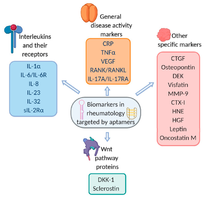
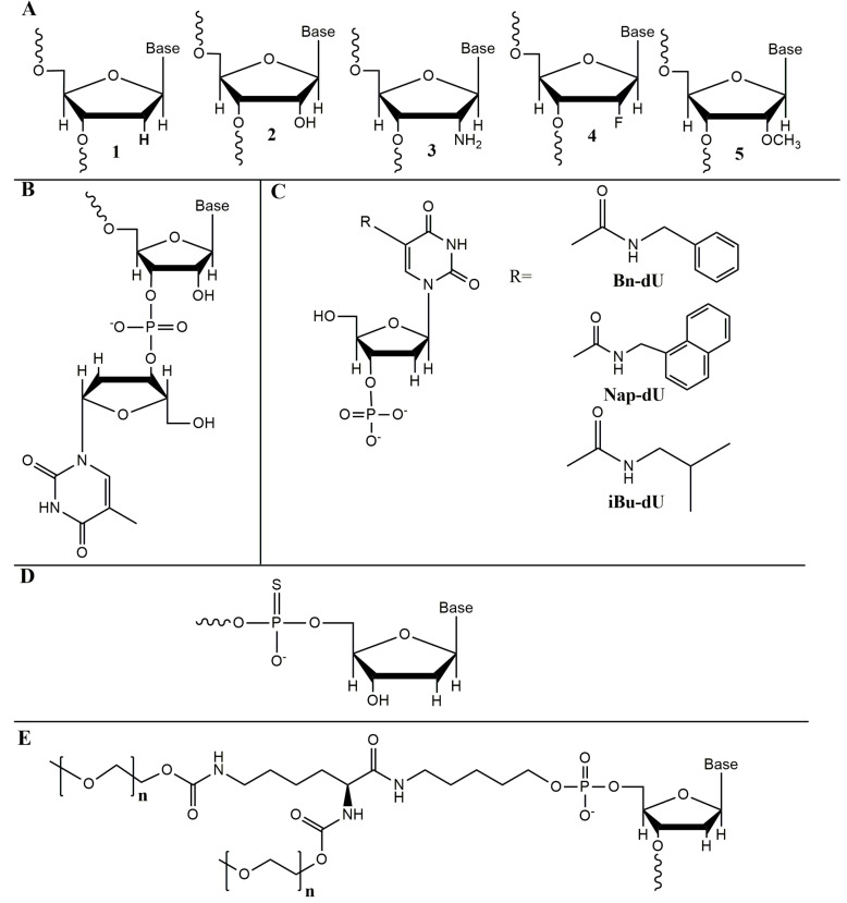
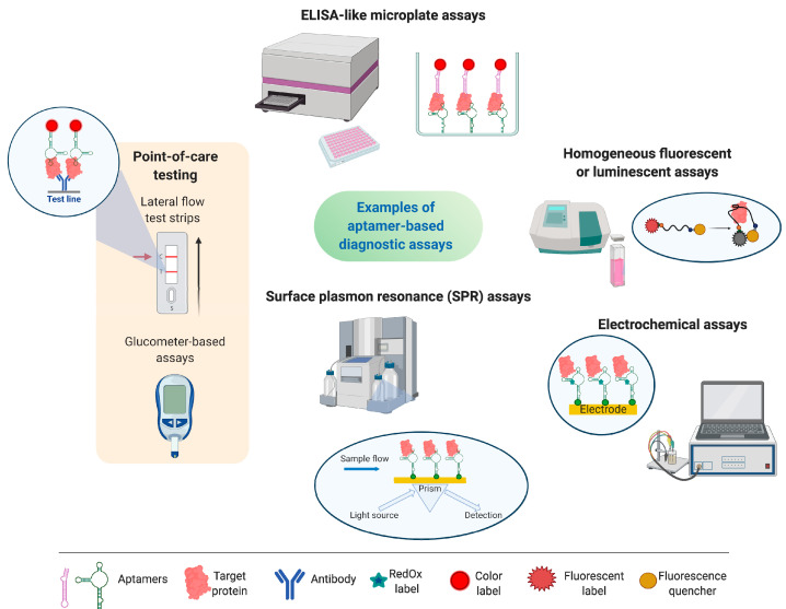
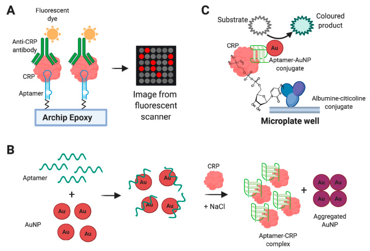
Nucleic acid aptamers capable of affine and specific binding to their molecular targets have now established themselves as a very promising alternative to monoclonal antibodies for diagnostic and therapeutic applications. Although the main focus in aptamers' research and development for biomedicine is made on cardiovascular, infectious, and malignant diseases, the use of aptamers as therapeutic or diagnostic tools in the context of rheumatic diseases is no less important. In this review, we consider the main features of aptamers that make them valuable molecular tools for rheumatologists, and summarize the studies on the selection and application of aptamers for protein biomarkers associated with rheumatic diseases. We discuss the progress in the development of aptamer-based diagnostic assays and targeted therapeutics for rheumatic disorders, future prospects in the field, and issues that have yet to be addressed.

Keywords: aptamer therapeutics; aptamers; aptasensors; protein biomarkers; rheumatic diseases.


Conflict of interest statement

The authors declare no conflict of interest. The funders had no role in the design of the study; in the collection, analyses, or interpretation of data; in the writing of the manuscript, or in the decision to publish the results.

Figures

Figure 1
Figure 1
Schematic representation of aptamer’s folding and target recognition, and the main advantages of nucleic acid aptamers.
Figure 2
Figure 2
Protein biomarkers in rheumatology currently targeted by aptamers.
Figure 3
Figure 3
Chemical modifications employed for the aptamers under review. (A) Natural deoxyribo (1) and ribonucleosides (2), 2′-amino-2′-deoxyribonucleoside (3), 2′-fluoro-2′-deoxyribonucleotide (4), and 2′-O-methylribonucleotide (5). (B) An “inverted” 3′-thymidine attached by 3′-3′-phosphodiester linkage. (C) Examples of hydrophobic modifications of heterocyclic bases used in SOMAmers [29]. (D) Phosphorothioate analogs of oligodeoxyribonucleotides. (E) 5′-PEG-modified (PEGylated) aptamer.
Figure 4
Figure 4
Examples of aptamer-based diagnostic assays and corresponding devices.
Figure 5
Figure 5
Examples of aptasensors for C-reactive protein: aptamer-based chip for fluorescent sandwich immunoassay (A) [111], colorimetric assay based on AuNPs aggregation (B) [112], and ELISA-like system employing citicoline for CRP capture and peroxidase-mimicking AuNPs [113] (C).
Figure 6
Figure 6
FRET-based optical aptasensor for TNFα based on the VR11 aptamer [122].
Figure 7
Figure 7
Aptasensing systems for VEGF: colorimetric aptasensor based on strand displacement amplification principle [127] (A), aptazyme-based system [129] (B), and aptasensor employing two concatemeric oligonucleotides and glucose oxidase [131] (C).
Figure 8
Figure 8
Aptasensor for IL-6 detection based on the sandwich pair of aptamers and gold nanoparticles [136].
Figure 9
Figure 9
Schematic representation of the aptamer/antibody based fluorescent IL-8 detection with signal enhancement by rolling circle amplification [93].
Figure 10
Figure 10
CTGF aptasensing system based on biolayer interferometry and enzyme-linked aptamer sandwich assay [97]. The precipitation of non-soluble substrate crystals at the final step causes a significant spectral shift and amplifies the detection signal.
Figure 11
Figure 11
Lateral flow aptasensor for osteopontin detection based on aptamer/antibody sandwich pair and gold nanoparticles [139].
Figure 12
Figure 12
Aptasensors for HNE detection: fluorescent aptasensor employing molecular beacon (A) [140] and colorimetric ELISA-like system with a chromogenic peptide substrate [141] (B).

References

    1. Tuerk C., Gold L. Systematic evolution of ligands by exponential enrichment: RNA ligands to bacteriophage T4 DNA polymerase. Science. 1990;249:505–510. - PubMed
    1. Robertson D.L., Joyce G.F. Selection in vitro of an RNA enzyme that specifically cleaves single-stranded DNA. Nature. 1990;344:467–468. doi: 10.1038/344467a0. - DOI - PubMed
    1. Ellington A.D., Szostak J.W. In vitro selection of RNA molecules that bind specific ligands. Nature. 1990;346:818–822. - PubMed
    1. Nimjee S.M., White R.R., Becker R.C., Sullenger B.A. Aptamers as Therapeutics. Annu. Rev. Pharmacol. Toxicol. 2017;57:61–79. doi: 10.1146/annurev-pharmtox-010716-104558. - DOI - PMC - PubMed
    1. Zhang Y., Lai B., Juhas M. Recent advances in aptamer discovery and applications. Molecules. 2019;24:941. doi: 10.3390/molecules24050941. - DOI - PMC - PubMed

LinkOut - more resources