. 2020 Dec 2;21(1):856.
doi: 10.1186/s12864-020-07233-2.

Detection of CRISPR-mediated genome modifications through altered methylation patterns of CpG islands

Affiliations

Detection of CRISPR-mediated genome modifications through altered methylation patterns of CpG islands

M Heath Farris et al. BMC Genomics. .

Abstract

Background: The development and application of CRISPR technologies for the modification of the genome are rapidly expanding. Advances in the field describe new CRISPR components that are strategically engineered to improve the precision and reliability of CRISPR editing within the genome sequence. Genome modification using induced genome breaks that are targeted and mediated by CRISPR components leverage cellular mechanisms for repair like homology directed repair (HDR) to incorporate genomic edits with increased precision.

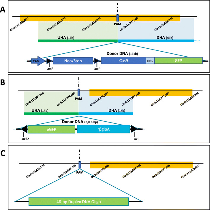
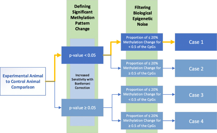
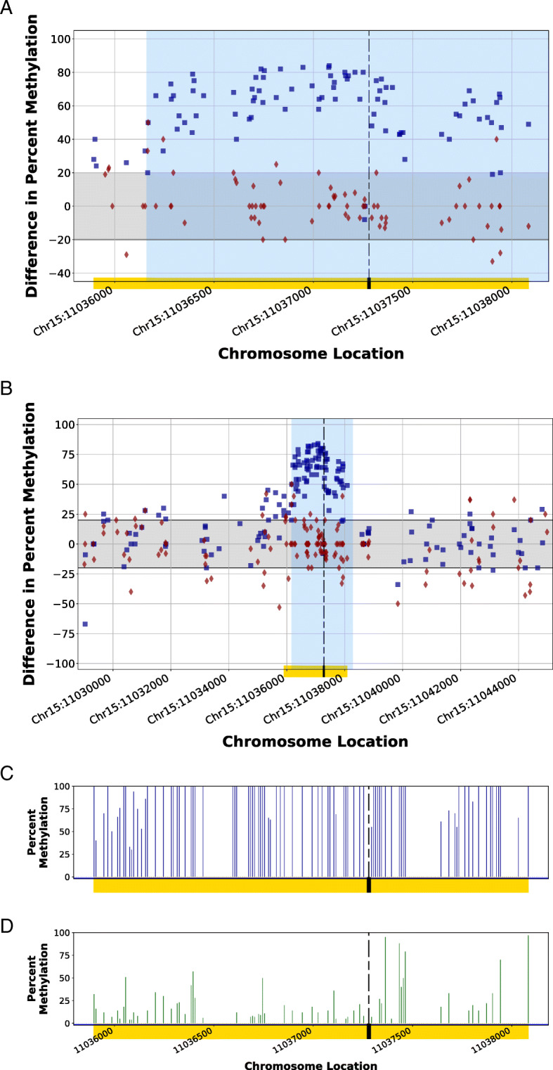
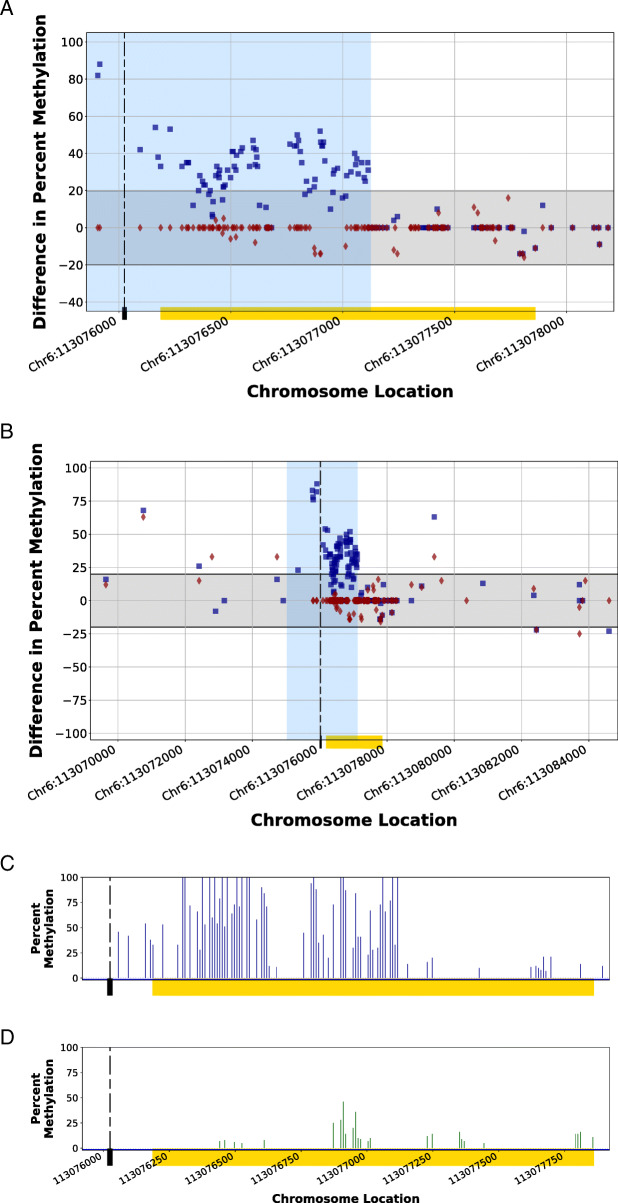
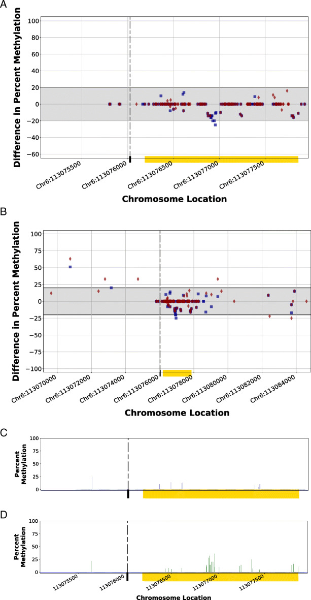
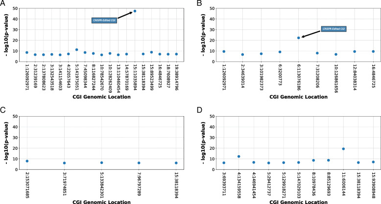
Results: In this report, we describe the gain of methylation at typically hypomethylated CpG island (CGI) locations affected by the CRISPR-mediated incorporation of donor DNA using HDR mechanisms. With characterization of CpG methylation patterns using whole genome bisulfite sequencing, these CGI methylation disruptions trace the insertion of the donor DNA during the genomic edit. These insertions mediated by homology-directed recombination disrupt the generational methylation pattern stability of the edited CGI within the cells and their cellular lineage within the animal strain, persisting across generations. Our approach describes a statistically based workflow for indicating locations of modified CGIs and provides a mechanism for evaluating the directed modification of the methylome of the affected CGI at the CpG-level.

Conclusions: With advances in genome modification technology comes the need to detect the level and persistence of methylation change that modifications to the genomic sequence impose upon the collaterally edited methylome. Any modification of the methylome of somatic or germline cells could have implications for gene regulation mechanisms governed by the methylation patterns of CGI regions in the application of therapeutic edits of more sensitively regulated genomic regions. The method described here locates the directed modification of the mouse epigenome that persists over generations. While this observance would require supporting molecular observations such as direct sequence changes or gene expression changes, the observation of epigenetic modification provides an indicator that intentionally directed genomic edits can lead to collateral, unintentional epigenomic changes post modification with generational persistence.

Keywords: CRISPR genome editing; CpG island; Epigenetic modification; Homology-directed repair; Methylation variance; Non-homologous end-joining; Statistical variance detection.


Conflict of interest statement

The authors declare that they have no competing interests.

Figures

Fig. 1
Fig. 1
Mouse strain edit sites. The methylation patterns of three CRISPR-edited animal strains were evaluated in this study, describe in detail in Table 1. Strain HDR1 (a) contains an 11-kb insert and was recombinantly inserted into the Rxfp3 locus of the mouse genome using a 1-kb upstream homology arm (UHA) and a 4-kb downstream homology arm (DHA). The protospacer adjacent motif (PAM) site was located at Chr15:11,037,280. Strain HDR2 (b) contains a 2005-bp insert and was recombinantly inserted into the Rosa26 locus of the mouse genome using a 1-kb UHA and a 1-kb DHA. The PAM site was located at Chr6:113,076,025. Strain NHEJ1 (c) contains a 48-bp duplex DNA oligonucleotide fragment incorporated into the Rosa26 locus by non-homologous end-joining. The PAM site was located at Chr6:113,076,025. For each figure, the yellow bar indicates the CGI location
Fig. 2
Fig. 2
Genome-wide sequence depth of CpG locations. WGBS was used to evaluate CpG methylation profiles within the genome of each edited and control animal. The distribution of the number of CpG calls observed is shown for HDR1 Animal (a) with 10,835,815 CpG locations with ≥5 calls, HDR2 Animal (b) with 10,109,462 CpG locations with ≥5 calls, NHEJ1 Animal (c) with 6,382,801 CpG locations with ≥5 calls, Control 1 Animal (d) with 11,948,365 CpG locations with ≥5 calls, and Control 2 Animal (e) with 8,754,364 CpG locations with ≥5 calls. The numbers of CpG locations are reported in millions (M)
Fig. 3
Fig. 3
Genomic and CRISPR-edited CGI methylation depth of call percentage. The percent of CpG calls within the respective CRISPR-edited sites for the modified animals exceeds that observed within the genome. For each call depth, the percentage of CpG calls are plotted for the CRISPR-edited CGI (light blue) as related to the genome (dark blue) for HDR1 Animal (a), HDR2 Animal (b), and NHEJ1 Animal (c)
Fig. 4
Fig. 4
Statistical filter process. The stepwise process for ranking and categorizing the CpG methylation patterns of genomic CGIs permitted the ranking of CGI changes between edited and control animal CGIs. Significantly changed CGIs followed a path (yellow bars) toward Case 1 based on significant p-value changes with Bonferroni correction and filtered beyond biological epigenetic noise
Fig. 5
Fig. 5
Methylomic difference of a completely edited CpG island within the genome of the HDR1 Animal using homology-directed repair in a CRISPR-mediated edit. Homology arms of a synthetic DNA fragment (region shaded in blue) in a homology-directed repair of a CRISPR-mediated genomic cut spanned a CpG island (CGI; Chr15:11,035,894–11,038,084; yellow bar) within the genome of HDR1 Animal, resulting in a destabilized methylation pattern (blue squares) as compared to the Control 1 Animal methylation patterns at the same location (red diamonds). Figure 4a illustrates the localized methylation variance introduced at the CGI, while Fig. 4b illustrates the comparison of the modified region to flanking endogenous genomic regions by displaying the variance of methylation patterns for 7000 bp upstream and downstream of the CGI. The methylation patterns of regions not influenced by the incorporation of donor DNA during the CRISPR edit displayed a methylation pattern similar to the control. The gray band indicates considered biological epigenetic variance (+/− 20% change). The dashed line indicates the protospacer adjacent motif (PAM) location of the targeted CRISPR cut site. Synthetic homology arms corresponded to genomic sequence 1000 bp upstream and downstream of the PAM site. The comparison of the percent methylation observed at the localized CGI region for the CRISPR-edited animal (Fig. 4c) and the unedited control animal (Fig. 4d) demonstrate the introduced methylation variance as a result of the introduction of the donor DNA. Blue squares (■) indicate the percent differences in CpG methylation for HDR1 Animal from Control 2 Animal at given chromosome locations. Red diamonds (♦) indicate the percent differences in CpG methylation for Control 1 Animal from Control 2 Animal at given chromosome locations
Fig. 6
Fig. 6
Methylomic difference of a partially edited CpG island within the genome of the HDR2 Animal using homology-directed repair in a CRISPR-mediated edit. The downstream homology arm of a synthetic DNA fragment (region shaded in blue) in a homology-directed repair of a CRISPR-mediated genomic cut spanned a portion of a CpG island (CGI; Chr6:113,076,186–113,077,861; yellow bar) within the genome of the HDR2 Animal, resulting in a destabilized methylation pattern (blue squares) as compared to the Control 1 Animal methylation patterns at the same location (red diamonds). The regions with methylation patterns modified by the incorporation of homology arms within the CGI and upstream of the CGI are shaded in a light blue background. While Fig. 5a illustrates the localized methylation variance introduced at the CGI, Fig. 5b illustrates the comparison of the modified region to flanking endogenous genomic regions by displaying the variance of methylation patterns for 7000 bp upstream and downstream of the CGI. The methylation patterns of the unmodified downstream portion of the CGI (unshaded) for the edited CGI as well as the flanking regions of endogenous genome displayed a methylation pattern similar to the control. The gray band indicates considered biological epigenetic variance (+/− 20% change). The dashed line indicates the protospacer adjacent motif (PAM) location of the targeted CRISPR cut site. The comparison of the percent methylation of the CpG sites observed at the localized CGI region for the CRISPR-edited animal (Fig. 5c) and the unedited control animal (Fig. 5d) demonstrates the introduced methylation variance as a result of the adoption of the donor DNA. Blue squares (■) indicate the percent differences in CpG methylation for HDR2 Animal from Control 2 Animal at given chromosome locations. Red diamonds (♦) indicate the percent differences in CpG methylation for Control 1 Animal from Control 2 Animal at given chromosome locations
Fig. 7
Fig. 7
Methylomic variance of a CpG island within the genome of the NHEJ1 Animal flanking a non-homologous end-joining repair in a CRISPR-mediated edit. A non-homologous end-joining repair of a CRISPR-mediated genomic cut flanking a CpG island (CGI; Chr6:113,076,186–113,077,861; yellow bar) was inserted within the genome of the NHEJ1 Animal, resulting in a methylation pattern of the edited animal (blue squares) that was similar to that of the Control 1 Animal at the same location (red diamonds) localized around the CGI edit site (Fig. 6a) as well as 7000 bp upstream and downstream of the edit (Fig. 6b). The gray band indicates considered biological epigenetic variance (+/− 20% change). The dashed line indicates the protospacer adjacent motif (PAM) location of the targeted CRISPR cut site. The comparison of the percent methylation of the CpG sites observed at the localized CGI region for the CRISPR-edited animal (Fig. 5c) and the unedited control animal (Fig. 5d) demonstrates a lack of variance in methylation pattern as a result of non-homologous end-joining insertion of the 48-bp duplex DNA oligo fragment. Blue squares (■) indicate the percent differences in CpG methylation for NHEJ1 Animal from Control 2 Animal at given chromosome locations. Red diamonds (♦) indicate the percent differences in CpG methylation for Control 1 Animal from Control 2 Animal at given chromosome locations
Fig. 8
Fig. 8
Edited CGIs affected by HDR show statistically significant p-values. In evaluating the methylation variance between CRISPR-edited and control mouse epigenomes, top CGIs for each comparison were determined by ranking of p-values observed and removing those CGIs that did not display greater than 50% of observed CpGs with methylation increases above 20%. Those top CGIs containing HDR edits had more significant p-values for methylation increase above biological variance (20% change) as compared to other CGIs across the mouse genome for HDR1 Animal (a) and HDR2 Animal (b). A CGI with a complete modification of its methylome displayed a significant increase of p-value over the control (a), while a CGI with only a partial modification displayed an increase of p-value over the control at a lesser significance (b). The top CGIs with methylation change within NHEJ1 Animal containing a NHEJ edit had no significant increase of p-value above biological variance, and the edited CGI was not among the top CGIs with methylation change (c). Comparison of CGIs in unedited Control 2 Animals displayed a low significance of change for the top CGIs (d)

References

    1. Doudna JA, Charpentier E. Genome editing. The new frontier of genome engineering with CRISPR-Cas9. Science. 2014;346(6213):1258096. doi: 10.1126/science.1258096. - DOI - PubMed
    1. Williams A, Henao-Mejia J, Flavell RA. Editing the mouse genome using the CRISPR-Cas9 system. Cold Spring Harb Protoc. 2016;2016(2):pdb top087536. doi: 10.1101/pdb.top087536. - DOI - PMC - PubMed
    1. Wang H, Yang H, Shivalila CS, Dawlaty MM, Cheng AW, Zhang F, et al. One-step generation of mice carrying mutations in multiple genes by CRISPR/Cas-mediated genome engineering. Cell. 2013;153(4):910–918. doi: 10.1016/j.cell.2013.04.025. - DOI - PMC - PubMed
    1. Hosur V, Low BE, Avery C, Shultz LD, Wiles MV. Development of humanized mice in the age of genome editing. J Cell Biochem. 2017;118(10):3043–3048. doi: 10.1002/jcb.26002. - DOI - PMC - PubMed
    1. Felgner PL, Gadek TR, Holm M, Roman R, Chan HW, Wenz M, et al. Lipofection: a highly efficient, lipid-mediated DNA-transfection procedure. Proc Natl Acad Sci U S A. 1987;84(21):7413–7417. doi: 10.1073/pnas.84.21.7413. - DOI - PMC - PubMed

LinkOut - more resources