. 2020 Dec 2;20(1):1178.
doi: 10.1186/s12885-020-07548-z.

Bladder cancer-derived interleukin-1 converts the vascular endothelium into a pro-inflammatory and pro-coagulatory surface

Affiliations

Bladder cancer-derived interleukin-1 converts the vascular endothelium into a pro-inflammatory and pro-coagulatory surface

A John et al. BMC Cancer. .

Abstract

Background: Bladder cancer cells orchestrate tumour progression by pro-inflammatory cytokines. Cytokines modulate the local tumour microenvironment and increase the susceptibility of tumour distant tissues for metastasis. Here, we investigated the impact of human bladder cancer cell derived factors on the ability to modulate and activate human vascular endothelial cells.

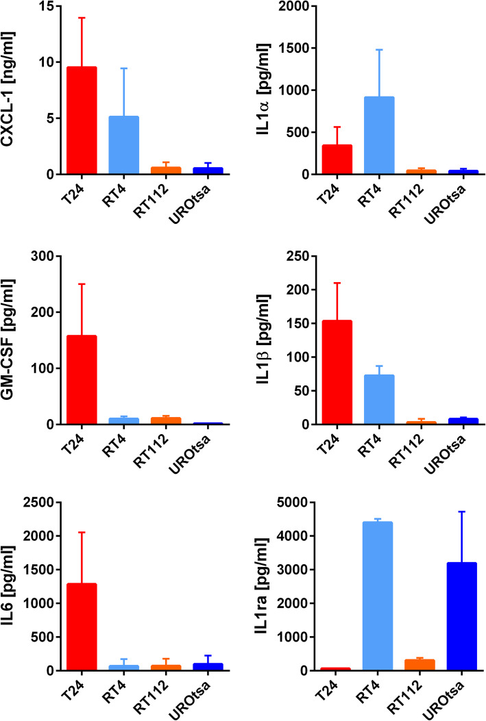
Methods: The pro-inflammatory and pro-coagulatory potential of four different bladder cancer cell lines was accessed by qRT-PCR arrays and ELISA. Modulation and activation of endothelial cells was studied in microfluidic devices. Clinical relevance of our findings was confirmed by immune histology in tissue samples of bladder cancer patients and public transcriptome data.

Results: The unbalanced ratio between interleukin (IL)-1 and IL-1 receptor antagonist (IL-1ra) in the secretome of bladder cancer cells converted the quiescent vascular endothelium into a pro-adhesive, pro-inflammatory, and pro-coagulatory surface. Microfluidic experiments showed that tumour cell induced endothelial cell activation promoted leukocyte recruitment and platelet adhesion. Human bladder cancer tissue analysis confirmed that loss of IL-1ra and elevated IL-1 expression was associated with enhanced cancer progression.

Conclusions: Our data indicate that IL-1 and IL-1ra were dysregulated in bladder cancer and could facilitate tumour dissemination through endothelial cell activation. Targeting the IL-1/IL-1ra axis might attenuate tumour-mediated inflammation and metastasis formation.

Keywords: Coagulation; Endothelial cells; Inflammation; Tumour microenvironment; von Willebrand factor.


Conflict of interest statement

The authors declare no conflict of interest.

Figures

Fig. 1
Fig. 1
Cytokine release profile of distinct UBC cell lines. ELISA measurement of cytokine release by UBC cells after 24 h cultivation in starvation medium. Except for IL1α, cytokine release was highest in T24 cells. By contrast, secretion of inhibitory IL1ra was almost absent in T24 cells. IL: Interleukin; IL1ra: IL1 receptor antagonist; CXCL-1: C-X-C motif ligand-1; GM-CSF: Granulocyte-macrophage colony-stimulating factor; n = 3
Fig. 2
Fig. 2
qRT-PCR gene profiling of HUVECs after 12 h exposure to tumour secretome. Genes promoting angiogenesis, immune cell recruitment, inflammation, cell adhesion, extracellular matrix remodelling and haemostasis (CXCL-1, IL-6, IL-8, VCAM-1, ICAM-1, VEGF-A, VEGF-C, MMP-9, TF, PAI-1) were up-regulated. Conversely, mRNA levels of genes inhibiting blood coagulation (thrombomodulin, PROCR) were reduced. Values were normalized to β-Actin (red dashed line). ICAM-1: Intercellular adhesion molecule 1, VCAM-1: vascular cell adhesion molecule 1, VEGF: Vascular Endothelial Growth Factor, MMP: Matrix metalloproteinase; n = 4 *P ≤ 0.05
Fig. 3
Fig. 3
Pro-inflammatory activation of endothelial cells after stimulation with tumour cell SN. a After incubation with T24 cell SN, CXCL-1, IL-6, IL-8, GM-CSF and PAI-1 were significantly released from HUVECs (HUVECs + T24 cell SN). The SN of UROtsa was not able to stimulate HUVECs (HUVECs + UROtsa SN). HUVECs: baseline cytokine levels produced by HUVECs; T24 and UROTsa cell SN: baseline levels of cytokine produced by the tumour cells; (n = 8–12) ** P ≤ 0.01. b Incubation of HUVECs for 6 h with T24 cell SN promoted the trans-localization of NF-kB (red) into the nucleus (blue) of the endothelial cells. Treatment of HUVECs with the SN of RT112 and UROtsa cells had no effect. RT4 cell SN induced a weak trans-localization of NF-kB. Addition of IL-1ra blocked NF-kB translocation upon treatment with T24 cell SN. The bar diagram shows the fluorescence intensity of nuclear NF-kB. Scale bar corresponds to 50 μm; (n = 10 images) ** P ≤ 0.01. c IL-1ra inhibited the T24 cell SN induced release of the indicated pro-inflammatory cytokines from HUVECs. Cytokine levels were shown in pg/ml; (n = 3) * P ≤ 0.05
Fig. 4
Fig. 4
Impact of UBC cell SN on the integrity of the endothelial cell layer. a HUVECs grown on gelatine coated ECIS electrodes were treated with starvation medium (control) or UBC cell SN. The impedance was measured continuously up to 12 h after treatment. T24 cell SN led to marked breakdown of the endothelial impedance. RT4 cell SN had a lower impact whereas RT112 cell SN, UROtsa cell SN or starvation medium (control) did not cause significant alterations after 12 h; n = 5; ** P ≤ 0.01. b HUVEC cells were incubated for 12 h with T24 cell SN (T24) or starvation medium (control) and then analysed by immunofluorescence staining of β-Actin (red) and CD31/PECAM-1 (green). In comparison to the control (left), treatment with T24 cell SN (right) induced the disintegration of adherent junctions (CD31/PECAM-1) indicating a weakening of the endothelial barrier. c Inhibition of cytokine signalling partially conserved endothelial barrier function. Antibodies against IL-6, CXCL-1 slightly mitigated impedance breakdown. In contrast, IL-1ra strongly attenuated tumour mediated endothelial dysfunction, whereas IL-8 neutralization had no impact; n = 4; ** P ≤ 0.01
Fig. 5
Fig. 5
Microfluidic experiments indicated the pro-inflammatory and pro-coagulatory activation of endothelial cells by tumour cell SN. HUVECs were perfused at 6 dyn/cm2 with hirudinated whole blood for 15 min. a, b In comparison to the SN of UROtsa cells, pre-treatment of HUVECs with T24 cell SN for 6 h promoted platelet binding (green) to gaps between the endothelial cells (white). CD31 was used as endothelial marker, vWF was used to identify platelets. (Additional experiments performed with washed and fluorescent platelets are shown in the data supplement.) b Magnified region of platelets bound to HUVECs treated withT24 cell SN. Quantitative analysis of the platelet covered area is shown as bar diagram. n = 3; * P ≤ 0.05 a, c Pre-treatment of HUVECs with T24 cell SN mediated the recruitment of leukocytes (red). c Magnified region of leukocytes attached to HUVECs treated withT24 cell SN. CD45 was used as leukocyte marker. The number of recruited leukocytes is shown as bar diagram; n = 3; * P ≤ 0.05. a-c Nuclei (blue) were stained with DAPI, scale bars correspond to 50 μm
Fig. 6
Fig. 6
Impact of IL-1 on bladder cancer progression. a IL-1ra staining intensity in a cohort of 105 patients with UBC measured semiquantitatively by IHC. Controls (benign urothelium) showed a higher IL1-ra expression than in patients with cancer. No significant difference was found between low grade and high grade tumours. The staining intensity score was defined as follows: no staining = 1, low intensity = 2, moderate intensity = 3 and high intensity = 4. * P ≤ 0.05. n.s. = not significant. Representative IHC images are shown in b-c. Scale bar corresponds to 500 μm. b IHC very low staining intensity (score = 1). c Intermediate staining intensity (score = 3). d High staining intensity (score = 4). e High IL-1β mRNA levels were linked to adverse clinicopathological features high grade disease, muscle invasiveness and tumour progression. MIBC: muscle invasive bladder cancer, NMIBC: non-muscle invasive bladder cancer; * P ≤ 0.05

References

    1. von der Maase H, Sengelov L, Roberts JT, Ricci S, Dogliotti L, Oliver T et al. Long-term survival results of a randomized trial comparing gemcitabine plus cisplatin, with methotrexate, vinblastine, doxorubicin, plus cisplatin in patients with bladder cancer. J Clin Oncol. 2005;23(21):4602–8. - PubMed
    1. Felsenstein KM, Theodorescu D. Precision medicine for urothelial bladder cancer: update on tumour genomics and immunotherapy. Nat Rev Urol. 2017; nrurol. 2017.2179. - PubMed
    1. Lima LG, Monteiro RQ. Activation of blood coagulation in cancer: implications for tumour progression. Biosci Rep. 2013;33(5):e00064. doi: 10.1042/BSR20130057. - DOI - PMC - PubMed
    1. Young A, Chapman O, Connor C, Poole C, Rose P, Kakkar AK. Thrombosis and cancer. Nat Rev Clin Oncol. 2012;9(8):437–49. - PubMed
    1. Kalagara T, Moutsis T, Yang Y, Pappelbaum KI, Farken A, Cladder-Micus L, et al. The endothelial glycocalyx anchors von Willebrand factor fibers to the vascular endothelium. Blood Adv. 2018;2(18):2347–2357. doi: 10.1182/bloodadvances.2017013995. - DOI - PMC - PubMed

MeSH terms