. 2020 Dec 2;17(1):368.
doi: 10.1186/s12974-020-02016-8.

Electrostimulation of the carotid sinus nerve in mice attenuates inflammation via glucocorticoid receptor on myeloid immune cells

Affiliations

Electrostimulation of the carotid sinus nerve in mice attenuates inflammation via glucocorticoid receptor on myeloid immune cells

Aidan Falvey et al. J Neuroinflammation. .

Abstract

Background: The carotid bodies and baroreceptors are sensors capable of detecting various physiological parameters that signal to the brain via the afferent carotid sinus nerve for physiological adjustment by efferent pathways. Because receptors for inflammatory mediators are expressed by these sensors, we and others have hypothesised they could detect changes in pro-inflammatory cytokine blood levels and eventually trigger an anti-inflammatory reflex.

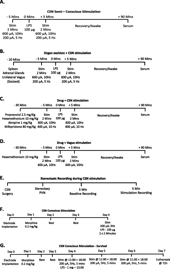
Methods: To test this hypothesis, we surgically isolated the carotid sinus nerve and implanted an electrode, which could deliver an electrical stimulation package prior and following a lipopolysaccharide injection. Subsequently, 90 min later, blood was extracted, and cytokine levels were analysed.

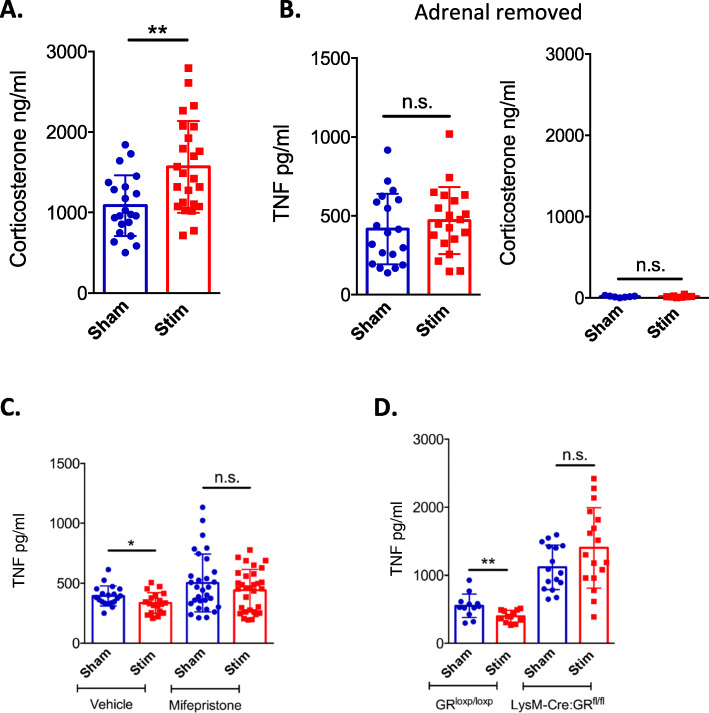
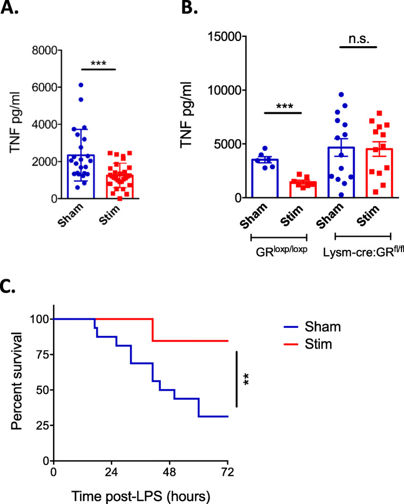
Results: Here, we found that carotid sinus nerve electrical stimulation inhibited lipopolysaccharide-induced tumour necrosis factor production in both anaesthetised and non-anaesthetised conscious mice. The anti-inflammatory effect of carotid sinus nerve electrical stimulation was so potent that it protected conscious mice from endotoxaemic shock-induced death. In contrast to the mechanisms underlying the well-described vagal anti-inflammatory reflex, this phenomenon does not depend on signalling through the autonomic nervous system. Rather, the inhibition of lipopolysaccharide-induced tumour necrosis factor production by carotid sinus nerve electrical stimulation is abolished by surgical removal of the adrenal glands, by treatment with the glucocorticoid receptor antagonist mifepristone or by genetic inactivation of the glucocorticoid gene in myeloid cells. Further, carotid sinus nerve electrical stimulation increases the spontaneous discharge activity of the hypothalamic paraventricular nucleus leading to enhanced production of corticosterone.

Conclusion: Carotid sinus nerve electrostimulation attenuates inflammation and protects against lipopolysaccharide-induced endotoxaemic shock via increased corticosterone acting on the glucocorticoid receptor of myeloid immune cells. These results provide a rationale for the use of carotid sinus nerve electrostimulation as a therapeutic approach for immune-mediated inflammatory diseases.

Keywords: Bioelectronic medicine; Carotid body; Carotid sinus nerve; Corticosterone; Electrostimulation; Immunology.


Conflict of interest statement

The authors and this manuscript have no conflict of interest nor competing interests.

Figures

Fig. 1
Fig. 1
Timelines for experimental protocols. Eight to 10-week-old C57BL/6 mice were used in various experimental protocols. Prior to CSN surgery, some experiments had organ/nerve excisions (b), and in others, blocking drugs were administered (c, d). In all instances with recovery, mice recovered in a heated cage (ad, f, g). LPS was administered IP (200 μl) in all instances at 100 μg (ad, f, except g) when a lethal dose of LPS was used (1 mg–200 μl). When stimulation was required (ag), it was administered at 600 μA + 10 Hz, 400 μA + 10 Hz or 200 μA + 5 Hz and 0.1 ms in all instances. Stimulation occurred during unconsciousness (ae) and consciousness (f, g). Experiments were typically conducted at least twice with an n number of 6–8
Fig. 2
Fig. 2
Electrostimulation of the CSN increases breath rate and decreases arterial blood pressure in mice. Eight to 10-week-old C57BL/6 mice were obtained, and CSN isolation surgery was performed. a Breath rate was recorded for 5 min, and the average breath per minute was calculated at baseline, during stimulation (200 μA, 5 Hz) and immediately after stimulation. Breath rate in mice before, during and after stimulation. Each individual point represents an animal, and data is expressed as means ± SD. b The mean carotid pressure recorded in anaesthetised mice (n = 3) before and during CSN electrostimulation with either 1 mA (left), 600 μA (middle) or 200 μA (right) current amplitude. Traces are averaged values
Fig. 3
Fig. 3
CSN electrostimulation attenuates inflammation independently of the vagus nerve. ag Eight to 10-week-old C57BL/6 mice were anaesthetised; CSN was isolated and either cut (g) of left intact (af). Electrical stimulation was applied at 600 μA, 10 Hz (ae) or 200 μA, 5 Hz (f, g) 5 min before and after IP LPS injection (100 μg). Blood was collected 90 min after LPS injection for serum analysis by Meso Scale Discovery (ad) or ELISA (eg). Impact of electrical activation of the CSN on a LPS-induced serum TNF levels, b IL-1β, c IL-6 and d IL-12p70. e Impact of electrical activation of the CSN on LPS-induced serum TNF levels in the presence of oil. f Impact of unilateral left vagal removal on LPS-induced serum TNF levels following left CSN electrostimulation. All individual points represent one animal, and data is expressed as means ± SD. g Impact of afferent CSN stimulation on LPS-induced serum TNF levels
Fig. 4
Fig. 4
Attenuation of inflammation mediated via CSN stimulation does not utilise the vagal anti-inflammatory reflex. Eight to 10-week-old C57BL/6 mice were obtained, and vagus nerve (a) or CSN (be) isolation surgery was performed, followed by electrical stimulation 200–600 μA, 5–10 Hz. Thirty minutes prior to surgery, sham vehicle (PBS), hexamethonium (10 mg/kg) (a, c), atropine (1 mg/kg) (b) or propranolol (2.5 mg/kg) (a, d) were administered IP. Blood was collected 90 min after an IP LPS injection (100 μg) for serum analysis by ELISA. e The spleen was surgically removed, and the CSN electrostimulation was applied or not. LPS-induced serum TNF release was evaluated by ELISA. ae Each individual point represents an animal, and data is expressed as means ± SD
Fig. 5
Fig. 5
CSN electrostimulation attenuates inflammation via glucocorticoids signalling in myeloid cells. C57BL/6 mice (ac) or LysM-Cre:GRfl/fl mice (d) were obtained, aged 8–10 weeks, and CSN isolation and stimulation (600 μA, 10 Hz) were conducted. An LPS IP injection (100 μg) was administered in all animals; 90 min later, blood was collected for serum analysis by assay. a Corticosterone was measured in sham and CSN electrostimulated animals. b Impact of bilateral adrenal gland removal prior to CSN isolation and stimulation on LPS-induced serum TNF levels and serum corticosterone levels. c Impact of mifepristone (80 mg/kg) or vehicle (10% DMSO) IP administration prior to CSN isolation and stimulation on LPS-induced serum TNF levels. d LysM-Cre:GRfl/fl mice and their littermate controls, GRloxp/loxp, were obtained, and both received electrostimulation of the CSN. LPS-induced serum TNF levels were measured by ELISA. All individual traces, or points, represent one animal. The means are represented as ± SD
Fig. 6
Fig. 6
Increased PVN activity during CSN stimulation. Eight to 10-week-old C57BL/6 mice were obtained, and the CSN was isolated and stimulated (600 μA, 10 Hz). Electrical activity recordings of the PVN of the hypothalamus were taken using a stereotaxic frame. Impact of CSN electrostimulation on PVN activity was evaluated. a A representative trace of activity in the PVN during CSN stimulation. b The average discharge was acquired per minute for baseline recordings and during stimulation. All individual traces or points represent one animal. The means are represented as ± SD. c After recording, the frontal plane of a representative brain was injected with dye to show the location of the recording electrode and a zoomed in image of the PVN. Both images shown are representative of the whole
Fig. 7
Fig. 7
Conscious CSN stimulation is protective against LPS-induced shock. Eight to 12-week-old wild-type and transgenic C57BL/6 mice underwent surgery 4 days prior to stimulation (ac). a, b Stimulation (200 μA, 5 Hz, 2 × 2 min) was applied on freely moving mice, and LPS (5 mg/kg) was injected IP. All individual points represent one animal, and the means are expressed as ± SD. a Impact of CSN electrostimulation in conscious animals on LPS-induced serum TNF levels. b LysM-Cre:GRfl/fl mice and littermate controls underwent the same procedure as in a. c Wild-type C57BL/6 mice were implanted with an electrode on their CSN and stimulated (n = 16, 200 μA, 5 Hz, 5 min) or not (n = 13–16) twice a day for the next 3 days. A lethal dose of LPS (20 mg/kg) was administered to the mice (1 mg) IP. Animal survival was monitored

References

    1. Bradley JR. TNF-mediated inflammatory disease. J Pathol. 2008;214:149–160. doi: 10.1002/path.2287. - DOI - PubMed
    1. Pavlov VA, et al. The cholinergic anti-inflammatory pathway: a missing link in neuroimmunomodulation. Mol Med. 2003;9:125–134. doi: 10.1007/BF03402177. - DOI - PMC - PubMed
    1. Turnbull AV, Rivier CL. Regulation of the hypothalamic-pituitary-adrenal axis by cytokines: actions and mechanisms of action. Physiol Rev. 1999;79:1–71. doi: 10.1152/physrev.1999.79.1.1. - DOI - PubMed
    1. Andersson U, Tracey KJ. Neural reflexes in inflammation and immunity. J Exp Med. 2012;209:1057–1068. doi: 10.1084/jem.20120571. - DOI - PMC - PubMed
    1. Bonaz B, et al. Chronic vagus nerve stimulation in Crohn’s disease: a 6-month follow-up pilot study. Neurogastroenterol Motil. 2016;28:948–953. doi: 10.1111/nmo.12792. - DOI - PubMed

Substances

LinkOut - more resources