. 2020 Dec 2;6(49):eabc9207.
doi: 10.1126/sciadv.abc9207. Print 2020 Dec.

Histone H3.3 beyond cancer: Germline mutations in Histone 3 Family 3A and 3B cause a previously unidentified neurodegenerative disorder in 46 patients

Laura Bryant  1 Dong Li  1 Samuel G Cox  2 Dylan Marchione  3 Evan F Joiner  4 Khadija Wilson  3 Kevin Janssen  3 Pearl Lee  5 Michael E March  1 Divya Nair  1 Elliott Sherr  6 Brieana Fregeau  6 Klaas J Wierenga  7 Alexandrea Wadley  7 Grazia M S Mancini  8 Nina Powell-Hamilton  9 Jiddeke van de Kamp  10 Theresa Grebe  11 John Dean  12 Alison Ross  12 Heather P Crawford  13 Zoe Powis  14 Megan T Cho  15 Marcia C Willing  16 Linda Manwaring  16 Rachel Schot  8 Caroline Nava  17   18 Alexandra Afenjar  19 Davor Lessel  20   21 Matias Wagner  22   23   24 Thomas Klopstock  25   26   27 Juliane Winkelmann  22   24   27   28 Claudia B Catarino  25 Kyle Retterer  15 Jane L Schuette  29 Jeffrey W Innis  29 Amy Pizzino  30   31 Sabine Lüttgen  32 Jonas Denecke  32 Tim M Strom  22   24 Kristin G Monaghan  15 DDD StudyZuo-Fei Yuan  3 Holly Dubbs  30   31 Renee Bend  33 Jennifer A Lee  33 Michael J Lyons  33 Julia Hoefele  24 Roman Günthner  34   35 Heiko Reutter  36 Boris Keren  18 Kelly Radtke  37 Omar Sherbini  30   31 Cameron Mrokse  37 Katherine L Helbig  37 Sylvie Odent  38 Benjamin Cogne  39   40 Sandra Mercier  39   40 Stephane Bezieau  39   40 Thomas Besnard  39   40 Sebastien Kury  39   40 Richard Redon  40 Karit Reinson  41   42 Monica H Wojcik  43   44 Katrin Õunap  41   42 Pilvi Ilves  45 A Micheil Innes  46 Kristin D Kernohan  47   48 Care4Rare Canada ConsortiumGregory Costain  49 M Stephen Meyn  49   50 David Chitayat  49   51 Elaine Zackai  52 Anna Lehman  53 Hilary Kitson  54 CAUSES StudyMartin G Martin  55   56 Julian A Martinez-Agosto  57   58 Undiagnosed Diseases NetworkStan F Nelson  57   59 Christina G S Palmer  57   60 Jeanette C Papp  57 Neil H Parker  61 Janet S Sinsheimer  62 Eric Vilain  63 Jijun Wan  57 Amanda J Yoon  57 Allison Zheng  57 Elise Brimble  64 Giovanni Battista Ferrero  65 Francesca Clementina Radio  66 Diana Carli  65 Sabina Barresi  66 Alfredo Brusco  67 Marco Tartaglia  66 Jennifer Muncy Thomas  68 Luis Umana  69 Marjan M Weiss  10 Garrett Gotway  69 K E Stuurman  8 Michelle L Thompson  70 Kirsty McWalter  15 Constance T R M Stumpel  71 Servi J C Stevens  71 Alexander P A Stegmann  71 Kristian Tveten  72 Arve Vøllo  73 Trine Prescott  72 Christina Fagerberg  74 Lone Walentin Laulund  75 Martin J Larsen  74 Melissa Byler  76 Robert Roger Lebel  76 Anna C Hurst  77 Joy Dean  77 Samantha A Schrier Vergano  78 Jennifer Norman  79 Saadet Mercimek-Andrews  49 Juanita Neira  80 Margot I Van Allen  53   81 Nicola Longo  82 Elizabeth Sellars  83 Raymond J Louie  33 Sara S Cathey  33 Elly Brokamp  84 Delphine Heron  18 Molly Snyder  85 Adeline Vanderver  30   31 Celeste Simon  4 Xavier de la Cruz  86   87 Natália Padilla  86 J Gage Crump  2 Wendy Chung  88 Benjamin Garcia  2   3 Hakon H Hakonarson  1 Elizabeth J Bhoj  89
Affiliations

Histone H3.3 beyond cancer: Germline mutations in Histone 3 Family 3A and 3B cause a previously unidentified neurodegenerative disorder in 46 patients

Laura Bryant et al. Sci Adv. .

Abstract

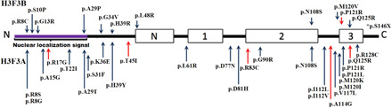
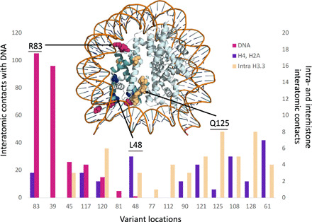
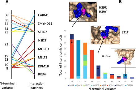
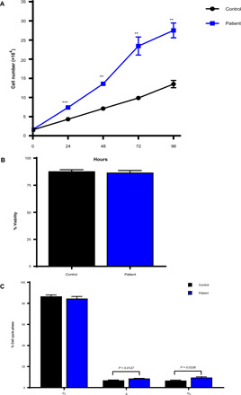
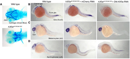
Although somatic mutations in Histone 3.3 (H3.3) are well-studied drivers of oncogenesis, the role of germline mutations remains unreported. We analyze 46 patients bearing de novo germline mutations in histone 3 family 3A (H3F3A) or H3F3B with progressive neurologic dysfunction and congenital anomalies without malignancies. Molecular modeling of all 37 variants demonstrated clear disruptions in interactions with DNA, other histones, and histone chaperone proteins. Patient histone posttranslational modifications (PTMs) analysis revealed notably aberrant local PTM patterns distinct from the somatic lysine mutations that cause global PTM dysregulation. RNA sequencing on patient cells demonstrated up-regulated gene expression related to mitosis and cell division, and cellular assays confirmed an increased proliferative capacity. A zebrafish model showed craniofacial anomalies and a defect in Foxd3-derived glia. These data suggest that the mechanism of germline mutations are distinct from cancer-associated somatic histone mutations but may converge on control of cell proliferation.


Figures

Fig. 1
Fig. 1. H3F3A mutations (NM_002107.4) and H3F3B mutations (NM_005324.4).
N, 1, 2, and 3 refer to the N-Helix, Helix-1, Helix-2, and Helix-3 of H3.3, respectively. Upper mutations are encoded by H3F3B, and lower mutations are encoded by H3F3A. Red arrows indicate mutations found in two or more unrelated patients. A few variants, p.N108S, p.P121R, and p.Q125R, were found in the same position in both H3F3A and H3F3B. *p.S146X is only present in an H3F3B alternate transcript not shown here.
Fig. 2
Fig. 2. Variant locations in the nucleosome.
At the top of the figure, we show the structure of the nucleosome with the H3.3 variants identified with spheres; the H3.3 monomer carrying them is colored in dark emerald green. The coloring of the variants reflects the predominating interactions at each location: DNA binding (magenta), intramonomer contacts (light orange), and contacts with other histones (dark blue). The same color code is used in the histogram below the structure, where we show the amount of the three interaction types at each location. Note that we use different y axis for these interactions: The y axis to the left corresponds to the H3.3-DNA binding contacts (magenta bars), and the y axis to the right corresponds to the intramonomer (light orange bars) and intermonomer contacts (dark blue bars).
Fig. 3
Fig. 3. Variant locations in H3.3-epigenetic regulator complexes.
Variant locations could be mapped to the experimental structure of different complexes involving H3.3. In (A), we show the variants mapped and the gene names of the H3.3 partners in the corresponding complex. The same color is used for the lines originating in the same variant These complexes include CARM1 (Coactivator Associated Arginine Methyltransferase 1), ZMYND11, SETD2, NSD3 (Histone-lysine N-methyltransferase NSD3), MORC3 (MORC Family CW-Type Zinc Finger 3), MLLT3 (MLLT3 Super Elongation Complex Subunit), KDM1B (Lysine Demethylase 1B), and BRD4. In (B), we show the total amount of interatomic interactions at each location, for each H3.3-epigenetic regulator complex. To help interpretation, we give three examples where we can see the histone tail (blue spheres) interacting with its partner (continuous surface in light orange); the histone residue at the variant location is shown in magenta.
Fig. 4
Fig. 4. qMS analysis of patient samples.
(A) Average profile of PTMs on canonical histones H3 and H4 across control lymphoblasts. Error bars represent SD (n = 9 donors; 3 biological replicates each). (B) Tukey boxplot depicting the coefficients of variation of 73 modified histone H3 and H4 peptides detected by nano–LC-MS/MS (biological variance: across all 14 donors; patient variance: across five patients; control variance: across nine controls). (C) Average histone H3.3 protein abundance (relative to total histone H3) in patient and control lymphoblasts. Error bars represent SD. (D) Volcano plot demonstrating significantly altered histone PTMs in patients versus controls. Dotted line represents P < 0.05 significance threshold. (E) K9 and K14 PTM abundances were compared between (i) protein transcribed from the mutant p.A15G allele from patient cells, (ii) protein transcript from the wild-type (WT) allele from the same patient cells, and (iii) protein transcribed from the WT alleles from a control. Note that the peptide from amino acids 9 to 17 is indistinguishable between canonical H3 and H3.3, so the WT peptide encompasses both. (F) A29P is the only mutation occurring on the same peptide that distinguishes H3.3 from H3. PTMs that fall on this peptide are compared across the mutant peptide, the WT peptide from the mutant sample, and the average profile of the peptide from control samples. *P < 0.05, **P < 0.01, and ***P < 0.001. This shows notable local deregulation of PTMs on the mutant peptide. qMS, quantitative mass spectrometry.
Fig. 5
Fig. 5. Cellular dynamics of patient fibroblasts.
(A) Five H3F3A/B patient fibroblast lines (H3F3B: p.G34V; H3F3A: p.R17G; H3F3A: p.G90R; H3F3A: p.T45I; and H3F3B: p.V117V or p.S146X in alternate transcript) demonstrated increased proliferation over six matched controls. **P < 0.005 and ***P < 0.0005. Data represent means ± SEM of three biological replicates using three technical replicates each. (B) The same five H3F3A/B patient fibroblasts and six controls show no major differences in cell viability. The data represent the means ± SEM of four biological replicates using two technical replicates each. (C) Cell cycle analysis showed differences in the S (P = 0.0127) and G2 (P = 0.0338) phase in the same five patient cell line compared to the six control fibroblast lines. Data represent the means ± SEM of four biological replicates using two technical replicates each.
Fig. 6
Fig. 6. Requirement of H3.3A for neural crest–derived glia and pigment cells.
(A) Ventral whole-mount views of larval zebrafish heads at 5 dpf stained with Alcian Blue. Homozygous h3f3adb1092 mutants display complete loss of neural crest–derived jaw cartilages (n = 10/10). (B to D) In situ hybridization of zebrafish embryos for markers of glia (foxd3; 24 hpf), melanocytes (dct; 27 hpf), and xanthophores (xdh; 27 hpf). Homozygous h3f3adb1092 mutants injected at the one-cell stage with a control mCherry RNA show partial reductions in cranial glia (n = 5), melanocytes (n = 4), and xanthophores (n = 3), while those injected with dominant-negative H3f3a RNA to further reduced H3.3A function show complete loss of melanocytes (n = 5) and severe reductions of glia (n = 6) and xanthophores (n = 4) throughout cranial and trunk regions.

References

    1. Zhao Z., Shilatifard A., Epigenetic modifications of histones in cancer. Genome Biol. 20, 245 (2019). - PMC - PubMed
    1. Kim J. H., Lee J. H., Lee I. S., Lee S. B., Cho K. S., Histone lysine methylation and neurodevelopmental disorders. Int. J. Mol. Sci. 18, 1404 (2017). - PMC - PubMed
    1. Peter C. J., Akbarian S., Balancing histone methylation activities in psychiatric disorders. Trends Mol. Med. 17, 372–379 (2011). - PMC - PubMed
    1. Bagchi R. A., Weeks K. L., Histone deacetylases in cardiovascular and metabolic diseases. J. Mol. Cell. Cardiol. 130, 151–159 (2019). - PubMed
    1. Wickramasekara R. N., Stessman H. A. F., Histone 4 lysine 20 methylation: A case for neurodevelopmental disease. Biology 8, 11 (2019). - PMC - PubMed

Publication types