. 2020 Dec 2;6(49):eabd0561.
doi: 10.1126/sciadv.abd0561. Print 2020 Dec.

BRAF inhibition protects against hearing loss in mice

Affiliations

BRAF inhibition protects against hearing loss in mice

Matthew A Ingersoll et al. Sci Adv. .

Abstract

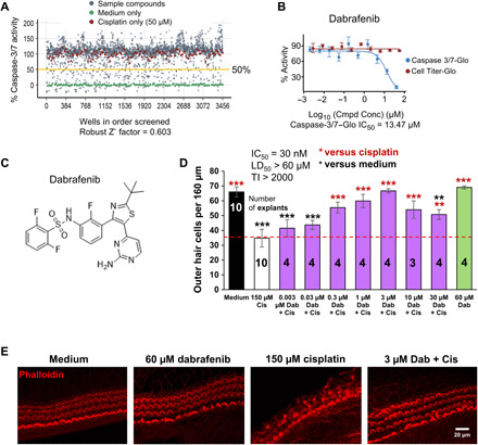
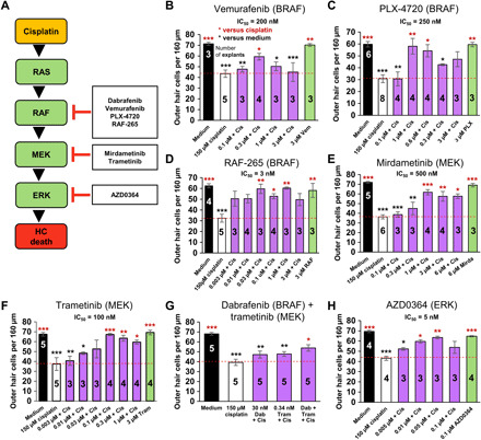
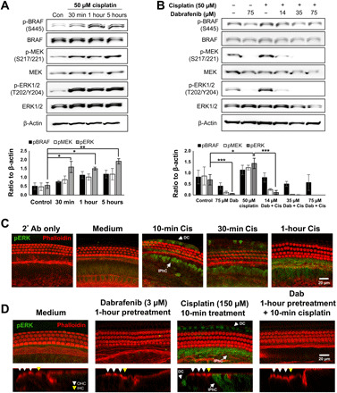
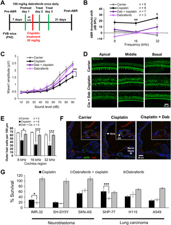
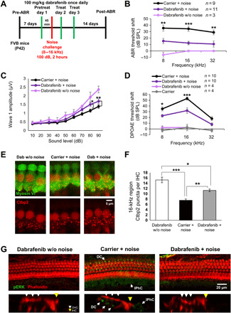
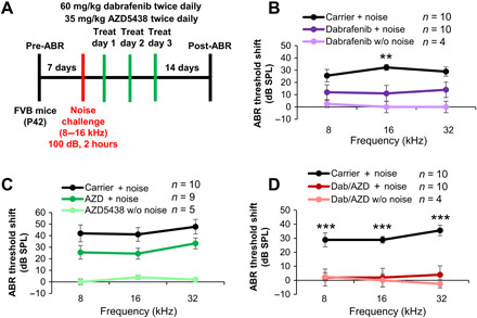
Hearing loss caused by noise, aging, antibiotics, and chemotherapy affects 10% of the world population, yet there are no Food and Drug Administration (FDA)-approved drugs to prevent it. Here, we screened 162 small-molecule kinase-specific inhibitors for reduction of cisplatin toxicity in an inner ear cell line and identified dabrafenib (TAFINLAR), a BRAF kinase inhibitor FDA-approved for cancer treatment. Dabrafenib and six additional kinase inhibitors in the BRAF/MEK/ERK cellular pathway mitigated cisplatin-induced hair cell death in the cell line and mouse cochlear explants. In adult mice, oral delivery of dabrafenib repressed ERK phosphorylation in cochlear cells, and protected from cisplatin- and noise-induced hearing loss. Full protection was achieved in mice with co-treatment with oral AZD5438, a CDK2 kinase inhibitor. Our study explores a previously unidentified cellular pathway and molecular target BRAF kinase for otoprotection and may advance dabrafenib into clinics to benefit patients with cisplatin- and noise-induced ototoxicity.


Figures

Fig. 1
Fig. 1. Dabrafenib protects from cisplatin-induced cell death in cell line and cochlear explant models.
(A) Cell-based small-molecule screen in dose-response mode using HEI-OC1 cell line; hits were defined as compounds that reduce caspase-3/7 activity by 50% or more in the presence of 50 μM cisplatin. Medium alone (green dot), cisplatin alone (red dot), and compound + cisplatin (gray dot). (B) Dabrafenib dose-response curve from caspase-3/7 assay (A) in the presence of 50 μM cisplatin (blue), and Cell Titer-Glo dose-response curve of dabrafenib alone in HEI-OC1 cells (red). (C) Molecular structure of dabrafenib. (D) Dose-response of dabrafenib (Dab) in P3 FVB mouse cochlear explants treated with or without cisplatin. Medium alone (black), cisplatin alone (white), Dab alone (green), or Dab added 1 hour before cisplatin (150 μM) to P3 FVB cochlear explants treated for 24 hours (purple). Numbers inside each bar indicate number of explants counted per treatment. Number of outer hair cells (OHCs) per 160 μm of middle turn regions of the cochlea were counted by phalloidin staining, means ± SEM, **P < 0.01, ***P < 0.001 compared to cisplatin alone (red) and medium alone (black) by one-way analysis of variance (ANOVA) with Bonferroni post hoc test. (E) Representative confocal images of phalloidin-stained whole-mount middle turn cochlear explants treated with medium alone, 60 μM Dab, 150 μM cisplatin, or 3 μM Dab and 150 μM cisplatin for 24 hours are shown.
Fig. 2
Fig. 2. Additional top-hit BRAF and MAPK inhibitors protect against cisplatin ototoxicity in explant.
(A) The putative BRAF/MEK/ERK cellular pathway and small-molecule protein kinase inhibitors in this pathway that were top hits in our inner ear cell line screen for protection from cisplatin-induced cell death. BRAF inhibitors vemurafenib (B), PLX-4720 (C), and RAF-265 (D); MEK1/2 inhibitors mirdametinib (E), trametinib (F), and combined dabrafenib and trametinib (G); and ERK1/2 inhibitor AZD-0364 (H) tested in the cochlear explant culture assay for protection against cisplatin-induced OHC death. Medium alone (black), cisplatin alone (white), compound alone (green), or compound added 1 hour before cisplatin (150 μM) to P3 FVB cochlear explants treated for 24 hours (purple). Numbers inside each bar indicate number of explants counted per treatment. Number of OHCs per 160 μm of middle turn regions of the cochlea were counted by phalloidin staining, means ± SEM, *P < 0.05, **P < 0.01, ***P < 0.001 compared to cisplatin alone (red) and medium alone (black) by one-way ANOVA with Bonferroni post hoc test.
Fig. 3
Fig. 3. Dabrafenib mitigates cisplatin-activated BRAF signaling cascade.
(A) Representative Western blot images of BRAF, ERK, and MEK phosphorylation in HEI-OC1 cells upon 50 μM cisplatin treatment for 30 min, 1 hour, and 5 hours. Phosphorylated protein bands were normalized to β-actin and averaged, means ± SEM, *P < 0.05, **P < 0.01 by one-way ANOVA with Bonferroni post hoc test. n = 4. (B) Representative Western blot images (n = 3) of BRAF, ERK, and MEK phosphorylation upon combined dabrafenib (14, 35, or 75 μM) and cisplatin (50 μM) treatment in HEI-OC1 cells. Cells are pretreated with dabrafenib for 1 hour before 1-hour cisplatin treatment. Medium alone, cisplatin alone, and 75 μM dabrafenib alone used as controls. Phosphorylated protein bands were normalized to β-actin and averaged, means ± SEM, *P < 0.05, ***P < 0.001 by one-way ANOVA with Bonferroni post hoc test. n = 3. (C) Representative phalloidin (red) and phosphorylated ERK (pERK) (green) stained confocal images of P3 FVB whole-mount middle turn mouse cochlea explants pretreated with 3 μM dabrafenib (Dab) for 1 hour before 10 min cisplatin (150 μM) exposure. Deiters’ cells (DC) and inner phalangeal cells (IPhC) with labeled arrows. n = 6 cochlea. (D) Representative phalloidin (red)– and pERK (green)–stained confocal images of P3 FVB whole-mount middle turn mouse cochlea explants pretreated with 3 μM dabrafenib (Dab) for 1 hour before 10 min cisplatin (150 μM) exposure. Ortho section shown below in which OHCs are identified with white arrows, inner HCs (IHCs) are identified with yellow arrows, and pERK-positive DCs and IPhCs are identified with labeled arrows. n = 6 cochlea.
Fig. 4
Fig. 4. Dabrafenib protects against cisplatin-induced hearing loss in adult mice and does not inhibit cisplatin tumor-killing efficacy.
(A) Schedule of administration of dabrafenib and cisplatin to adult P42 FVB mice. (B) ABR threshold shifts following protocol from (A). Untreated controls (gray), cisplatin alone (black), dabrafenib alone (light purple), and dabrafenib and cisplatin (dark purple). (C) Amplitudes of ABR wave 1 at 16 kHz from (B). (B and C) Means ± SEM, *P < 0.05, **P < 0.01, compared to cisplatin alone by two-way ANOVA with Bonferroni post hoc test. (D) Representative myosin VI–stained confocal images of 8-, 16-, and 32-kHz cochlear regions from carrier alone, cisplatin alone, and combined dabrafenib (Dab) and cisplatin-treated mice. (E) Number of OHCs per 160 μm in 8-, 16-, and 32-kHz cochlear regions were counted by myosin VI staining; data shown as means ± SEM, *P < 0.05, ***P < 0.001 by two-way ANOVA with Bonferroni post hoc test. (F) Representative 4′,6-diamidino-2-phenylindole (DAPI) (blue), pERK (green), and Tuj1 (red) immunofluorescence-stained cochlear sections from P42 FVB mice treated with carrier alone, 1-hour cisplatin (30 mg/kg), or dabrafenib (100 mg/kg) 45 min pretreatment followed by 1-hour cisplatin. Higher-magnification images of the organ of Corti region are inlaid in the upper right corner. n = 3 mice. (G) Cell Titer-Glo percent cell survival of neuroblastoma and lung carcinoma cell lines pretreated 1 hour with dabrafenib followed by 48-hour combined cisplatin and dabrafenib treatment. Data shown as means ± SEM, *P < 0.05, ***P < 0.001 compared to cisplatin alone by one-way ANOVA with Bonferroni post hoc test. n = 6.
Fig. 5
Fig. 5. Dabrafenib pretreatment protects against noise-induced hearing loss in adult mice.
(A) Schedule of administration of dabrafenib and noise exposure to adult P42 FVB mice. (B) ABR threshold shifts following protocol from (A). Dabrafenib treatment without noise exposure (light purple), noise alone (black), and dabrafenib treatment with noise exposure (dark purple). (C) Amplitudes of ABR wave 1 at 16 kHz from (B). (D) DPOAE threshold shifts following protocol from (A). Carrier without noise exposure (gray), dabrafenib treatment without noise exposure (light purple), noise alone (black), and dabrafenib treatment with noise exposure (dark purple). (B to D) Means ± SEM, *P < 0.05, **P < 0.01, ***P < 0.001 compared to noise alone by two-way ANOVA with Bonferroni post hoc test. (E) Representative Ctbp2 puncta (red)– and myosin VI (green)–stained confocal images of IHCs in whole-mount mouse cochlea at the 16-kHz region. (F) Number of Ctbp2 puncta per IHC at the 16-kHz cochlear region were counted; data shown as means ± SEM, *P < 0.05, **P < 0.01, ***P < 0.001 by one-way ANOVA with Bonferroni post hoc test. n = 4 cochlea. (G) Representative phalloidin (red) and pERK (green) immunofluorescence-stained adult FVB mice cochlea. Mice were exposed to noise following protocol from (A), then euthanized, and cochlea-fixed 3 hours after noise exposure. Ortho section below shows OHCs identified with white arrows, IHCs identified with yellow arrows, and pERK-positive DCs and IPhCs identified with labeled arrows. n = 2 cochlea.
Fig. 6
Fig. 6. Dabrafenib and AZD5438 posttreatment protect against noise-mediated hearing loss in adult mice.
(A) Schedule of administration of compound and noise exposure to adult P42 FVB mice. (B) ABR threshold shifts recorded 14 days after 100-dB 8- to 16-kHz octave band noise for 2 hours for control and dabrafenib (60 kg/mg) twice daily treated mice by oral gavage. Dabrafenib treatment without noise exposure (light purple), noise alone (black), and dabrafenib treatment with noise exposure (dark purple). Means ± SEM, **P < 0.01 compared to noise alone by two-way ANOVA with Bonferroni post hoc test. (C) ABR threshold shifts recorded 14 days after 100-dB 8- to 16-kHz octave band noise for 2 hours for control and AZD5438 (35 kg/mg) twice daily treated mice by oral gavage. AZD5438 treatment without noise exposure (light green), noise alone (black), and dabrafenib treatment with noise exposure (dark green). The values were not significant compared to noise alone by two-way ANOVA with Bonferroni post hoc test. (D) ABR threshold shifts recorded 14 days after 100-dB 8- to 16-kHz octave band noise for 2 hours for control and combined dabrafenib (60 kg/mg) and AZD5438 (35 mg/kg) twice daily treated mice by oral gavage. Dabrafenib and AZD5438 treatment without noise exposure (light red), noise alone (black), dabrafenib, and AZD5438 treatment (dark red). Means ± SEM, ***P < 0.001 compared to noise alone by two-way ANOVA with Bonferroni post hoc test.

References

    1. Hazlitt R. A., Min J., Zuo J., Progress in the development of preventative drugs for cisplatin-induced hearing loss. J. Med. Chem. 61, 5512–5524 (2018). - PMC - PubMed
    1. Rybak L. P., Dhukhwa A., Mukherjea D., Ramkumar V., Local drug delivery for prevention of hearing loss. Front. Cell. Neurosci. 13, 300 (2019). - PMC - PubMed
    1. Xu H., Robinson G. W., Huang J., Lim J. Y.-S., Zhang H., Bass J. K., Broniscer A., Chintagumpala M., Bartels U., Gururangan S., Hassall T., Fisher M., Cohn R., Yamashita T., Teitz T., Zuo J., Onar-Thomas A., Gajjar A., Stewart C. F., Yang J. J., Common variants in ACYP2 influence susceptibility to cisplatin-induced hearing loss. Nat. Genet. 47, 263–266 (2015). - PMC - PubMed
    1. Dasari S., Tchounwou P. B., Cisplatin in cancer therapy: Molecular mechanisms of action. Eur. J. Pharmacol. 740, 364–378 (2014). - PMC - PubMed
    1. Ding D., Allman B. L., Salvi R., Review: Ototoxic characteristics of platinum antitumor drugs. Anat. Rec. (Hoboken). 295, 1851–1867 (2012). - PubMed

Publication types