. 2020 Dec 2;6(49):eabc5705.
doi: 10.1126/sciadv.abc5705. Print 2020 Dec.

Resurrecting the ancient glow of the fireflies

Affiliations

Resurrecting the ancient glow of the fireflies

Y Oba et al. Sci Adv. .

Abstract

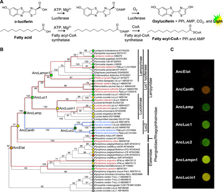
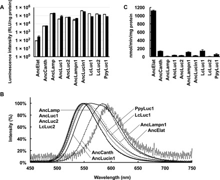
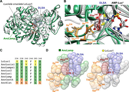
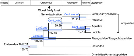
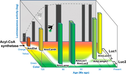
The color of firefly bioluminescence is determined by the structure of luciferase. Firefly luciferase genes have been isolated from more than 30 species, producing light ranging in color from green to orange-yellow. Here, we reconstructed seven ancestral firefly luciferase genes, characterized the enzymatic properties of the recombinant proteins, and determined the crystal structures of the gene from ancestral Lampyridae. Results showed that the synthetic luciferase for the last common firefly ancestor exhibited green light caused by a spatial constraint on the luciferin molecule in enzyme, while fatty acyl-CoA synthetic activity, an original function of firefly luciferase, was diminished in exchange. All known firefly species are bioluminescent in the larvae, with a common ancestor arising approximately 100 million years ago. Combined, our findings propose that, within the mid-Cretaceous forest, the common ancestor of fireflies evolved green light luciferase via trade-off of the original function, which was likely aposematic warning display against nocturnal predation.


Figures

Fig. 1
Fig. 1. Firefly bioluminescence and color evolution.
(A) Coleopteran bioluminescence reaction. (B) Molecular phylogeny of luciferases and related enzymes. The leaf nodes are labeled with species name, protein name, and GenBank accession number. Branches are labeled with bootstrap probability (1000 reconstructions). The resurrected ancestral nodes are shown as a square. The leaf nodes are indicated with in vitro luminescent colors (green, yellow-green, yellow, orange, or red) judged by the luminescence maximum values in references (table S3). The resurrected ancestral nodes are indicated with in vitro luminescence colors judged by the combination of luminescence maximum (Table 1) and perceived coloration. Luciferases without spectral data and fatty acyl-CoA synthetase (nonluciferase) are denoted with white and gray circles, respectively. The 18S rRNA–based species trees (fig. S6) are superimposed onto the subtrees of Luc1 type (red lines) and Luc2 type (blue lines) to confirm consistency between the gene/protein phylogeny and the species phylogeny. (C) Photographs of the luminescence of seven ancestral luciferases in 96-well plate. The camera exposure time for each well is uneven to avoid the changes in coloration by overexposure. Luminescence of AncElat was not photographed even by longer exposure. Luminescence of AncCanth was categorized as yellow on the basis of λmax value but appears in orange on the photograph probably because of its broad spectrum.
Fig. 2
Fig. 2. Biochemical properties of ancestral luciferases.
(A) Integration of luminescence intensities from 2 to 32 s. The values are shown by relative light unit per nanogram of protein (RLU/ng). White and black bars represent the mean with standard deviation values at 50 and 200 μM of d-luciferin, respectively (n = 3). (B) Luminescent spectra of seven recombinant ancestral luciferases and three extant firefly luciferases. (C) ACS activities of seven ancestral luciferases and three extant firefly luciferases to lauric acid are shown. Bars represent SE of the means (n = 3 to 6).
Fig. 3
Fig. 3. Structure of AncLamp.
(A) The structure of AncLamp-DLSA complex (green) is superposed on that of Luciola cruciata LcLuc1-DLSA complex (white). DLSA is represented as stick model. The all ligand omitting Fo-Fc map (contoured at 6 σ) is shown in blue. The positions of N and C termini are indicated. (B) Detail of the substrate-binding site of superposed AncLamp-DLSA (green), LcLuc1-DLSA (white), and AncLamp-substrates (d-luciferin/ATP) (light green) complex structures. The residues differing between AncLamp and LcLuc1, Ile236 (Val239 in LcLuc1), Ile240 (Val243), and Leu285 (Ile288), and those in close contact with these residues, Phe246 (Phe249) and Thr238 (Thr241), are shown in stick models. The molecules of the DLSA and probable intermediate, luciferyl-AMP (AMP-Luc*), are shown as stick models colored by element (carbon atoms are colored in white, gray, and light gray for AncLamp-DLSA, LcLuc1-DLSA, and AncLamp-substrates complex structures, respectively). (C) Amino acids of the ancestral luciferases and LcLuc1 at the sites discussed in the text. The residue numbers of AncLamp are indicated above the alignment. See fig. S1 for original alignment. (D) Close-up view of the substrate-binding site of AncLamp (left) and LcLuc1 (right) in van der Waals model. The different residues between two proteins, i.e., Ile236 (Val239 in LcLuc1), Ile240 (Val243), and Leu285 (Ile288), and those in close contact, Phe246 (Phe249) and Thr238 (Thr241), are shown. The distances from Cε1 atoms of Phe246 (Phe249) to DLSA are indicated in red.
Fig. 4
Fig. 4. Dating of ancestral species.
Geological dating of the ancestral species (concestors) (50) bearing the ancestral luciferases based on the molecular clock analysis of 18S rRNA genes of Elateriformia species. The nodes corresponding to the hypothesized concestors, which genomes encoded AncElat, AncCanth, AncLampn1, and AncLucin1 are indicated as ConElat, ConCanth, ConLampn1, and ConLucin1, respectively. ConLuc1/2 is the concestor of Lampyridae, which had the duplicated genes of AncLuc1 and AncLuc2 as the first time and AncLamp as the last time (Fig. 1B). The median of estimated geological age and 95% HPD (light blue bar) from the MCMC estimations (fig. S6) are indicted on each node. The geohistorical positions of the oldest firefly fossil with abdominal photophore structure (6) and gene duplication between Luc1-type and Luc2-type genes are indicated on the phylogeny or geological time scale.
Fig. 5
Fig. 5. Scheme of the evolution of firefly’s bioluminescence.
Horizontal and vertical axes represent the approximate age and luminescence activity, respectively. Z axis shows the color of ancestral luciferases.

References

    1. S. Lewis, Silent Sparks: The Wondrous World of Fireflies (Princeton Univ. Press, 2016).
    1. Martin G. J., Branham M. A., Whiting M. F., Bybee S. M., Total evidence phylogeny and the evolution of adult bioluminescence in fireflies (Coleoptera: Lampyridae). Mol. Phylogenet. Evol. 107, 564–575 (2017). - PubMed
    1. Martin G. J., Stanger-Hall K. F., Branham M. A., Da Silveira L. F. L., Lower S. E., Hall D. W., Li X.-Y., Lemmon A. R., Lemmon E. M., Bybee S. M., Higher-level phylogeny and reclassification of Lampyridae (Coleoptera: Elateroidea). Insect Syst. Diver. 3, 11 (2019).
    1. Fallon T. R., Lower S. E., Chang C.-H., Bessho-Uehara M., Martin G. J., Bewick A. J., Behringer M., Debat H. J., Wong I., Day J. C., Suvorov A., Silva C. J., Stanger-Hall K. F., Hall D. W., Schmitz R. J., Nelson D. R., Lewis S. M., Shigenobu S., Bybee S. M., Larracuente A. M., Oba Y., Weng J.-K., Firefly genomes illuminate parallel origins of bioluminescence in beetles. eLife 7, e36495 (2018). - PMC - PubMed
    1. Seliger H. H., Lall A. B., Lloyd J. E., Biggley W. H., The colors of firefly bioluminescence—I. Optimization model. Photochem. Photobiol. 36, 673–680 (1982).

Publication types

LinkOut - more resources