Review
. 2021 Jan-Jun:296:100135.
doi: 10.1074/jbc.REV120.015980. Epub 2020 Dec 6.

Priming of SARS-CoV-2 S protein by several membrane-bound serine proteinases could explain enhanced viral infectivity and systemic COVID-19 infection

Affiliations
Review

Priming of SARS-CoV-2 S protein by several membrane-bound serine proteinases could explain enhanced viral infectivity and systemic COVID-19 infection

Pablo Fuentes-Prior. J Biol Chem. 2021 Jan-Jun.

Abstract

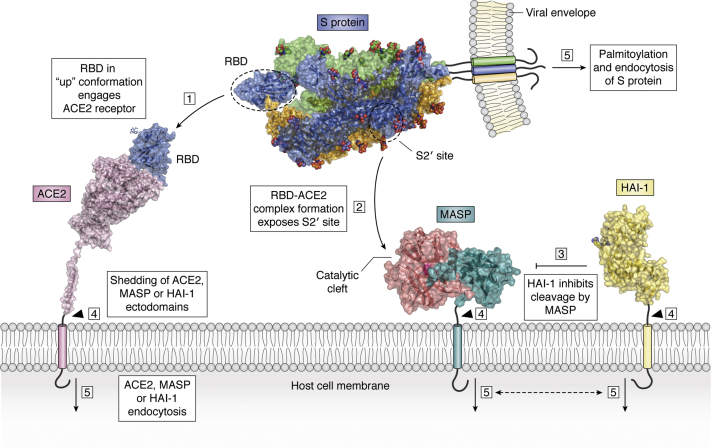
The ongoing COVID-19 pandemic has already caused over a million deaths worldwide, and this death toll will be much higher before effective treatments and vaccines are available. The causative agent of the disease, the coronavirus SARS-CoV-2, shows important similarities with the previously emerged SARS-CoV-1, but also striking differences. First, SARS-CoV-2 possesses a significantly higher transmission rate and infectivity than SARS-CoV-1 and has infected in a few months over 60 million people. Moreover, COVID-19 has a systemic character, as in addition to the lungs, it also affects the heart, liver, and kidneys among other organs of the patients and causes frequent thrombotic and neurological complications. In fact, the term "viral sepsis" has been recently coined to describe the clinical observations. Here I review current structure-function information on the viral spike proteins and the membrane fusion process to provide plausible explanations for these observations. I hypothesize that several membrane-associated serine proteinases (MASPs), in synergy with or in place of TMPRSS2, contribute to activate the SARS-CoV-2 spike protein. Relative concentrations of the attachment receptor, ACE2, MASPs, their endogenous inhibitors (the Kunitz-type transmembrane inhibitors, HAI-1/SPINT1 and HAI-2/SPINT2, as well as major circulating serpins) would determine the infection rate of host cells. The exclusive or predominant expression of major MASPs in specific human organs suggests a direct role of these proteinases in e.g., heart infection and myocardial injury, liver dysfunction, kidney damage, as well as neurological complications. Thorough consideration of these factors could have a positive impact on the control of the current COVID-19 pandemic.

Keywords: COVID-19; HAI-1/SPINT1; TMPRSS2; cell tropism; coronaviruses; membrane-associated serine proteinases (MASPs); serpins; spike (S) protein; structure–function relationship; viral fusion.


Conflict of interest statement

Conflict of interest The authors declare that they have no conflicts of interest with the contents of this article.

Figures

Figure 1
Figure 1
The SARS-CoV-2 spike (S) protein is responsible for viral attachment to host cells and membrane fusion.A, schematic representation of domain organization. B, 3D structure of trimeric S glycoprotein in the prefusion conformation (after PDB 6VSB). The RBD of one monomer is in the “up” conformation, i.e., exposed for interaction with the human ACE2 ectodomain (blue), while the two other monomers are depicted as green and light-orange solid surfaces. Note that carbohydrate chains (color-coded spheres) are distributed all over the surface of the trimer. C, postfusion conformation of the viral S protein. After priming, the heptad repeats and central helix from each S2’ monomer adopt extended, tightly packed continuous α-helical structures that generate a 180-Å-long, cone-shaped arrangement with a prominent triple-helical core. (Here, the structure of the highly related murine hepatitis virus is represented; PDB 6B3O, ref. (161)). Residues topologically equivalent to Leu922 and Ser1196 of each monomer are located less than 20 Å apart. In this manner, the rearranged S trimer brings viral and host cell membranes into close proximity to allow their fusion and infection. D, S2’ cleavage site (Arg815-Ser816, red arrow), highlighting the solvent-protected conformation of these residues, which is incompatible with proteinase binding and proteolysis: the Arg815 side chain is clamped by acidic residues (Asp820, Asp867) and contacts with Phe823, while the side chain of Ser816 points inward and is fixed by those of Glu819 and Gln1054. E, comparison of S2’ priming sites in SARS-CoV-1 and 2. Several critical residues around the Arg815 side chain (Arg797 in SARS-CoV-1) are color-coded (carbon atoms are orange and green in SARS-CoV-1 and 2, respectively). Different conformations for most of the activation loop (upper right part of the panel) leads to significant changes in the shielding of the Arg815 side chain; Ser813→Thr797 and Glu868→Asp850 replacements in particular generate a “cage” better suited to hold the Arg815 side chain in its protected conformation. F, structure of the Arg815-binding pocket in MERS-CoV S protein (after PDB 5X59). As a major difference with SARS-CoV-1 and 2, the connector topologically equivalent to Val826-Gln836 in SARS-CoV-2 is defined and forms a lid that would sterically clash with an approaching proteinase. This lid projects the side chains of Ile828, Pro831 and Tyr833 toward Arg815, which, together with the replacement of Asp867 by an aliphatic valine, makes the Arg815 cage much more apolar in MERS-CoV. Conceivably, these subtle sequence and structure variations facilitate exposure and cleavage of the Arg815-Ser816 peptide bond SARS-CoV-2 and thus contribute to its increase infectivity compared with other related coronaviruses. (To facilitate comparisons, residues are numbered according to their topological equivalences in SARS-CoV-2).
Figure 2
Figure 2
Membrane-associated serine proteinases (MASPs): mosaic proteinases for cleavage of transmembrane and membrane-proximal substrates. Structural domains (approximate scale) are color-coded. Note the variety of noncatalytic scaffolds found in these mosaic proteinases, most notably in the matriptases (TMPRSS6, -7 and -14), corin/TMPRSS10, and enterokinase/TMPRSS15. In addition to type II transmembrane serine proteinases (TTSPs), type I or glycosylphosphatidylinositol (GPI)-anchored proteins that possess similar catalytic domains are also included (TMPRSS12/TMPSD, PRSS8/prostasin, PRSS21/testisin, PRS41/TESSP-1, and PRS55/T-SP1). CUB, complement C1r/C1s, Uegf, Bmp1; FRZ, frizzled; LDLR, low-density lipoprotein receptor; MAM, meprin, A-5 protein, and receptor protein-tyrosine phosphatase mu; SEA, sea urchin sperm protein, enterokinase, agrin; SRCR, scavenger receptor cysteine-rich.
Figure 3
Figure 3
Expression patterns and structure of membrane-associated serine proteinases.A, organs with highest expression levels of specific MASPs. Data for some organs are indicated only for males or females, although there are no known sex-associated differences in MASP expression in these organs. Data from the GTEx Portal (gtexportal.org) (see also Fig. 4A). BC, 3D crystal structure of human hepsin/TMPRSS1 ectodomain, represented as a cartoon highlighting major secondary structure elements; loops that shape the active-site cleft are noted. (After PDB entry 1P57: N-terminal SRCR module (deep-teal cyan); serine proteinase domain (deep salmon-red)). A small-molecule inhibitor bound in the S1 specificity pocket of the serine proteinase domain (2-{5-[amino(iminio)methyl]-1H-benzimidazol-2-YL}benzenolate) is shown as color-coded spheres (carbon, pink; oxygen, red; nitrogen, blue; and hydrogen, gray). B, side view, highlighting the proximity of the N-terminal residue of the SRCR domain, Pro50, to the transmembrane helix, Gly24-Leu44. This locates also the rigidly attached catalytic domain of the proteinase essentially flat against the cell membrane (74). A similar localization should be expected for the catalytic domains of other MASPs, which implies that the Arg815-Ser816 bond in the S protein would be cleaved close to the cell membrane, facilitating rapid interaction with the exposed viral fusion peptide and escape from immune surveillance. C, view of the proteinase in the “standard orientation”, e.g., with active-site residues (given with all their nonhydrogen atoms, color-coded) facing the viewer and substrates running from left to right. The N and C termini of the catalytic chain are noted (Ile16 and Thr253, respectively) as well as Asp189 at the bottom of the S1 pocket, which is largely responsible for the recognition and cleavage of substrates after a basic Arg/Lys residue.
Figure 4
Figure 4
Expression patterns of human ACE2, MASPs and their endogenous inhibitors.A, comparison of the expression levels of human ACE2 and a selection of those MASPs with restricted expression patterns. Spinesin/TMPRSS5 is predominantly expressed in the brain and the tibial nerve, corin in the heart, and TMPRSS12, PRSS21, PRSS41 as well as PRSS51 in the testis. Further, matriptase-2 and hepsin are expressed at highest levels in the liver or in liver and kidney, respectively. The members of the airway tract subgroup, TMPRSS11A–11F are predominantly expressed in the mucosa of the esophagus and in the vagina. B, schematic representation of domain organization in the endogenous Kunitz-type inhibitors of membrane-associated serine proteinases, HAI-1/SPINT1 and HAI-2/SPINT2. CD, expression patterns of human HAI-1 (C) and HAI-2 (D). Note the overall complementarity of expression profiles in different organs (e.g., HAI-2 but not its paralog is expressed in the brain and arteries), although both inhibitors are similarly expressed in several organs, including kidneys, prostate, and vagina. The extremely low expression of both Kunitz inhibitors in the heart is also noteworthy, which suggests that the proteolytic activity of the heart-specific MASP, corin/TMPRSS10, would only be controlled by circulating serpins. The expression values are given as Transcripts per Million (TPM), as reported in the GTex Portal (gtexportal.org). HAI, hepatocyte growth factor activator inhibitor; MANEC, motif at N-terminus with eight-cysteines; SPINT, serine protease inhibitor Kunitz type.
Figure 5
Figure 5
Mechanism of S protein activation and viral–host cell membrane fusion. The SARS-CoV-2 spike protein is found in the virions in a metastable prefusion state, in which the S1/S2 site has been cleaved by furin within the secretory pathway. (1) The RBD within the N-terminal S1 subunit of the S protein is exposed in the so-called “up” conformation and engages the host cell receptor, ACE2. (In the more stable “down” conformation, the RBD is not accessible to ACE2, which probably helps to evade immune surveillance, ref. (7)). A single ACE2 molecule is shown bound to the RBD of an S monomer, but two trimeric S proteins could simultaneously engage an ACE2 dimer in vivo (12). Also omitted for simplicity is the B0AT1 molecule that binds to the long C-terminal helix of ACE2 (after PDB 6M17). (2) RBD–ACE2 complex formation stabilizes a metastable conformation of the spike protein, stimulating “breathing” of the S2’ site and eventual exposure of the Arg815-Ser816 peptide bond to proteolytic cleavage by TMPRSS2 or another MASP. (3) MASP-mediated cleavage and activation of the S protein are controlled by endogenous inhibitors, most notably HAI-1 and/or HAI-2, but also by circulating serpins. Here, the HAI-1 ectodomain is shown, according to the reported crystal structure (PDB 5H7V; ref. (162)); the side chain of the P1 residue, Arg260, is highlighted. Other cellular processes that coregulate membrane fusion and viral uptake are (4) shedding of ACE2, MASPs and/or HAI-1/HAI-2 ectodomains, either by MASPs in trans or by membrane-bound metalloproteinases, and (5) endocytosis, which could be modulated by phosphorylation of cytosolic peptides in the human factors, or by palmitoylation of the Cys-rich endodomain of SARS-CoV-2. The scheme focuses on the “early”, endocytosis-independent pathway of virus cell entry. Endocytic trafficking (the “late” pathway) seems to play a minor role.

Similar articles

Cited by

References

    1. Li W., Moore M.J., Vasilieva N., Sui J., Wong S.K., Berne M.A., Somasundaran M., Sullivan J.L., Luzuriaga K., Greenough T.C., Choe H., Farzan M. Angiotensin-converting enzyme 2 is a functional receptor for the SARS coronavirus. Nature. 2003;426:450–454. - PMC - PubMed
    1. Belouzard S., Chu V.C., Whittaker G.R. Activation of the SARS coronavirus spike protein via sequential proteolytic cleavage at two distinct sites. Proc. Natl. Acad. Sci. U. S. A. 2009;106:5871–5876. - PMC - PubMed
    1. Heurich A., Hofmann-Winkler H., Gierer S., Liepold T., Jahn O., Pöhlmann S. TMPRSS2 and ADAM17 cleave ACE2 differentially and only proteolysis by TMPRSS2 augments entry driven by the severe acute respiratory syndrome coronavirus spike protein. J. Virol. 2014;88:1293–1307. - PMC - PubMed
    1. Kirchdoerfer R.N., Cottrell C.A., Wang N., Pallesen J., Yassine H.M., Turner H.L., Corbett K.S., Graham B.S., McLellan J.S., Ward A.B. Pre-fusion structure of a human coronavirus spike protein. Nature. 2016;531:118–121. - PMC - PubMed
    1. Yuan Y., Cao D., Zhang Y., Ma J., Qi J., Wang Q., Lu G., Wu Y., Yan J., Shi Y., Zhang X., Gao G.F. Cryo-EM structures of MERS-CoV and SARS-CoV spike glycoproteins reveal the dynamic receptor binding domains. Nat. Commun. 2017;8:15092. - PMC - PubMed

Publication types

MeSH terms