Review
. 2020 Dec;216(4):837-878.
doi: 10.1534/genetics.120.303565.

Starvation Responses Throughout the Caenorhabditiselegans Life Cycle

Affiliations
Review

Starvation Responses Throughout the Caenorhabditiselegans Life Cycle

L Ryan Baugh et al. Genetics. 2020 Dec.

Abstract

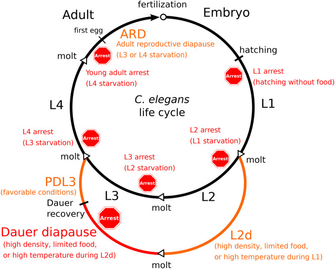
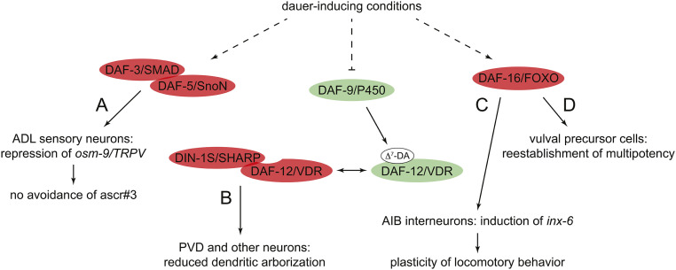
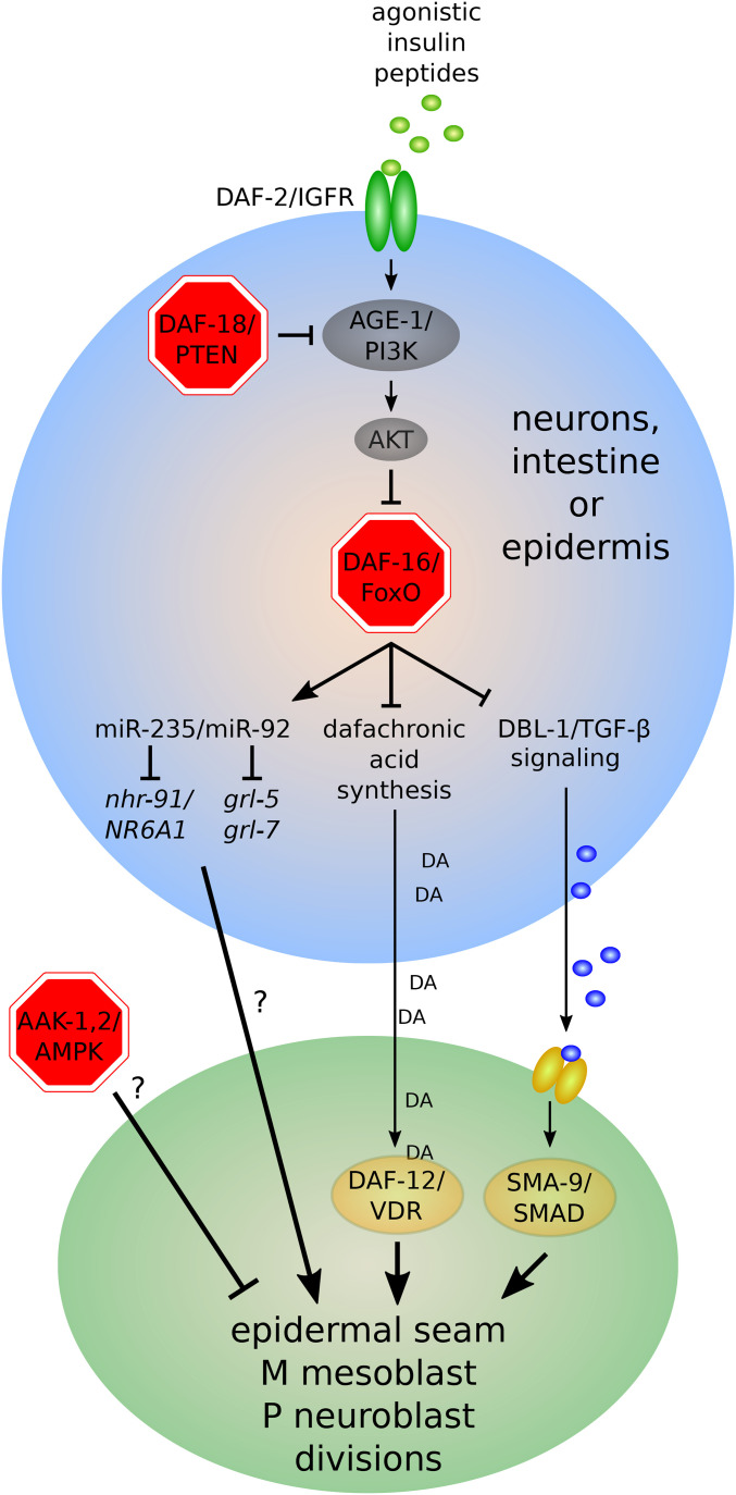
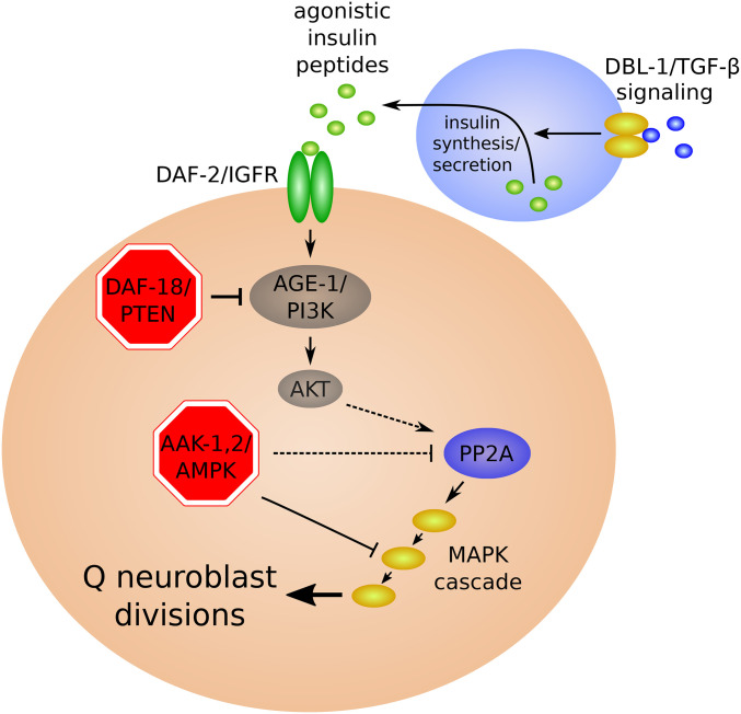
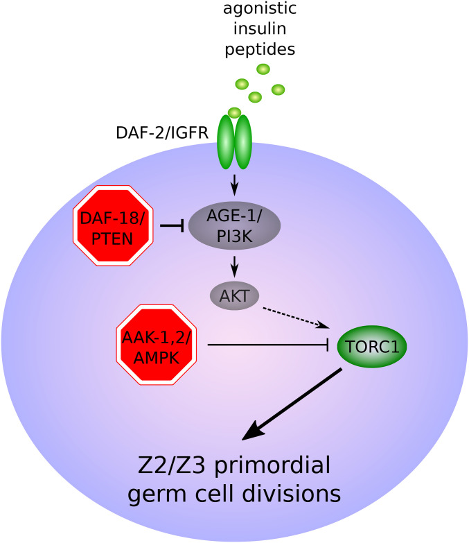
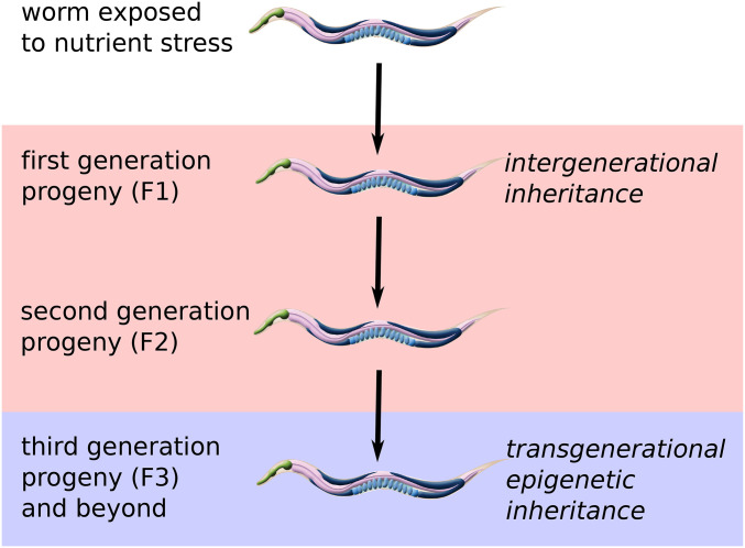
Caenorhabditis elegans survives on ephemeral food sources in the wild, and the species has a variety of adaptive responses to starvation. These features of its life history make the worm a powerful model for studying developmental, behavioral, and metabolic starvation responses. Starvation resistance is fundamental to life in the wild, and it is relevant to aging and common diseases such as cancer and diabetes. Worms respond to acute starvation at different times in the life cycle by arresting development and altering gene expression and metabolism. They also anticipate starvation during early larval development, engaging an alternative developmental program resulting in dauer diapause. By arresting development, these responses postpone growth and reproduction until feeding resumes. A common set of signaling pathways mediates systemic regulation of development in each context but with important distinctions. Several aspects of behavior, including feeding, foraging, taxis, egg laying, sleep, and associative learning, are also affected by starvation. A variety of conserved signaling, gene regulatory, and metabolic mechanisms support adaptation to starvation. Early life starvation can have persistent effects on adults and their descendants. With its short generation time, C. elegans is an ideal model for studying maternal provisioning, transgenerational epigenetic inheritance, and developmental origins of adult health and disease in humans. This review provides a comprehensive overview of starvation responses throughout the C. elegans life cycle.

Keywords: L1 arrest; WormBook; dauer; quiescence; starvation.


Figures

Figure 1
Figure 1
Developmental responses to starvation throughout the life cycle. The progression of developmental stages in well-fed conditions is shown by the black arrow. L2d (predauer) development, PDL3 (postdauer L3) development, and ARD (adult reproductive diapause) are depicted in orange. Dauer diapause, L1 arrest, and other examples of developmental arrest are in red, with a stop sign indicating approximately when they occur relative to stage-specific molts. Specific conditions leading to each developmental response are indicated in parentheses below the response. Molts are depicted by open arrow heads. Hatching, dauer recovery, and onset of reproduction are also indicated with black lines. ARD can occur in response to L3 (Gerisch et al. 2020) or L4 (Angelo and Van Gilst 2009; Seidel and Kimble 2011) starvation, depending on conditions. In contrast, absolute starvation of L3 larvae results in L4 arrest (Schindler et al. 2014). Analogous to L3 and L4 arrest, an L2 arrest potentially results from starving previously fed L1 larvae, although this has not been demonstrated.
Figure 2
Figure 2
Schematic model depicting integration of environmental cues in the ASI sensory neurons. Molecules that promote reproductive development are green, and those that promote dauer arrest are red. A generic transmembrane ascaroside receptor is portrayed as a thick red line. DAF-1 and DAF-4 are homologs of the human type I and type II TGFβ receptor, respectively. See text for details. IGFR, insulin-like growth factor receptor; ILP, insulin-like peptide.
Figure 3
Figure 3
Signal transduction pathways regulated by DAF-7/TGFβ (left) and insulin-like peptides (right). Core pathway components are depicted in gray. More recently identified signaling modulators that promote reproductive development or dauer arrest are shown in green and red, respectively. Multiple mechanisms of cross-talk between these pathways are not depicted. See text for details.
Figure 4
Figure 4
Hypothetical model of pathways involved in dafachronic acid (DA) biosynthesis by enzymes acting in intestine, XXX cells, and hypodermis. Solid arrows denote steps supported by experimental data. See text for details.
Figure 5
Figure 5
Examples of dauer execution programs (A–D). Molecules depicted in green and red promote reproductive development and dauer arrest, respectively. See text for details.
Figure 6
Figure 6
Regulation of lateral epidermal seam cell, M mesoblast, and P neuroblast divisions during L1 arrest. Factors required to arrest cell divisions are shown in red. See text for details.
Figure 7
Figure 7
Regulation of Q neuroblast divisions during L1 arrest. ins-3, ins-4, and presumably other agonistic ILPs secreted from chemosensory neurons and possibly other tissues in response to food activate IIS, leading to the activation of PP2A and the RAF-MEK-ERK MAP kinase cascade and Q neuroblast divisions (Zheng et al. 2018b). Factors required to arrest cell divisions are shown in red. See text for details.
Figure 8
Figure 8
Regulation of Z2/Z3 primordial germ cell divisions during L1 arrest. IIS is activated in response to feeding, presumably by unknown agonistic ILPs, which is hypothesized to result in activation of TORC1 and Z2/Z3 divisions (Fukuyama et al. 2012). Factors required to arrest cell divisions are shown in red. See text for details.
Figure 9
Figure 9
Functions of octopamine during starvation. Octopamine functions as a neurotransmitter to mediate several of the effects of starvation on behavior, functions as a neurohormone to alter lipid metabolism in support of starvation survival, and is used to decorate an ascaroside-based pheromone in starved larvae to produce an alarm pheromone. References: 1 (Horvitz et al. 1982), 2 (Bayer and Hobert 2018), 3 (Rengarajan et al. 2019), 4 (Artyukhin et al. 2013b), and 5 (Tao et al. 2016).
Figure 10
Figure 10
Factors affecting starvation resistance. Genes, molecules, and aspects of life history that impinge on starvation resistance are depicted on the outside, and processes that mediate effects on resistance are on the inside. Regulatory interactions between processes are also indicated with arrows. All known factors are not included. See also Tables 3 and 4.
Figure 11
Figure 11
Nongenetic inheritance of starvation responses. Multigenerational effects of environmental conditions are classified by the number of generations they are inherited. Phenotypic effects that persist for only one or two generations (F1 or F2 progeny) are “intergenerational.” In contrast, effects that persist for three or more generations (F3 and beyond) indicate “transgenerational epigenetic inheritance.” This distinction is made since direct effects on germ cells can theoretically persist for two generations, whereas epigenetic inheritance requires germline mechanisms that actively maintain a regulatory state. Worm images were borrowed with permission from WormAtlas (Altun and Hall 2009).
Figure 12
Figure 12
Global regulation of gene expression in response to nutrient availability. AMPK restrains H3K4 methylation activity of the COMPASS complex during starvation, limiting transcription (Demoinet et al. 2017). RNA Polymerase II is recruited to promoters of housekeeping genes during starvation, but initiation and elongation are inhibited without feeding (Maxwell et al. 2014). Alternative mRNA isoforms are expressed in fed and starved larvae, in particular those encoding splicing factors themselves (Maxwell et al. 2012). Translation is repressed during starvation but dramatically upregulated in response to feeding, with ribosomal proteins being synthesized immediately (Stadler and Fire 2013).

References

    1. Abrahante J. E., Daul A. L., Li M., Volk M. L., Tennessen J. M. et al. , 2003. The Caenorhabditis elegans hunchback-like gene lin-57/hbl-1 controls developmental time and is regulated by microRNAs. Dev. Cell 4: 625–637. 10.1016/S1534-5807(03)00127-8 - DOI - PubMed
    1. Adachi T., Kunitomo H., Tomioka M., Ohno H., Okochi Y. et al. , 2010. Reversal of salt preference is directed by the insulin/PI3K and Gq/PKC signaling in Caenorhabditis elegans. Genetics 186: 1309–1319. 10.1534/genetics.110.119768 - DOI - PMC - PubMed
    1. Ahmadi M., and Roy R., 2016. AMPK acts as a molecular trigger to coordinate glutamatergic signals and adaptive behaviours during acute starvation. eLife 5: e16349 10.7554/eLife.16349 - DOI - PMC - PubMed
    1. Ailion M., and Thomas J. H., 2000. Dauer formation induced by high temperatures in Caenorhabditis elegans. Genetics 156: 1047–1067. - PMC - PubMed
    1. Alam H., Williams T. W., Dumas K. J., Guo C., Yoshina S. et al. , 2010. EAK-7 controls development and life span by regulating nuclear DAF-16/FoxO activity. Cell Metab. 12: 30–41. 10.1016/j.cmet.2010.05.004 - DOI - PMC - PubMed

Publication types

MeSH terms