Clinical Trial
. 2020 Dec 2;12(572):eabb5413.
doi: 10.1126/scitranslmed.abb5413.

Slowing late infantile Batten disease by direct brain parenchymal administration of a rh.10 adeno-associated virus expressing CLN2

Affiliations
Clinical Trial

Slowing late infantile Batten disease by direct brain parenchymal administration of a rh.10 adeno-associated virus expressing CLN2

Dolan Sondhi et al. Sci Transl Med. .

Abstract

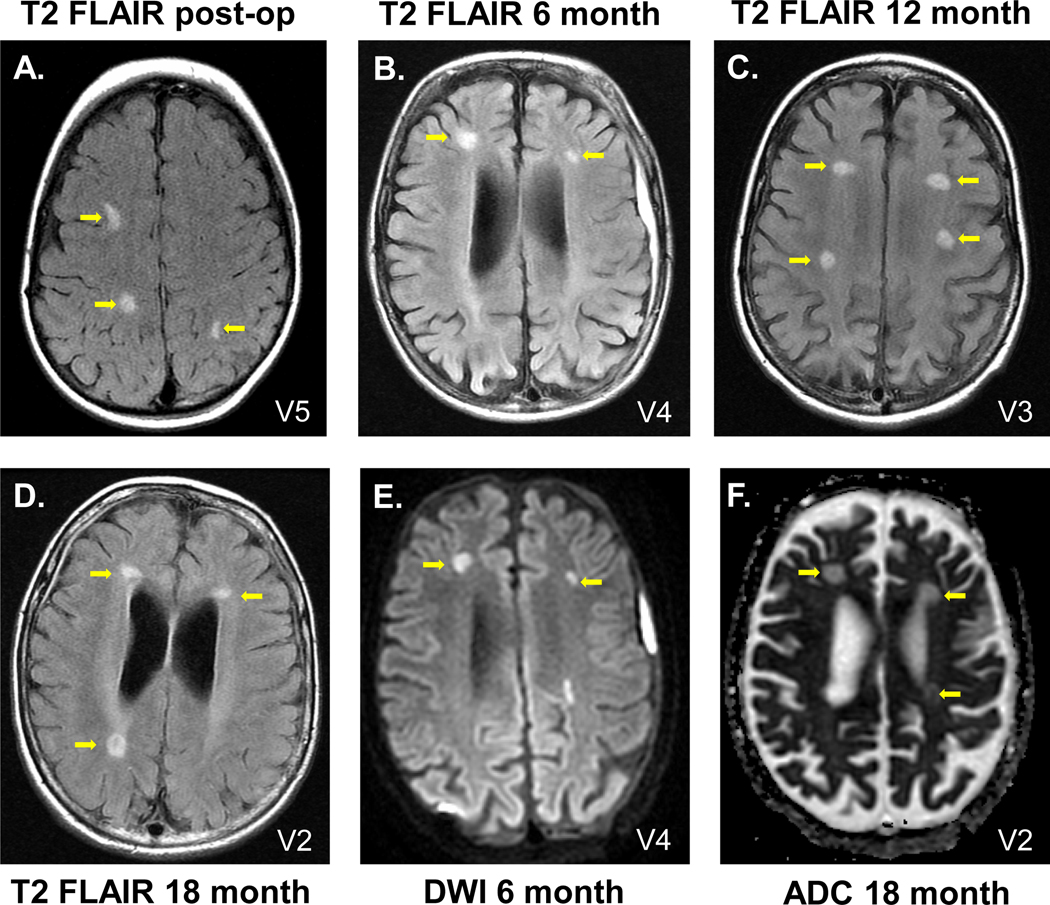
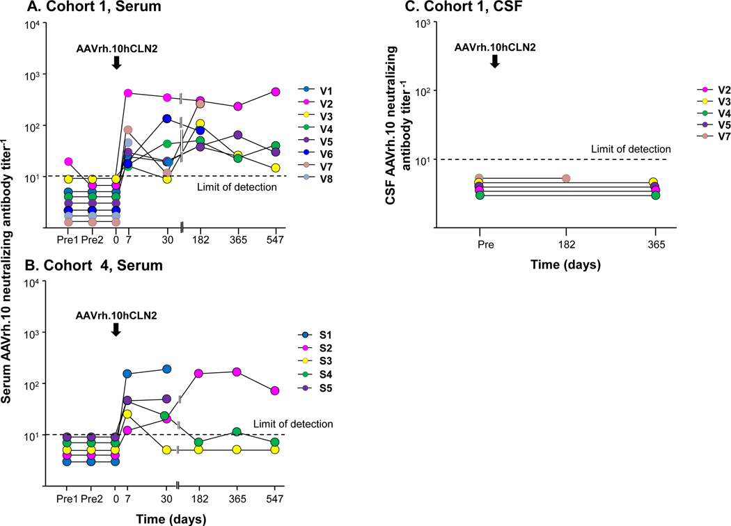
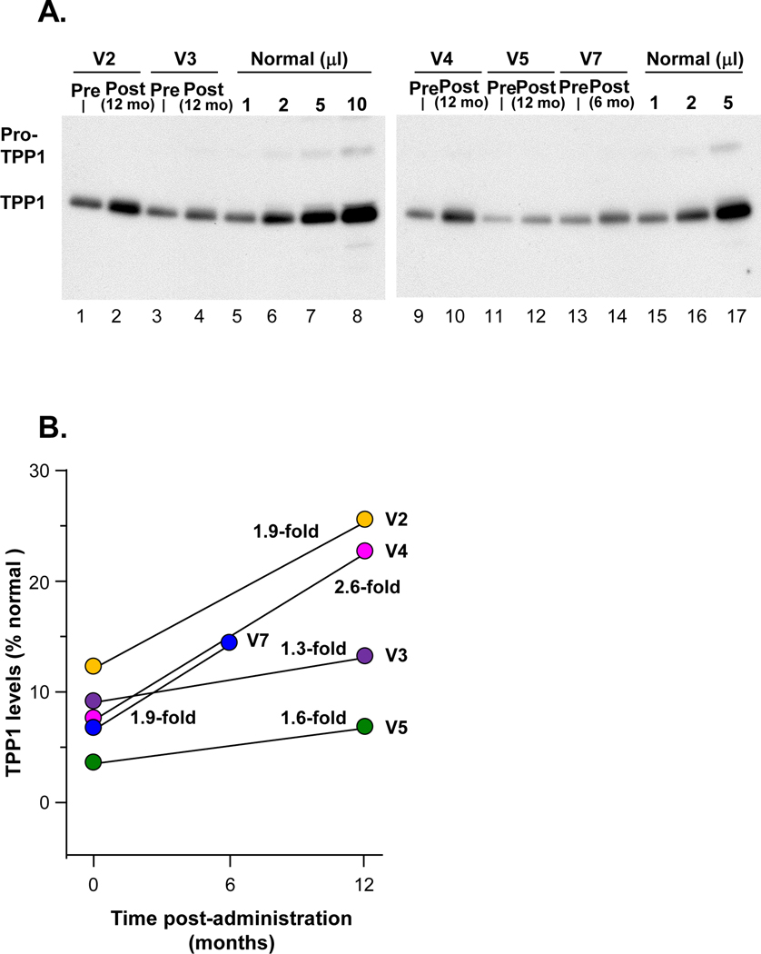
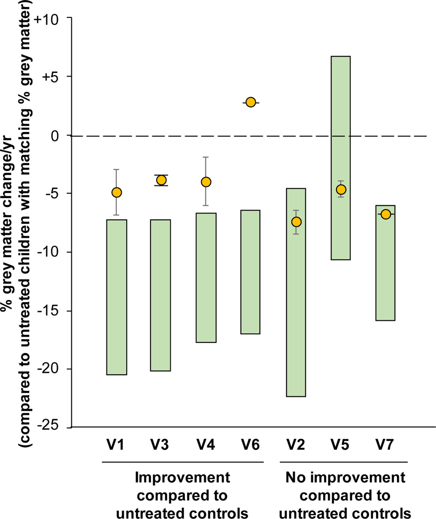
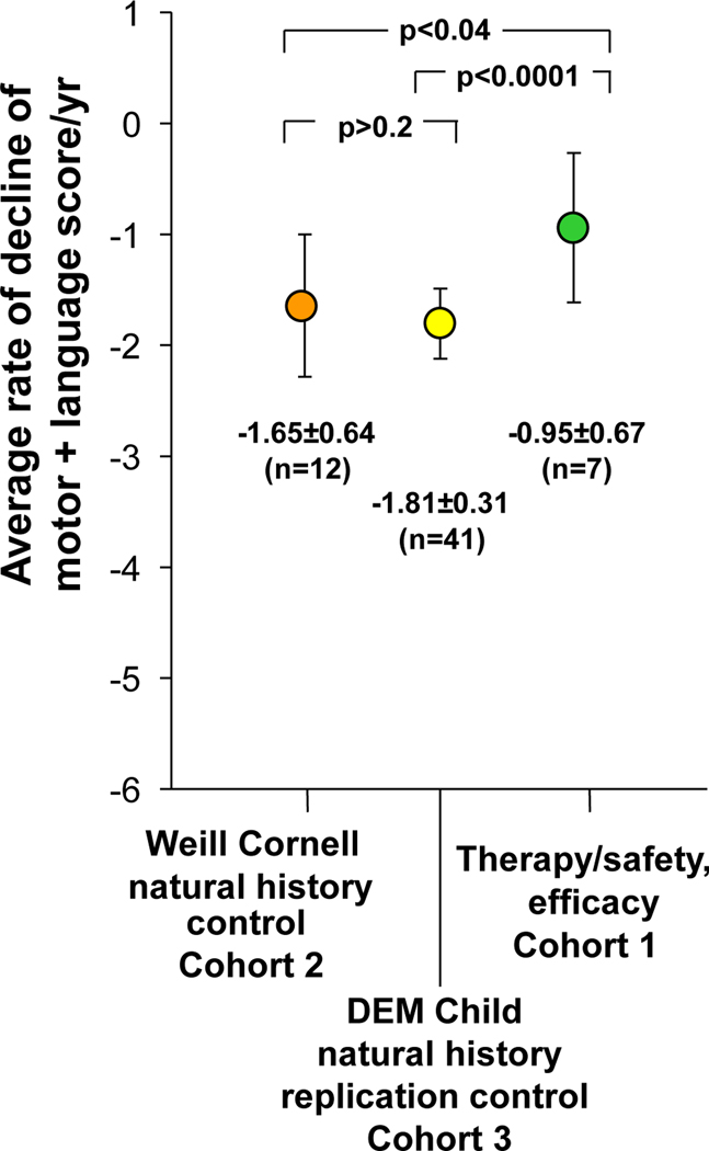
Late infantile Batten disease (CLN2 disease) is an autosomal recessive, neurodegenerative lysosomal storage disease caused by mutations in the CLN2 gene encoding tripeptidyl peptidase 1 (TPP1). We tested intraparenchymal delivery of AAVrh.10hCLN2, a nonhuman serotype rh.10 adeno-associated virus vector encoding human CLN2, in a nonrandomized trial consisting of two arms assessed over 18 months: AAVrh.10hCLN2-treated cohort of 8 children with mild to moderate disease and an untreated, Weill Cornell natural history cohort consisting of 12 children. The treated cohort was also compared to an untreated European natural history cohort of CLN2 disease. The vector was administered through six burr holes directly to 12 sites in the brain without immunosuppression. In an additional safety assessment under a separate protocol, five children with severe CLN2 disease were treated with AAVrh.10hCLN2. The therapy was associated with a variety of expected adverse events, none causing long-term disability. Induction of systemic anti-AAVrh.10 immunity was mild. After therapy, the treated cohort had a 1.3- to 2.6-fold increase in cerebral spinal fluid TPP1. There was a slower loss of gray matter volume in four of seven children by MRI and a 42.4 and 47.5% reduction in the rate of decline of motor and language function, compared to Weill Cornell natural history cohort (P < 0.04) and European natural history cohort (P < 0.0001), respectively. Intraparenchymal brain administration of AAVrh.10hCLN2 slowed the progression of disease in children with CLN2 disease. However, improvements in vector design and delivery strategies will be necessary to halt disease progression using gene therapy.


Figures

Figure 1.
Figure 1.. Axial T2 FLAIR (T2 FLAIR), diffusion weighted imaging (DWI) and apparent diffusion coefficient (ADC) MRI assessment of participants post-therapy.
MRI abnormalities were localized at the sites of the catheter tips, where there is the highest concentration of the administered vector. A-D. Examples of T2 FLAIR. A. Participant V5, 1 day post-administration; B. V4, 6 months; C. V3, 12 months; D. V2, 18 months. E. Example of DWI, participant V4, 6 months. F. Example of ADC, participant V2, 18 months. Yellow arrows identify the abnormalities. See Table 4 for the complete dataset of T2 FLAIR, DWI and ADC abnormalities observed.
Figure 2.
Figure 2.. AAVrh.10 neutralizing antibody titers.
Titers are expressed as the reciprocal of dilution at which 50% inhibition of an AAVrh.10Luc reporter gene expression in 293 ORF6 cells in vitro. Samples were not available for some time points for some participants. Vector administration is indicated by an arrow. Dashed black line represents the limit of assay detection. A. Cohort 1 (V1-V8), serum. Participant V8 only had 1 time point (7 days) and then dropped out of the 18-month follow-up study. Participant V7 received the lower dose (2.85×10¹¹ gc). B. Cohort 4 (S1-S5), serum. Participants S3-S5 received the lower dose (2.85×10¹¹ gc). C. Cohort 1, cerebral spinal fluid (CSF). CSF samples were available from subjects V2-V5, V7.
Figure 3.
Figure 3.. Human TPP1 in CSF following AAVrh.10hCLN2 administration to cohort 1.
CSF was analyzed from TPP1 by Western analysis. The IRB approved protocol allowed for CSF sampling before therapy and only 1 time after therapy. A. Western analysis. Lanes 1–2, participant V2; lanes 3–4, participant V3; lanes 5–8, 1, 2, 5 and 10 μl, respectively, of combined CSFs of three healthy children (1:1:1 volume mix) as a positive control. Lanes 9–10, participant V4; lanes 11–12, participant V5; lanes 13–14, participant V7; lanes 15–17, 1, 2 and 5 ml, respectively, of combined CSFs of three healthy subjects (1:1:1 volume mix) as a positive control. B. Quantitation of TPP1 in CSF before and after therapy expressed as percent normal TPP1 in CSF following AAVrh.10hCLN2 administration compared to pre-administration (pre vs post % normal, p<0.03, paired two-tailed t-test). V7 received the lower dose (2.85×10¹¹ gc).
Figure 4.
Figure 4.. Quantitative MRI assessment of grey matter decline in treated vs untreated CLN2 children.
The reduction in the % grey matter, assessed by MRI, is shown for each treated participant (yellow circle) above the range of grey matter decline (green bar) for untreated CLN2 children matched by % grey matter. The range of the grey matter decline is derived from the data in figures S5 and S6. Treated children with grey matter decline above the range for the untreated cohort indicates a decline that is slower and outside the 95% confidence interval for untreated children. Participants V2 and V5 were the youngest trial participants and had slow rates of decline at the time of treatment such that the effect of therapy was not yet apparent. Participant V6 had only one post-treatment scan and therefore error bars could not be calculated. Participant V7 received the lower dose (2.85×10¹¹ gc).
Figure 5.
Figure 5.. Quantitation of the rate of decline of motor and language assessment in the therapy cohort (cohort 1) compared to Weill Cornell natural history control cohort (cohort 2) and the DEM Child natural history replication control (cohort 3).
For cohorts 1 and 2, linear regression was taken for each participant’s motor and language assessment over time to calculate the individual rate of decline. The individual rates of decline for all children within a cohort were then averaged to calculate the rate of decline/year for each individual cohort. The rates of decline/year for each cohort are plotted as a mean rate of decline with the error bars representing ± one SD from the mean. The raw data for cohorts 1 and 3 are in Table 2 and table S7. For cohort 3, the sample size, mean, and standard deviation were derived from Nickel et al (5); p values were determined using a two-tailed unpaired Student t-test.

Similar articles

Cited by

References

    1. Sleat DE, Donnelly RJ, Lackland H, Liu CG, Sohar I, Pullarkat RK, Lobel P, Association of mutations in a lysosomal protein with classical late-infantile neuronal ceroid lipofuscinosis. Science 277, 1802–1805 (1997); published online EpubSep 19 (10.1126/science.277.5333.1802). - DOI - PubMed
    1. Williams RE, Gottlob I, Lake BD, Goebel HH, Wheeler W, Wheeler RB, in The Neuronal Ceroid Lipofuscinoses (Batten Disease), Goebel HH, Mole SE, Lake BD, Eds. (IOS Press, Amsterdam, 1999), pp. 37–54.
    1. Haltia M, The neuronal ceroid-lipofuscinoses. J Neuropathol Exp Neurol 62, 1–13 (2003); published online EpubJan (10.1093/jnen/62.1.1). - DOI - PubMed
    1. Worgall S, Kekatpure MV, Heier L, Ballon D, Dyke JP, Shungu D, Mao X, Kosofsky B, Kaplitt MG, Souweidane MM, Sondhi D, Hackett NR, Hollmann C, Crystal RG, Neurological deterioration in late infantile neuronal ceroid lipofuscinosis. Neurology 69, 521–535 (2007); published online EpubAug 7 (10.1212/01.wnl.0000267885.47092.40). - DOI - PubMed
    1. Nickel M, Simonati A, Jacoby D, Lezius S, Kilian D, Van de Graaf B, Pagovich OE, Kosofsky B, Yohay K, Downs M, Slasor P, Ajayi T, Crystal RG, Kohlschutter A, Sondhi D, Schulz A, Disease characteristics and progression in patients with late-infantile neuronal ceroid lipofuscinosis type 2 (CLN2) disease: an observational cohort study. Lancet Child Adolesc Health 2, 582–590 (2018); published online EpubAug (10.1016/S2352-4642(18)30179-2). - DOI - PMC - PubMed

Publication types

Substances