Vaccines That Reduce Viral Shedding Do Not Prevent Transmission of H1N1 Pandemic 2009 Swine Influenza A Virus Infection to Unvaccinated Pigs
- PMID: 33268518
- PMCID: PMC7851569
- DOI: 10.1128/JVI.01787-20
Vaccines That Reduce Viral Shedding Do Not Prevent Transmission of H1N1 Pandemic 2009 Swine Influenza A Virus Infection to Unvaccinated Pigs
Abstract
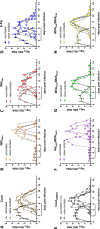
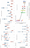
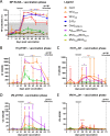
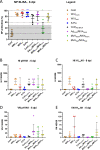
Swine influenza A virus (swIAV) infection causes substantial economic loss and disease burden in humans and animals. The 2009 pandemic H1N1 (pH1N1) influenza A virus is now endemic in both populations. In this study, we evaluated the efficacy of different vaccines in reducing nasal shedding in pigs following pH1N1 virus challenge. We also assessed transmission from immunized and challenged pigs to naive, directly in-contact pigs. Pigs were immunized with either adjuvanted, whole inactivated virus (WIV) vaccines or virus-vectored (ChAdOx1 and MVA) vaccines expressing either the homologous or heterologous influenza A virus hemagglutinin (HA) glycoprotein, as well as an influenza virus pseudotype (S-FLU) vaccine expressing heterologous HA. Only two vaccines containing homologous HA, which also induced high hemagglutination inhibitory antibody titers, significantly reduced virus shedding in challenged animals. Nevertheless, virus transmission from challenged to naive, in-contact animals occurred in all groups, although it was delayed in groups of vaccinated animals with reduced virus shedding.IMPORTANCE This study was designed to determine whether vaccination of pigs with conventional WIV or virus-vectored vaccines reduces pH1N1 swine influenza A virus shedding following challenge and can prevent transmission to naive in-contact animals. Even when viral shedding was significantly reduced following challenge, infection was transmissible to susceptible cohoused recipients. This knowledge is important to inform disease surveillance and control strategies and to determine the vaccine coverage required in a population, thereby defining disease moderation or herd protection. WIV or virus-vectored vaccines homologous to the challenge strain significantly reduced virus shedding from directly infected pigs, but vaccination did not completely prevent transmission to cohoused naive pigs.
Keywords: influenza A; pH1N1; pig; transmission; vaccine.
© Crown copyright 2021.
Figures


Similar articles
-
Age at Vaccination and Timing of Infection Do Not Alter Vaccine-Associated Enhanced Respiratory Disease in Influenza A Virus-Infected Pigs.Clin Vaccine Immunol. 2016 Jun 6;23(6):470-482. doi: 10.1128/CVI.00563-15. Print 2016 Jun. Clin Vaccine Immunol. 2016. PMID: 27030585 Free PMC article.
-
Poly I:C adjuvanted inactivated swine influenza vaccine induces heterologous protective immunity in pigs.Vaccine. 2015 Jan 15;33(4):542-8. doi: 10.1016/j.vaccine.2014.11.034. Epub 2014 Nov 28. Vaccine. 2015. PMID: 25437101 Free PMC article.
-
Protection of pigs against pandemic swine origin H1N1 influenza A virus infection by hemagglutinin- or neuraminidase-expressing attenuated pseudorabies virus recombinants.Virus Res. 2015 Mar 2;199:20-30. doi: 10.1016/j.virusres.2015.01.009. Epub 2015 Jan 17. Virus Res. 2015. PMID: 25599604
-
A neuraminidase-based inactivated influenza virus vaccine significantly reduced virus replication and pathology following homologous challenge in swine.Vaccine. 2025 Feb 6;46:126574. doi: 10.1016/j.vaccine.2024.126574. Epub 2024 Dec 7. Vaccine. 2025. PMID: 39645432
-
A prime-boost concept using a T-cell epitope-driven DNA vaccine followed by a whole virus vaccine effectively protected pigs in the pandemic H1N1 pig challenge model.Vaccine. 2019 Jul 18;37(31):4302-4309. doi: 10.1016/j.vaccine.2019.06.044. Epub 2019 Jun 24. Vaccine. 2019. PMID: 31248687
Cited by
-
Vaccines based on the replication-deficient simian adenoviral vector ChAdOx1: Standardized template with key considerations for a risk/benefit assessment.Vaccine. 2022 Aug 19;40(35):5248-5262. doi: 10.1016/j.vaccine.2022.06.008. Epub 2022 Jun 14. Vaccine. 2022. PMID: 35715352 Free PMC article.
-
Heterologous prime-boost H1N1 vaccination exacerbates disease following challenge with a mismatched H1N2 influenza virus in the swine model.Front Immunol. 2023 Oct 20;14:1253626. doi: 10.3389/fimmu.2023.1253626. eCollection 2023. Front Immunol. 2023. PMID: 37928521 Free PMC article.
-
Aerosol immunization with influenza matrix, nucleoprotein, or both prevents lung disease in pig.NPJ Vaccines. 2024 Oct 13;9(1):188. doi: 10.1038/s41541-024-00989-8. NPJ Vaccines. 2024. PMID: 39397062 Free PMC article.
-
Reassortment incompetent live attenuated and replicon influenza vaccines provide improved protection against influenza in piglets.NPJ Vaccines. 2024 Jul 13;9(1):127. doi: 10.1038/s41541-024-00916-x. NPJ Vaccines. 2024. PMID: 39003272 Free PMC article.
-
Respiratory and Intramuscular Immunization With ChAdOx2-NPM1-NA Induces Distinct Immune Responses in H1N1pdm09 Pre-Exposed Pigs.Front Immunol. 2021 Nov 3;12:763912. doi: 10.3389/fimmu.2021.763912. eCollection 2021. Front Immunol. 2021. PMID: 34804053 Free PMC article.
References
-
- Lewis NS, Russell CA, Langat P, Anderson TK, Berger K, Bielejec F, Burke DF, Dudas G, Fonville JM, Fouchier RA, Kellam P, Koel BF, Lemey P, Nguyen T, Nuansrichy B, Peiris JM, Saito T, Simon G, Skepner E, Takemae N, Webby RJ, Van Reeth K, Brookes SM, Larsen L, Watson SJ, Brown IH, Vincent AL, ESNIP3 Consortium . 2016. The global antigenic diversity of swine influenza A viruses. Elife 5:e12217. doi:10.7554/eLife.12217. - DOI - PMC - PubMed
Publication types
MeSH terms
Substances
Grants and funding
LinkOut - more resources
Full Text Sources
Other Literature Sources
Medical

