. 2021 Mar 1;95(5):e02215-20.
doi: 10.1128/JVI.02215-20. Epub 2020 Dec 2.

Regulation of VP30-Dependent Transcription by RNA Sequence and Structure in the Genomic Ebola Virus Promoter

Affiliations

Regulation of VP30-Dependent Transcription by RNA Sequence and Structure in the Genomic Ebola Virus Promoter

Simone Bach et al. J Virol. .

Erratum in

Abstract

Viral transcription and replication of Ebola virus (EBOV) is balanced by transcription factor VP30, an RNA binding protein. An RNA hairpin at the transcription start site (TSS) of the first gene (NP hairpin) in the 3'-leader promoter is thought to mediate the VP30 dependency of transcription. Here, we investigated the constraints of VP30 dependency using a series of monocistronic minigenomes with sequence, structure and length deviations from the native NP hairpin. Hairpin stabilizations decreased while destabilizations increased transcription in the absence of VP30, but in all cases, transcription activity was higher in the presence versus absence of VP30. This also pertains to a mutant that is unable to form any RNA secondary structure at the TSS, demonstrating that the activity of VP30 is not simply determined by the capacity to form a hairpin structure at the TSS. Introduction of continuous 3'-UN5 hexamer phasing between promoter elements PE1 and PE2 by a single point mutation in the NP hairpin boosted VP30-independent transcription. Moreover, this point mutation, but also hairpin stabilizations, impaired the relative increase of replication in the absence of VP30. Our results suggest that the native NP hairpin is optimized for tight regulation by VP30 while avoiding an extent of hairpin stability that impairs viral transcription, as well as for enabling the switch from transcription to replication when VP30 is not part of the polymerase complex.IMPORTANCE A detailed understanding is lacking how the Ebola virus (EBOV) protein VP30 regulates activity of the viral polymerase complex. Here, we studied how RNA sequence, length and structure at the transcription start site (TSS) in the 3'-leader promoter influence the impact of VP30 on viral polymerase activity. We found that hairpin stabilizations tighten the VP30 dependency of transcription but reduce transcription efficiency and attenuate the switch to replication in the absence of VP30. Upon hairpin destabilization, VP30-independent transcription - already weakly detectable at the native promoter - increases, but never reaches the same extent as in the presence of VP30. We conclude that the native hairpin structure involving the TSS (i) establishes an optimal balance between efficient transcription and tight regulation by VP30, (ii) is linked to hexamer phasing in the promoter, and (iii) favors the switch to replication when VP30 is absent.


Figures

FIG 1
FIG 1
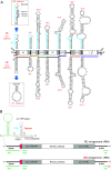
(A) Architecture of the negative-sense EBOV genome (vRNA). Potential secondary structures at the 3′-end and internal transcription start regions are depicted at the top. The transcription start sequence (TSS) of the first NP gene and gene start (GS) sequences of the six internal EBOV genes are in light blue. Corresponding hairpin structures are also predicted to form on the mRNA level (illustrated at the bottom). The previously reported NheI mutant of the NP HP (15) is illustrated above and below the native NP HP; in the genomic sequence of the NheI NP variant at the top, the mutations are indicated by asterisks and the NheI site generated on the cDNA levels is depicted next to the corresponding RNA sequence stretch (gray shaded). Schematic white boxes in the center mark the reading frames for EBOV proteins NP, VP35, VP40, GP, VP30, VP24, and L; 5′- and 3′-untranslated regions (UTRs) are depicted as light gray boxes, with dark gray areas marking the positions of the predicted secondary structures illustrated above and below the genome. The box representing the first NP hairpin structure is in red; mRNAs are illustrated as colored lines, with dots indicating the 5′-cap. p, genomic 3′-leader and antigenomic trailer promoters (26). The secondary structures are the minimum free energy (MFE) structures (predicted by RNAfold [27] using the default parameters; http://rna.tbi.univie.ac.at/cgi-bin/RNAWebSuite/RNAfold.cgi); their stabilities (ΔG values, in kilocalories per mole), depicted in red, were calculated with one single-stranded residue included on the 5′-side (genomic RNA) and on the 3′-side (genomic RNA and mRNA) of the stem base. (B) Illustration of the replication-competent (RC; top scheme) and replication-deficient (RD; bottom scheme) minigenome (MG) skeleton used in this study; the secondary structure potential at the genomic 3′ end is depicted above the top scheme, which was validated by structure probing of naked RNA in vitro (13, 15, 16). RD MGs lack the trailer promoter’s terminal 55 nt (depicted in red), thus restricting RNA synthesis to primary transcription and antigenome synthesis. PE, promoter elements (in green); the genomic replication promoter is bipartite, consisting of PE1 and PE2. Bold pink letters highlight the 3′-U residues of 3′-UN5 hexamers between PE1 and PE2 (17). The TSS and PE2 are separated by a spacer region (orange letters). Negative values are used for nucleotide numbering of the genomic negative-sense RNA, reflecting the genome’s 3′-to-5′ negative-sense orientation. The genomic 3′-terminal G is depicted in parentheses as its presence is not essential for initiation of antigenomic RNA synthesis by the EBOV RNA polymerase complex (5). Gray boxes in the schematic genome representation represent sequences antisense (as) to the 5′-UTR of the NP mRNA and antisense to the 3′-UTR of the L mRNA.
FIG 2
FIG 2
Illustration of MG variants analyzed in Fig. 3. The genomic (vRNA) 3′-leader encoding the native wt NP hairpin (HP) structure (area shaded in light blue) is shown in the center. The NP HP was either changed by mutation (variant NheI NP) or deletion (variant Δ5′ spacer) or replaced with (engineered) HP structures derived from GS regions of internal EBOV genes. For color coding, see the legend to Fig. 1B. Hexamer periodicity between PE1 and PE2 is depicted for each variant by highlighting of the pink 3′-U residues of 3′-UN5 hexamers and the nucleotide distance between positions −51 and −80, which is a multiple of six in all variants shown. Nucleotides of PE1 and PE2 are shaded in green. MG variants with HP structures derived from GS regions of internal EBOV genes (except for the VP40 HP) were adjusted to hexamer phasing by inserting (boxed nucleotides) or deleting nucleotides, either at the 5′-side of the hairpin stem or in its apical loop. ins., insertion; del., deletion. Underlined nucleotides in the NheI hairpin mark point mutations relative to the wt NP HP.
FIG 3
FIG 3
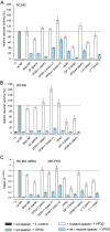
(A) Reporter gene activity of RC MG variants specified in Fig. 2 and transfected together with a plasmid expressing VP30 (gray and white bars) or omitting such a plasmid (light blue bars). The upper dotted line represents the respective activity value for the MG carrying the native 3′-leader (wt NP), which was set to 100% (gray column). −L, background control, i.e., MG transfection without the plasmid encoding the polymerase L. (B) Reporter gene activity of corresponding constructs as part of the RD MG. Values (± SEM) in panel A and B are based on at least 3 biological replicates with 2 or 3 technical replicates each. (C) qRT-PCR analysis of mRNA levels in selected samples obtained from the same kind of RC MG-transfected cells as analyzed in panel A. Mean 2−ΔΔCT values as a measure of mRNA levels relative to the wt NP MG, based on at least 3 independent experiments with a total of 2 or 3 technical replicates each. For details on the qRT-PCR setup, see Materials and Methods.
FIG 4
FIG 4
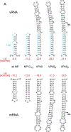
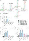
(A) RNA secondary structure potential of stabilized variants of the wt NP HP (variant NP G−72) or the VP40 HP (variants VP405 and VP406, constructed as part of a series of VP40 HP variants [19]). Predicted MFE structures (RNAfold; for more details, see the legend to Fig. 1A) and their ΔG values (in kilocalories per mole; red) are shown at the genomic (vRNA) or mRNA level. The TSS is in light blue, point mutations (relative to the parental native structure) are in pink, nucleotide insertions are in green, and deletions (del.) are shown by arrows. (B to E) Influence of RNA secondary structure stabilization at the TSS on reporter activity and viral RNA synthesis in the presence and absence of VP30. (B) Luciferase reporter gene assays of the RC MG constructs illustrated in panel A. The mean activity value for the wt NP MG was set to 100%. Mean relative reporter gene activity values ± SEM were derived from at least 3 independent experiments with 3 technical replicates each. (C to E) Corresponding qRT-PCR analyses for the quantification of mRNA, cRNA, and vRNA levels in total RNA samples derived from the MG-transfected cells that were analyzed for reporter activity in panel B. Values are based on 3 independent experiments with 3 technical replicates each. 2−ΔΔCT values were normalized to RNA levels measured for the wt NP MG (2−ΔΔCT = 1). *, P < 0.05; **, P < 0.01; ***, P < 0.001; ****, P < 0.0001 (unpaired t test with Welch’s correction). For more details, see Materials and Methods.
FIG 5
FIG 5
Effects of NP hairpin destabilization as well as continuous hexamer periodicity between PE1 and PE2 on reporter activity and viral RNA levels in the presence versus absence of VP30. (A) Predicted secondary structures on the genomic and mRNA level for the wt NP HP and variants NP G−72, NP U−75, and NP U−75/G−72. For details on color coding, see Fig. 1B and 2. ΔG values of the MFE structures predicted by RNAfold using the default parameters are indicated (for more details, see the legend to Fig. 1A). Exchanged nucleotides in the mutant structures are underlined. del., deletion. In the NP U−75 and U−75/G−72 variants, horizontal arrows additionally highlight the point mutation at position −75 (pink) and the stabilizing point mutations at −72 (black). For genomic RNA (vRNA), numbers with minus signs and for mRNA the same numbers with plus signs were used, counting from the genome 3′ end. (B and C) Luciferase reporter gene assays of RC (B) and RD (C) MG variants described for panel A and Fig. 2. Reporter activities relative to those of the wt NP MG (set to 100%) are given, based on at least 3 biological replicates (± SEM) with 2 or 3 technical replicates each. (D to F) Corresponding qRT-PCR analyses of total RNA samples derived from the cells analyzed in panel B, specific for (D) mRNA, (E) antigenomic/copy RNA (cRNA), and (F) genomic/viral RNA (vRNA). Red numbers in panels C and D represent mean n-fold activity increases relative to the wt NP MG, in the absence of VP30. Mean 2−ΔΔCT values ± SEM are derived from at least three independent experiments with two or three technical replicates each. *, P < 0.05; **, P < 0.01; ***, P < 0.001; ****, P < 0.0001 (unpaired t test with Welch’s correction). For more details, see Materials and Methods.
FIG 6
FIG 6
Effects of VP30 on MGs deviating from hexamer phasing between PE1 and PE2. (A) Predicted TSS-spacer structures of MG variants that either do [wt (NP) and VP35+1 (loop) v1] or do not [VP35, NP−1 (stem), and GP−2 (Δ2 nt PE1)] conform to hexamer phasing. In variant GP−2 (Δ2 nt PE1), genome nucleotides −54 and −55 of PE1 are deleted (17). Insertions or deletions of nucleotides are marked by boxes. ins., insertion; del., deletion. For the color code, see the legend to Fig. 1B. (B to D) Corresponding qRT-PCR analyses of total RNA obtained from cells transfected with the RC MGs illustrated in panel A, distinguishing between relative amounts of (B) viral mRNA, (C) cRNA, and (D) vRNA. Mean 2−ΔΔCT values ± SEM are derived from at least 3 independent experiments with 2 or 3 technical replicates each. **, P < 0.01; ****, P < 0.0001 (unpaired t test with Welch’s correction). The dotted lines mark the level of the −L controls. For more details, see Materials and Methods.
FIG 7
FIG 7
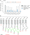
Influence of hairpin/spacer length on VP30-independent transcription. (A) Luciferase reporter gene assays of monocistronic RC MGs comprising the VP40 HP and mutant variants thereof. The VP40 hairpin was elongated in steps of 6 nt. The MG variants VP24−4, L+1 (stem), and L+1 (loop) were included as well to represent different sequence contexts. Numbers above the columns indicate the numbers of nucleotides that were inserted into the spacer relative to the wt NP MG. Numbers are shown in orange for variants with a different sequence context but the same spacer expansion of 48 nt. (B) Hairpin structures predicted to form at the mRNA 5′ ends of the MGs analyzed in panel A. ΔG values are those of the MFE structure predicted by RNAfold using the default parameters (for more details, see the legend to Fig. 1A). Nucleotides inserted into the EBOV-specific hairpin sequences are in green. Nucleotide positions in the lower hairpin stem regions (gray boxes) that deviate from the wt NP stem sequence are in red.
FIG 8
FIG 8
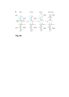
Schematic representation of primers used for qRT-PCR of viral RNA species. RT primers are in red. as, antisense. The box representing the first NP hairpin structure is pink (see also Fig. 1).

Similar articles

Cited by

References

    1. Rougeron V, Feldmann H, Grard G, Becker S, Leroy EM. 2015. Ebola and Marburg haemorrhagic fever. J Clin Virol 64:111–119. doi:10.1016/j.jcv.2015.01.014. - DOI - PMC - PubMed
    1. Burk R, Bollinger L, Johnson JC, Wada J, Radoshitzky SR, Palacios G, Bavari S, Jahrling PB, Kuhn JH. 2016. Neglected filoviruses. FEMS Microbiol Rev 40:494–519. doi:10.1093/femsre/fuw010. - DOI - PMC - PubMed
    1. Mehedi M, Falzarano D, Seebach J, Hu X, Carpenter MS, Schnittler H-J, Feldmann H. 2011. A new Ebola virus nonstructural glycoprotein expressed through RNA editing. J Virol 85:5406–5414. doi:10.1128/JVI.02190-10. - DOI - PMC - PubMed
    1. Mühlberger E. 2007. Filovirus replication and transcription. Future Virol 2:205–215. doi:10.2217/17460794.2.2.205. - DOI - PMC - PubMed
    1. Deflubé LR, Cressey TN, Hume AJ, Olejnik J, Haddock E, Feldmann F, Ebihara H, Fearns R, Mühlberger E. 2019. Ebolavirus polymerase uses an unconventional genome replication mechanism. Proc Natl Acad Sci U S A 116:8535–8543. doi:10.1073/pnas.1815745116. - DOI - PMC - PubMed

LinkOut - more resources