. 2021 Jan 27;41(4):711-725.
doi: 10.1523/JNEUROSCI.1497-20.2020. Epub 2020 Dec 2.

Cocaine-Dependent Acquisition of Locomotor Sensitization and Conditioned Place Preference Requires D1 Dopaminergic Signaling through a Cyclic AMP, NCS-Rapgef2, ERK, and Egr-1/Zif268 Pathway

Affiliations

Cocaine-Dependent Acquisition of Locomotor Sensitization and Conditioned Place Preference Requires D1 Dopaminergic Signaling through a Cyclic AMP, NCS-Rapgef2, ERK, and Egr-1/Zif268 Pathway

Sunny Zhihong Jiang et al. J Neurosci. .

Abstract

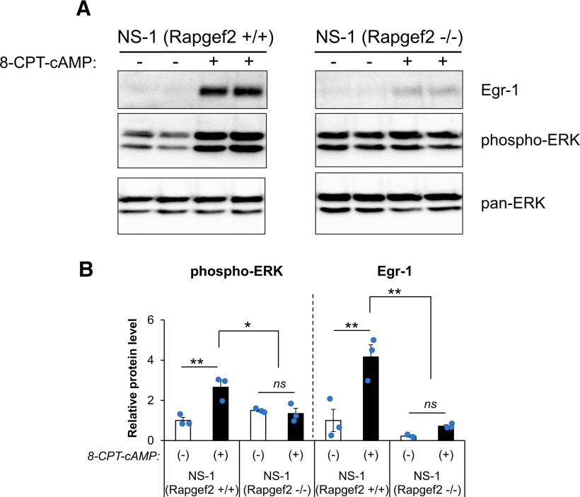
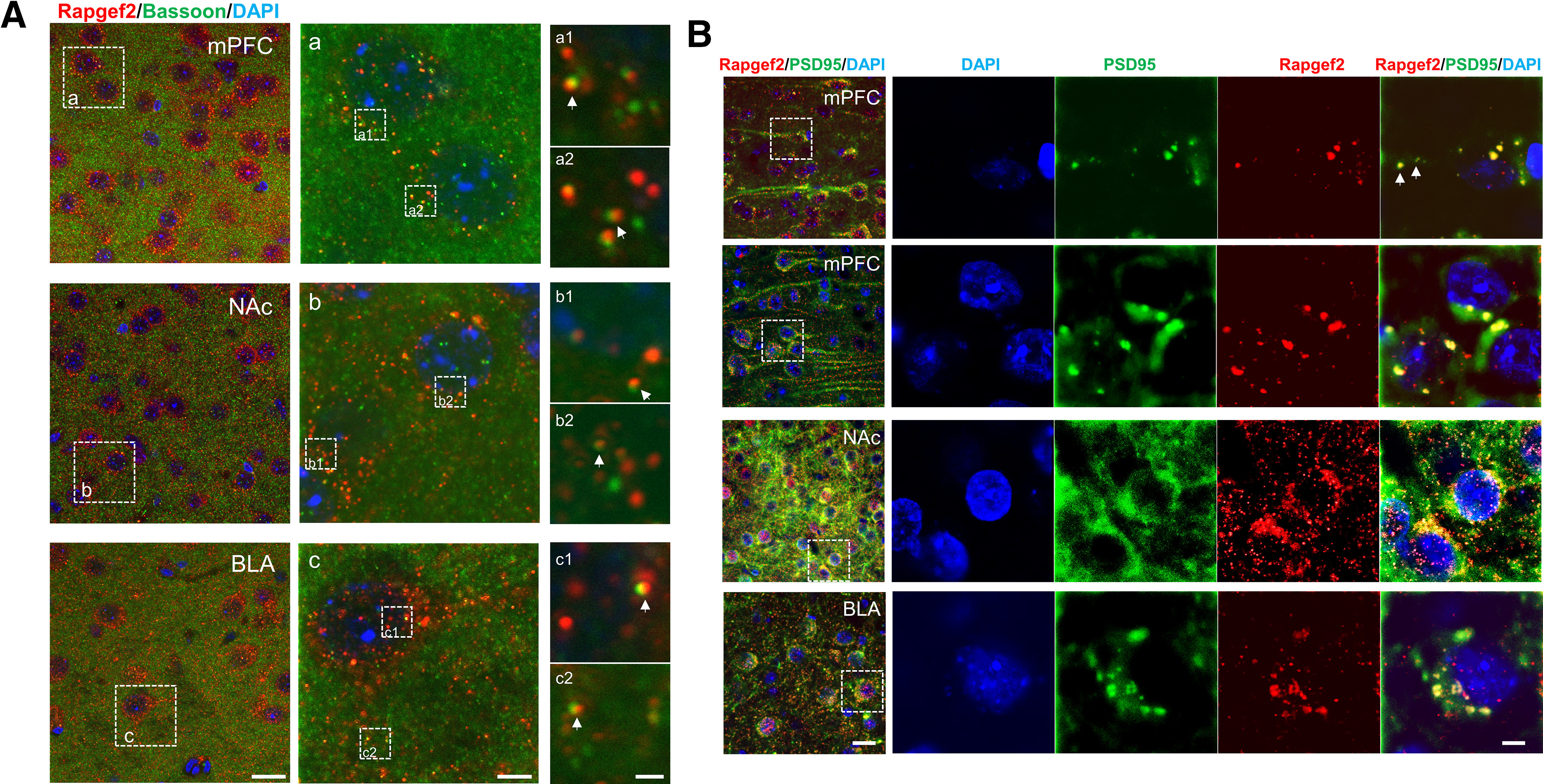
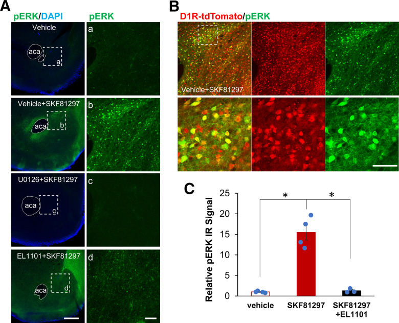
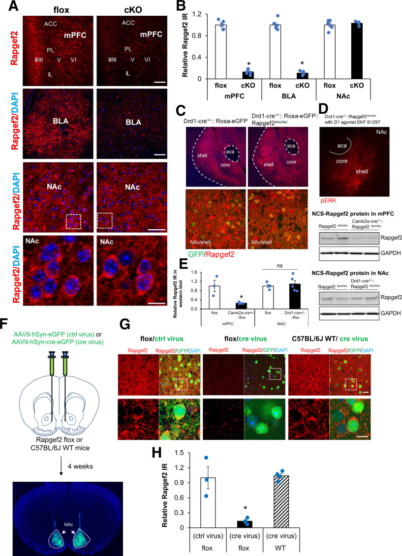
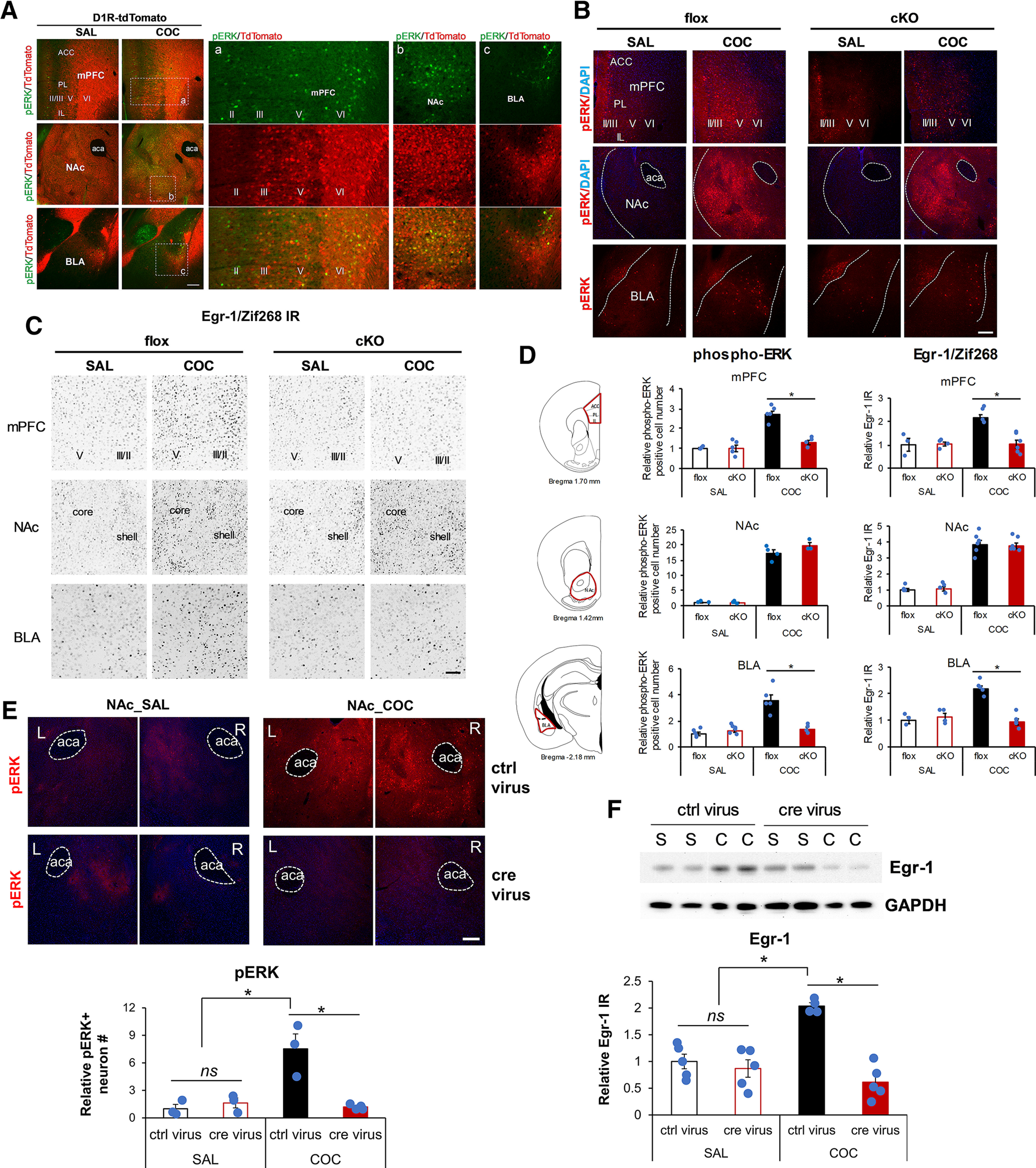
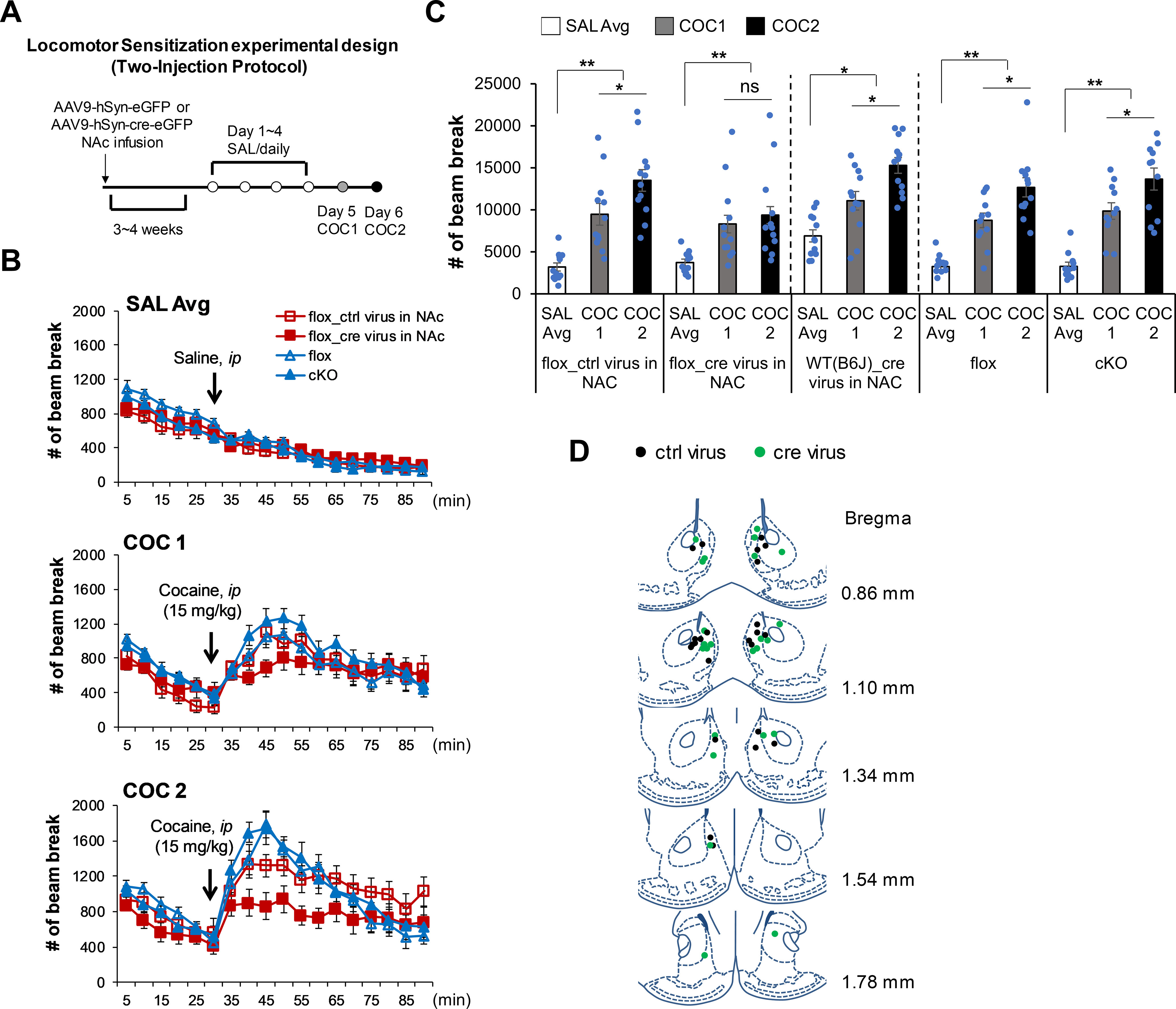
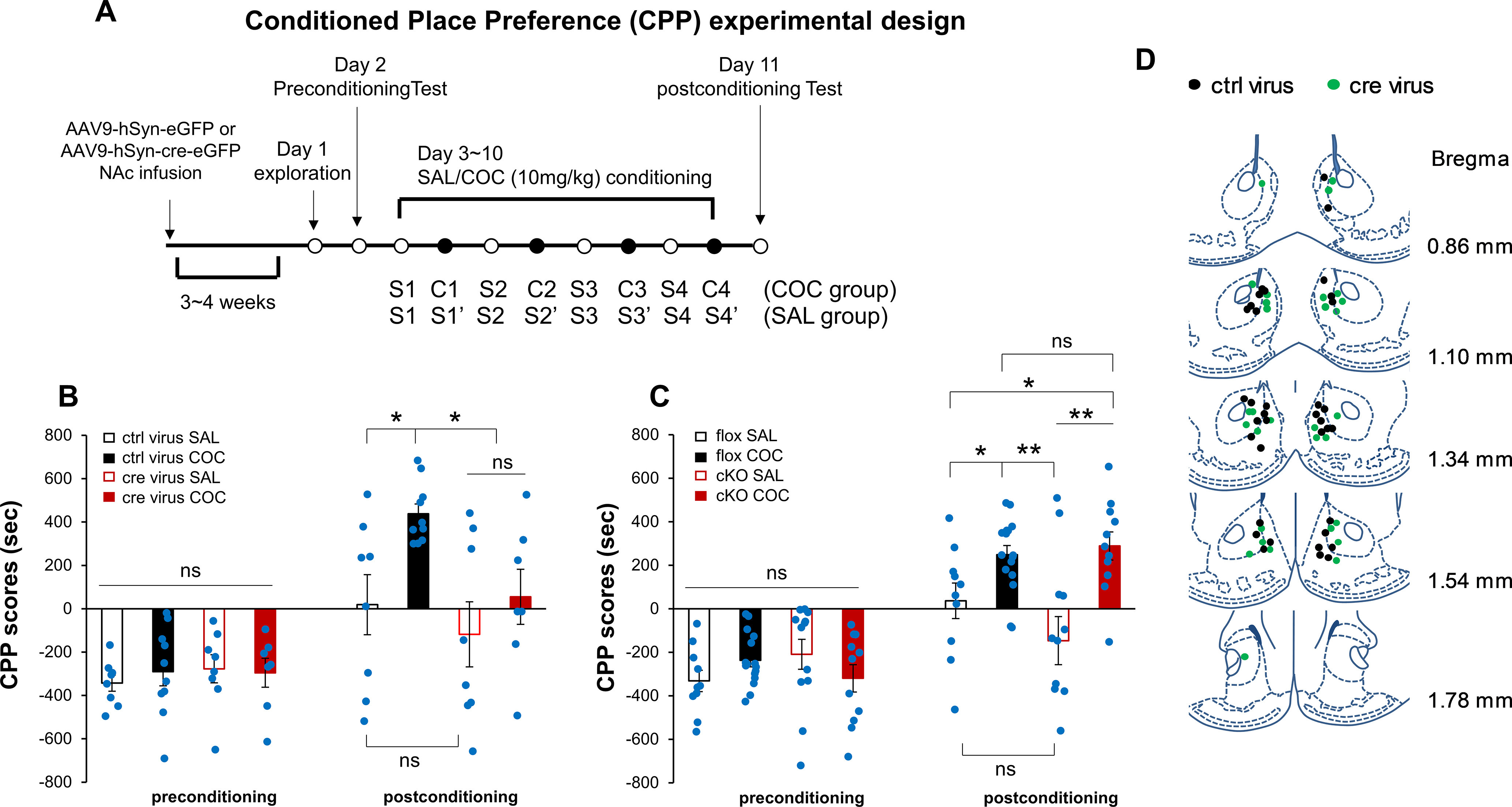
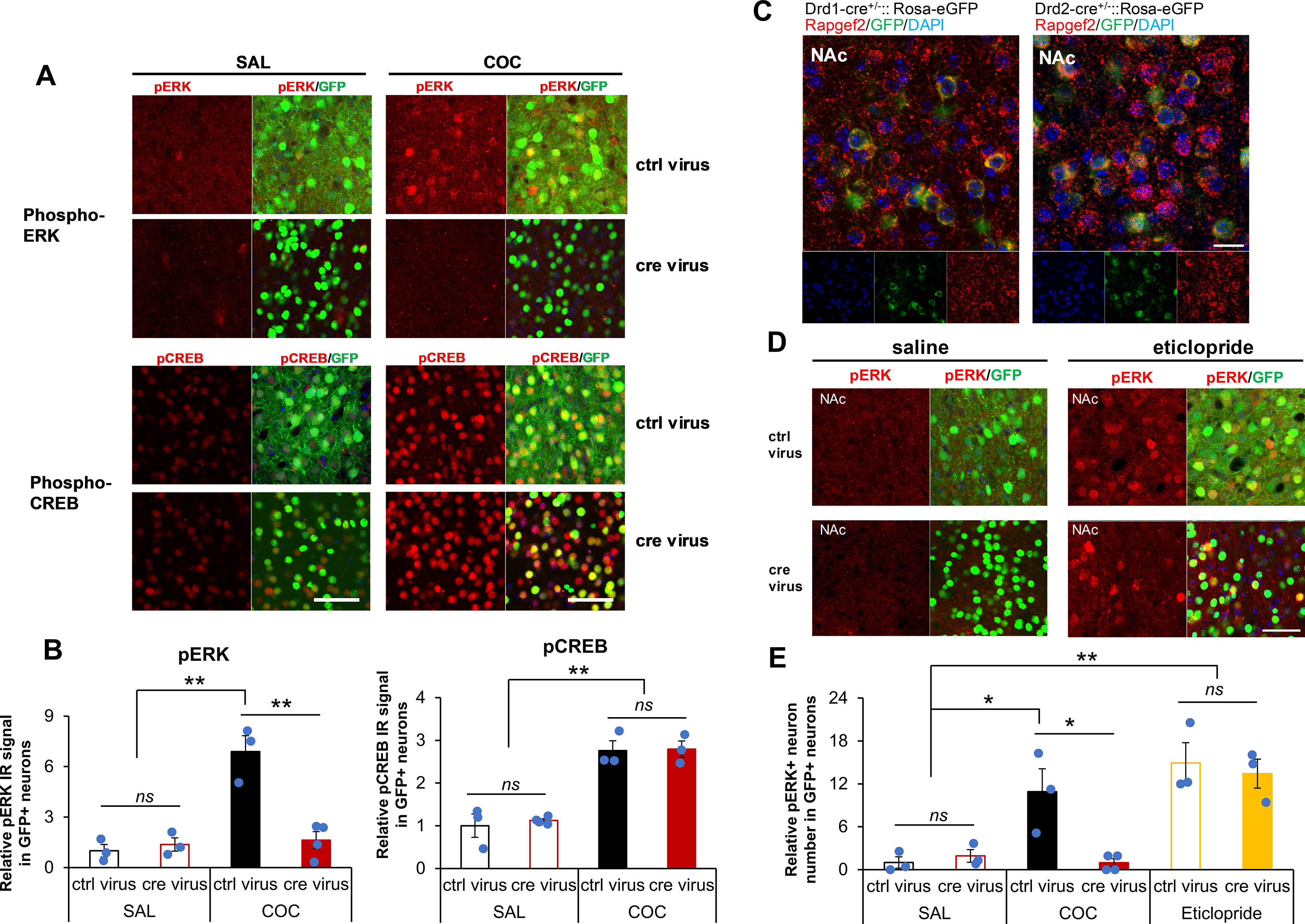
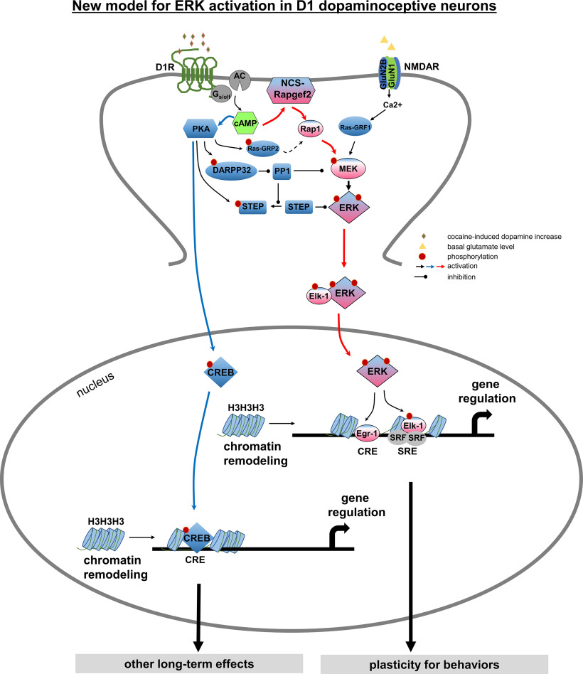
Elucidation of the mechanism of dopamine signaling to ERK that underlies plasticity in dopamine D1 receptor-expressing neurons leading to acquired cocaine preference is incomplete. NCS-Rapgef2 is a novel cAMP effector, expressed in neuronal and endocrine cells in adult mammals, that is required for D1 dopamine receptor-dependent ERK phosphorylation in mouse brain. In this report, we studied the effects of abrogating NCS-Rapgef2 expression on cAMP-dependent ERK→Egr-1/Zif268 signaling in cultured neuroendocrine cells; in D1 medium spiny neurons of NAc slices; and in either male or female mouse brain in a region-specific manner. NCS-Rapgef2 gene deletion in the NAc in adult mice, using adeno-associated virus-mediated expression of cre recombinase, eliminated cocaine-induced ERK phosphorylation and Egr-1/Zif268 upregulation in D1-medium spiny neurons and cocaine-induced behaviors, including locomotor sensitization and conditioned place preference. Abrogation of NCS-Rapgef2 gene expression in mPFC and BLA, by crossing mice bearing a floxed Rapgef2 allele with a cre mouse line driven by calcium/calmodulin-dependent kinase IIα promoter also eliminated cocaine-induced phospho-ERK activation and Egr-1/Zif268 induction, but without effect on the cocaine-induced behaviors. Our results indicate that NCS-Rapgef2 signaling to ERK in dopamine D1 receptor-expressing neurons in the NAc, but not in corticolimbic areas, contributes to cocaine-induced locomotor sensitization and conditioned place preference. Ablation of cocaine-dependent ERK activation by elimination of NCS-Rapgef2 occurred with no effect on phosphorylation of CREB in D1 dopaminoceptive neurons of NAc. This study reveals a new cAMP-dependent signaling pathway for cocaine-induced behavioral adaptations, mediated through NCS-Rapgef2/phospho-ERK activation, independently of PKA/CREB signaling.SIGNIFICANCE STATEMENT ERK phosphorylation in dopamine D1 receptor-expressing neurons exerts a pivotal role in psychostimulant-induced neuronal gene regulation and behavioral adaptation, including locomotor sensitization and drug preference in rodents. In this study, we examined the role of dopamine signaling through the D1 receptor via a novel pathway initiated through the cAMP-activated guanine nucleotide exchange factor NCS-Rapgef2 in mice. NCS-Rapgef2 in the NAc is required for activation of ERK and Egr-1/Zif268 in D1 dopaminoceptive neurons after acute cocaine administration, and subsequent enhanced locomotor response and drug seeking behavior after repeated cocaine administration. This novel component in dopamine signaling provides a potential new target for intervention in psychostimulant-shaped behaviors, and new understanding of how D1-medium spiny neurons encode the experience of psychomotor stimulant exposure.

Keywords: D1 dopaminergic signaling; Egr-1; MAPK; NCS-Rapgef2; cAMP; cocaine reward.


Figures

Figure 1.
Figure 1.
NCS-Rapgef2-dependent phospho-ERK activation and Egr-1 upregulation in neuroendocrine cell line NS-1 induced by 8-CPT-cAMP. A, NS-1 cells (NS-1(Rapgef2+/+)) or NS-1 cells with NCS-Rapgef2 KO by CRISPR (NS-1 (Rapgef2−/−)) were treated with 100 μm 8-CPT-cAMP or 0.01% DMSO (8-CPT-cAMP (–) group) for 1 h; then the cell lysate was collected for Western blot analysis of pan-ERK, phospho-ERK, and Egr-1. Equal amount of total protein (20 µg) was loaded in each lane. B, Phospho-ERK and Egr-1 protein levels were quantified with ImageJ and compared with NS1 (Rapgef2+/+)/8-CPT-cAMP (–) group (n = 3 per group). *p < 0.05; **p < 0.001; post hoc Bonferroni test following two-way ANOVA. Columns and bars represent mean ± sem and scattered points represent individual values. ns, not significant.
Figure 2.
Figure 2.
Postsynaptic localization of NCS-Rapgef2 in dopaminoceptive neurons in mPFC, NAc, and BLA. A, NCS-Rapgef2 IR signal (red) is not colocalized with Bassoon (green), a scaffolding protein of presynaptic active zone, but in close apposition to the Bassoon puncta, indicated by white arrows in a1 & a2 (mPFC), b1 & b2 (NAc), and c1 & c2 (BLA). Scale bars: Left, 20 µm; Middle, 5 µm; Right, 1 µm. B, NCS-Rapgef2 IR signal (red) is colocalized with postsynaptic marker PSD95 IR signal (green) in the soma and dendrites (indicated by white arrows) of neurons in mPFC pyramidal cells, NAc MSNs, and excitatory neurons in BLA. Right, At higher magnification, the images outlined by white boxes on the left. Scale bars: Left, 20 µm; Right, 5 µm.
Figure 3.
Figure 3.
NCS-Rapgef2 inhibitor EL1101 attenuated phospho-ERK activation in the NAc of mouse brain slice induced by D1 receptor agonist SKF81297. A, Brain slices (coronal sections in 250 µm) were incubated vehicle (DMSO final concentration 0.1%), 10 μm SKF81297, 10 μm U0126 plus 10 μm SKF81297, or 100 μm EL1101 plus 10 μm SKF81297 for 30 min. After fixation with 4% PFA, slices were stained with phospho-ERK antibody. Phospho-ERK activation in the NAc of mouse brain slice can be induced by D1 receptor agonist SKF81297, which was blocked by the MEK1 inhibitor U0126 or attenuated by NCS-Rapgef2 inhibitor EL1101. Right, At higher magnification, the images outlined by white boxes on the left. Scale bars: Left, 500 µm; Right, 100 µm. B, D1-specific activation of phospho-ERK in the NAc of D1d1a-tdTomato mouse indicated by colocalization of phospho-ERK IR signal (green) and D1 MSNs (tdTomato, red). Scale bar, 50 µm. C, Phospho-ERK IR signals in the NAc of mouse brain slices from multiple C57BL/6J animals in multiple experiments were quantified with National Institutes of Health ImageJ using the mean gray values of integrated density after being converted to grayscale. Relative phospho-ERK IR from 10 μm SKF81297-treated or 100 μm EL1101- plus 10 μm SKF81297-treated slices for each animal was normalized by phospho-ERK IR from vehicle (DMSO final concentration 0.1%)-treated slices. N = 3 or 4 animals per group. *p < 0.001 (post hoc Bonferroni test following one-way ANOVA). Columns and bars represent mean ± sem and scattered points represent individual values.
Figure 4.
Figure 4.
Region-specific ablation of NCS-Rapgef2 expression. A, Camk2α-cre+/−::Rapgef2cko/cko (cKO) was generated by crossing Rapgef2cko/cko (flox) with Camk2α-cre mice. NCS-Rapgef2 expression was largely ablated in mPFC and BLA of cKO but was unaffected in NAc of cKO. Scale bars: mPFC and BLA, 100 µm; NAc, 50 and 10 µm. B, NCS-Rapgef2 IR signals in mPFC, BLA, and NAc from cKO mice were quantified with National Institutes of Health ImageJ and compared with NCS-Rapgef2 IR signals from flox mice. N = 4-7 animals per group. *p < 0.001 (Student's t test for each region). C-E, Cre expression in D1d1a-cre mouse line is not strong enough to KO NCS-Rapgef2 in D1 MSNs in NAc. NCS-Rapgef2 expression is largely intact in the NAc (C, top panels), especially in D1-MSNs (bottom panels, green) of Drd1-cre+/−:: Rosa-eGFP:: Rapgef2cko/cko compared with Drd1-cre+/−::Rosa-eGFP, indicated by Rapgef2 immunohistochemistry (C). Phospho-ERK activation in the NAc of Drd1-cre+/−:: Rapgef2cko/cko was normally induced by D1 receptor agonist SKF81297 (2 mg/kg, i.p., 15 min) (D). Western blots using protein lysates from the NAc of Drd1-cre+/−:: Rapgef2cko/cko (Drd1-cre+/−::flox) further confirmed that NCS-Rapgef2 expression was not significantly decreased, compared with flox controls (n = 5 for each group; Student's t test). However, NCS-Rapgef2 expression in the mPFC from Camk2α-cre+/−::Rapgef2cko/cko (Camk2α-cre+/−::flox) was largely reduced (n = 3 or 4 for each group). *p < 0.05 (Student's t test) (E). F-H, NCS-Rapgef2 in NAc was abrogated by bilateral injection of AAV9-hSyn-cre-eGFP (cre virus) into the ventral striatum of Rapgef2cko/cko (flox) mice. Representative image of brain slice 4 weeks after AAV bilateral viral injection in NAc (F). NCS-Rapgef2 can be efficiently eliminated in NAc by injection of AAV9-hSyn-cre-eGFP (cre virus) into the ventral striatum of Rapgef2cko/cko (flox) mice. However, expression of NCS-Rapgef2 in the NAc was not affected by injection of AAV9-hSyn-eGFP (ctrl virus) into Rapgef2cko/cko (flox) mice or by injection of AAV9-hSyn-cre-eGFP (cre virus) into C57BL/6J WT mice. Bottom panels, At higher magnification, the images outlined by white boxes in top panels. Scale bars: Top, 20 µm; Bottom, 10 µm (G). NCS-Rapgef2 IR signals in GFP-positive neurons were quantified with ImageJ. N = 3 or 4 animals per group (H). *p < 0.05 (post hoc Bonferroni test following one-way ANOVA). Columns and bars represent mean ± sem and scattered points represent individual values. ns, not significant.
Figure 5.
Figure 5.
Expression of NCS-Rapgef2 is required for cocaine-induced phosphorylation of ERK and upregulation of Egr-1/Zif268 in mouse brain. A, A majority of cocaine-induced phospho-ERK (pERK) activation (10 min after administration of 20 mg/kg cocaine, i.p.) was in D1 receptor-positive neurons in mPFC, NAc, and BLA. In PFC, 56.7 ± 2.8% of pERK+ neurons in layer II/III, 78.1 ± 2.4% of pERK+ neurons in layer V, and 97.3 ± 0.7% pERK+ neurons in layer VI were also tdTomato-positive. In BLA, 72.2 ± 2.8% of pERK+ neurons were D1 receptor-positive; and in shell and core of NAc almost all (98.6 ± 0.1%) pERK+ neurons were D1-MSNs. Right panels (Aa,Ab,Ac) show the images at higher magnification, outlined by white boxes on the left. Scale bar, 200 µm. B, Phospho-ERK elevation observed 10 min after intraperitoneal administration of 20 mg/kg cocaine in Rapgef2cko/cko (flox) mice is attenuated in mPFC and BLA, but not in NAc, of Camk2α-cre+/−::Rapgef2cko/cko (cKO) mice. Scale bar, 200 µm. C, Egr-1/Zif268 elevation observed 1 h after 20 mg/kg cocaine treatment in Rapgef2cko/cko (flox) mice is attenuated in mPFC and BLA, but not in NAc, of Camk2α-cre+/−::Rapgef2cko/cko (cKO) mice. Scale bar, 100 µm. D, Quantification of phospho-ERK and Egr-1/Zif268 in Rapgef2cko/cko (flox) or Camk2α-cre+/−::Rapgef2cko/cko (cKO) mice after saline or cocaine treatment. Relative number of phospho-ERK-positive neurons or Egr-1 IR from mPFC, NAc, and BLA of each animal was normalized by the average value from age-matched saline-treated Rapgef2cko/cko (flox) mice. N = 3-6 animals per group. *p < 0.001 (post hoc Bonferroni test following two-way ANOVA). E, Bilateral ablation of NCS-Rapgef2 in NAc impaired phospho-ERK elevation in response to cocaine. AAV9-hSyn-eGFP (ctrl virus) or AAV9-hSyn-cre-eGFP (cre virus) was injected into the NAc of Rapgef2cko/cko mice. Four weeks later, phospho-ERK activation in both sides of NAc was examined by immunohistochemistry after acute cocaine (20 mg/kg, i.p., 10 min) injection. N = 3 or 4 animals per group. *p < 0.001 (post hoc Bonferroni test following two-way ANOVA). F, Cocaine-induced Egr-1/Zif268 increase in the NAc of Rapgef2cko/cko mice with ctrl virus was impaired in the NAc with cre virus. Four weeks after viral infusion, mice were killed 1 h after saline or cocaine (20 mg/kg) injection. The tissue from NAc was punched out from 0.5 mm coronal sections of mouse brains and protein lysate was used for Western blot with Egr-1/Zif268 antibody. Total protein loading of each sample was first normalized by GAPDH immunoreactivity (IR). Relative Egr-1/Zif268 IR was obtained by comparing to average of that from NAc of saline-treated Rapgef2cko/cko bilaterally injected with AAV9-hSyn-eGFP (ctrl virus). N = 4 or 5 animals per group. *p < 0.001 (post hoc Bonferroni test following two-way ANOVA). Columns and bars represent mean ± sem and scattered points represent individual values. ns, not significant.
Figure 6.
Figure 6.
Locomotor sensitization induced by cocaine was abolished in the mice with NCS-Rapgef2 ablation in the NAc. A, Experimental design for locomotor sensitization. Camk2α-cre+/−::Rapgef2cko/cko (cKO) mice, Rapgef2cko/cko (flox) mice with bilateral NAc AAV9-hSyn-cre-eGFP (cre virus) injection, and their corresponding controls were subjected to a two-injection protocol for cocaine locomotor sensitization. For the first 4 d, animals were habituated for 30 min in an activity chamber, then injected with saline, and allowed free movement within the chamber for 60 min. On day 5, after 30 min of habituation in the same box in the same room, animals received the first dose of cocaine. On day 6, in the same context (same box in same room), animals received a second dose of cocaine after a habituation trial. B, Animal locomotor activity in response to saline or cocaine administration. Activity was monitored in two trials each day: 30 min free running and 1 h after saline or cocaine (15 mg/kg) injection. C, Locomotor activity (total number of beam breaks) for 1 h following saline or cocaine administration. SAL Avg, Average of activity during 4 d of saline injection; COC1, activity after the first dose of cocaine (15 mg/kg) on day 5; COC2, activity after the second dose of cocaine (15 mg/kg) on day 6. N = 11 or 12 animals per group. Repeated treatments of SAL, COC1, and COC2 on each animal in each group: *p < 0.05; **p < 0.001; one-way repeated-measures ANOVA followed by post hoc Bonferroni test. Columns and bars represent mean ± sem and scattered points represent individual values. D, Schematic representation of the injection sites in the NAc for the mice (Rapgef2cko/cko (flox) with bilateral injection of AAV9-hSyn-eGFP (ctrl virus) or AAV9-hSyn-cre-eGFP (cre virus)) used for locomotor sensitization test. N = 12 animals for each group. ns, not significant.
Figure 7.
Figure 7.
Conditioned Place Preference (CPP) induced by cocaine was impaired in the mice with NCS-Rapgef2 KO in the NAc. A, Experimental design for CPP. A shuttle box with a door that can be opened to allow the animal free access to the two chambers was used for CPP. The two chambers differed on several sensory and environmental properties. Animals received 8 d/four sets of cocaine and saline-pairing: for cocaine group, pair one chamber with cocaine, pair opposite one with saline; for saline group, pair both sides with saline. Animals were tested for the place preference before drug pairing and postconditioning by recording the amount of time they spent in each chamber. B, CPP scores for mice with NCS-Rapgef2 KO in the NAc compared with the corresponding controls. N = 7-10 animals per group. *p < 0.05 (three-way ANOVA followed by post hoc all pairwise multiple comparison Bonferroni test). C, CPP scores for Camk2α-cre+/−::Rapgef2cko/cko (cKO) mice compared with the corresponding controls Rapgef2cko/cko (flox). N = 10-16 animals per group. *p < 0.05; **p < 0.001; three-way ANOVA followed by post hoc all pairwise multiple comparison Bonferroni test. Columns and bars represent mean ± sem and scattered points represent individual values. D, Schematic representation of the injection sites in the NAc for the mice used for CPP test. N = 18 animals for ctrl virus injection: n = 8 animals for saline group, n = 10 animals for cocaine group. N = 15 animals for cre virus injection: n = 8 animals for saline group, n = 7 animals for cocaine group. ns, not significant.
Figure 8.
Figure 8.
Cocaine treatment resulted in ERK phosphorylation in an NCS-Rapgef2-dependent manner, whereas CREB phosphorylation was unaffected by NCS-Rapgef2 ablation. A, AAV9-hSyn-eGFP (ctrl virus) or AAV9-hSyn-cre-eGFP (cre virus) was injected into both sides of NAc of Rapgef2cko/cko mice. After 4 weeks, saline or cocaine (20 mg/kg) was injected (i.p.), and animals were perfused 10 min later for phospho-ERK and phospho-CREB immunohistochemistry. The phospho-ERK activation was significantly increased in the NAc with ctrl virus after cocaine treatment, but not in the NAc with cre virus; phospho-CREB elevation induced by cocaine treatment was not affected by NCS-Rapgef2 ablation in the NAc. Scale bars: 50 µm. B, Phospho-ERK and phospho-CREB IR signals in AAV-targeted neurons in the NAc were quantified using Fiji ImageJ (detailed information in Methods and Materials, n = 3 or 4 animals per group). Mean gray value of phospho-ERK and phospho-CREB IR intensity in GFP-positive neurons was measured. Relative phospho-ERK IR signal of each animal was normalized by the average value from the control virus-injected group that was administered saline. **p < 0.001 (two-way ANOVA followed by post hoc Bonferroni test). C, NCS-Rapgef2 is expressed in both D1 and D2 MSNs in NAc. NCS-Rapgef2 immunohistochemistry was performed using brain sections from Drd1-cre+/−::Rosa-eGFP or Drd2-cre+/−::Rosa-eGFP mice. NCS-Rapgef2 IR signal (red) was seen in either D1 MSNs, indicated by GFP IR signal (green) in the NAc of Drd1-cre+/−::Rosa-eGFP mice, or in D2 MSNs indicated by GFP IR signal (green) in the NAc of Drd2-cre+/−::Rosa-eGFP mice. Scale bars, 20 µm. D, Phospho-ERK induction in presumptive D2-MSNs in NAc after in vivo eticlopride (eti) administration is not NCS-Rapgef2-dependent. Phospho-ERK activation in NAc was measured in NAc of Rapgef2cko/cko mice in which either AAV9-hSyn-eGFP (ctrl virus) or AAV9-hSyn-cre-eGFP (cre virus) was injected into NAc; and 4 weeks later, mice were treated with either saline or the D2R antagonist eticlopride (2 mg/kg, i.p., 15 min). Scale bars, 50 µm. E, Phospho-ERK-positive neurons with GFP-positive (pERK+ and GFP+) in NAc were quantified after saline or eticlopride treatment and compared with cocaine treatment. Relative number of phospho-ERK-positive neurons in NAc of each animal was normalized by the average value from saline-treated Rapgef2cko/cko (flox) mice with ctrl virus. N = 3 or 4 animals per group. *p < 0.05; **p <0.001; two-way ANOVA followed by post hoc Bonferroni test. Columns and bars represent mean ± sem and scattered points represent individual values. ns, not significant.
Figure 9.
Figure 9.
Proposed direct model for dopamine-dependent ERK activation in D1-dopaminoceptive neurons. Cocaine acts by increasing synaptic dopamine leading to ERK activation in NAc, required for locomotor sensitization and CPP. It has previously been proposed that D1 receptor activation affects ERK activity only indirectly, via PKA- and DARPP-32/STEP-mediated inhibition of ERK dephosphorylation (Svenningsson et al., 2004; Valjent et al., 2005), whereas direct ERK activation itself occurs in the D1 MSNs only via NMDAR-dependent glutamatergic signaling (Pascoli et al., 2011; Cahill et al., 2014) through calcium-sensitive Ras-guanine nucleotide releasing factor (Ras-GRF1) (Farnsworth et al., 1995; Fasano et al., 2009). We propose here a more parsimonious mechanism for ERK-dependent cocaine-induced dopaminergic signaling, in which cAMP elevation by dopamine in D1-MSNs results in parcellation of signaling between ERK and CREB with separate cellular consequences under the control of each pathway. An indirect modulatory role for PKA-dependent ERK phosphatase inhibition after psychomotor stimulant administration (Valjent et al., 2005; Sun et al., 2007), and additional ERK regulation by glutamatergic input to D1 dopaminoceptive neurons in the context of cellular plasticity underlying cocaine addiction (Valjent et al., 2000; Park et al., 2013; Cahill et al., 2014; Pascoli et al., 2014) is not contradicted by this model. We posit that D1 receptor activation, and cAMP elevation, in D1 MSNs likely results in parallel effects on ERK, both directly via NCS-Rapgef2 and indirectly via PKA with the effects of cocaine requiring multiple necessary, but perhaps individually insufficient inputs activated by dopamine, that converge on D1-MSN ERK phosphorylation. These include PKA/DARPP/STEP/PP1 (Svenningsson et al., 2004), PKA/RasGRP2/Rap1 (Nagai et al., 2016a,b), a NMDAR-dependent ras activation (Fasano et al., 2009; Pascoli et al., 2011), and NCS-Rapgef2/Rap1/B-Raf/MEK.

References

    1. Adams JP, Sweatt JD (2002) Molecular psychology: roles for the ERK MAP kinase cascade in memory. Annu Rev Pharmacol Toxicol 42:135–163. 10.1146/annurev.pharmtox.42.082701.145401 - DOI - PubMed
    1. Baik JH. (2013) Dopamine signaling in reward-related behaviors. Front Neural Circuits 7:152 10.3389/fncir.2013.00152 - DOI - PMC - PubMed
    1. Bertran-Gonzalez J, Bosch C, Maroteaux M, Matamales M, Herve D, Valjent E, Girault JA (2008) Opposing patterns of signaling activation in dopamine D1 and D2 receptor-expressing striatal neurons in response to cocaine and haloperidol. J Neurosci 28:5671–5685. 10.1523/JNEUROSCI.1039-08.2008 - DOI - PMC - PubMed
    1. Brami-Cherrier K, Valjent E, Herve D, Darragh J, Corvol JC, Pages C, Arthur SJ, Girault JA, Caboche J (2005) Parsing molecular and behavioral effects of cocaine in mitogen- and stress-activated protein kinase-1-deficient mice. J Neurosci 25:11444–11454. 10.1523/JNEUROSCI.1711-05.2005 - DOI - PMC - PubMed
    1. Briand LA, Blendy JA (2013) Not all stress is equal: CREB is not necessary for restraint stress reinstatement of cocaine-conditioned reward. Behav Brain Res 246:63–68. 10.1016/j.bbr.2013.02.026 - DOI - PMC - PubMed

Publication types

MeSH terms

LinkOut - more resources